大韓獸醫學會誌 (2015) 第 55 卷 第 3 號 Korean J Vet Res(2015) 55(3) : 181~184 http://dx.doi.org/10.14405/kjvr.2015.55.3.181
181
<Original Article>
Expression analysis of ciliary rootlet coiled coil protein mRNA during Xenopus development
Md. Mahfujur Rahman, In-Shik Kim, Dong-Choon Ahn, Ho-Seong Cho, Won-Il Kim, Bumseok Kim, Gee-Wook Shin, Jungkee Kwon, Md. Rashedunnabi Akanda, Byung-Yong Park*
College of Veterinary Medicine and Bio-Safety Research Institute, Chonbuk National University, Jeonju 561-756, Korea (Received: January 22, 2015; Revised: August 16, 2015; Accepted: August 18, 2015)
Abstract : Ciliary rootlet coiled coil protein (CROCC), the structural component that originates from the basal body at the proximal end of the ciliary rootlet, plays a crucial role in maintaining the cellular integrity of ciliated cells. In the current study, we cloned Xenopus CROCC and performed the expression analysis. The amino acid sequence of Xenopus laevis was related to those of Drosophila, cow, goat, horse, chicken, mouse and human. Reverse transcription polymerase chain reaction analysis revealed that CROCC mRNA encoding a coiled coil protein was present maternally, as well as throughout early development. In situ hybridization indicated that CROCC mRNA occurred in the animal pole of embryo during gastrulation and subsequently in the presumptive neuroectoderm at the end of gastrulation. At tailbud stages, CROCC mRNA expression was localized in the anterior roof plate of the developing brain, pharyngeal epithelium connected to gills, esophagus, olfactory placode, intestine and nephrostomes of the pronephric kidney. Our study suggests that CROCC may be responsible for control of the development of various ciliated organs.
Keywords : ciliary rootlet coiled coil protein, esophagus, nephrostomes, pharyngeal epithelium, Xenopus
Introduction
Cilia and flagella are highly ordered organelles in eukary- otes which are involved in generating directional movement.
Functional defects of these organelles are linked to several human diseases called ciliopathies [4]. Cilia also play an important role in generating directed fluid flow that is essen- tial for a variety of physiological and developmental processes, including cell migration and left-right asymmetry determina- tion of organ development [2]. Proper understanding of the function of ciliated cells would help to elucidate the molecu- lar basis of animal and human diseases. Xenopus embryo is a suitable model for the analysis of ciliated cell fate due to the development of ciliated cells in its surface ectoderm [8, 13].
Rootletin, the ciliary rootlet coiled coil protein (CROCC), is the structural component of ciliary rootlet that originates from the basal body at the proximal end of the cilium found in the cilia of the rootlet. CROCC has two different domains, a globular head domain and a tail domain consisting of extended coiled-coil structures. It is a ubiquitous long-sought component of the motile cilia. Knockdown of rootletin in mice ablates the formation of ciliary rootlets in ciliated cells, which is suggestive of a crucial role for the ciliary rootlet in maintaining the cellular integrity of ciliated cells [13]. In
cells with motile cilia such as epithelia lining the airways and brain ventricles, ciliary rootlets appear as a fibrous network [3].
Rootletin is related to C-Nap1, which is a structural com- ponent of the ciliary rootlet in murine photoreceptor cells [15]. Subsequently, near complete elimination of rootletin from mice was found to cause photoreceptor degeneration and impaired mucociliary clearance, which is consistent with a key function of this protein in rootlet structures [14].
Though CROCC is a component of the ciliary rootlet and there is some literature available regarding the structure and function of CROCC in other species, the expression pattern during early development remains unclear. We therefore con- ducted phylogenetic analysis of CROCC and determined the temporal-spatial expression pattern of CROCC mRNA dur- ing Xenopus early development.
Materials and Methods
Xenopus (X.) laevis husbandry
X. laevis were handled in accordance with animal welfare regulations of the Institutional Animal Care and Use Com- mittees (IACUC; CBU 2013-0010), Chonbuk National Uni- versity Laboratory Animal Centre, South Korea. X. laevis embryos were maintained according to standard protocols
*Corresponding author
Tel: +82-63-850-0961, Fax: +82-63-850-0910 E-mail: [email protected]
182
Md. Mahfujur Rahman, In-Shik Kim, Dong-Choon Ahn, Ho-Seong Cho, Won-Il Kim, Bumseok Kim, Gee-Wook Shin, Jungkee Kwon, Md. Rashedunnabi Akanda, Byung-Yong Park[10]. Embryos were staged [6] and raised in 0.1x normal amphibian medium [11]. All efforts were made to minimize the discomfort of animals used.
Reverse transcription polymerase chain reaction (RT- PCR) analysis
Total RNA was extracted from stage 1 to 45 embryos [6], digested with DNase I, and purified using an RNeasy cleanup kit (Qiagen, the Netherlands). The gene reference sequence for Xenopus CROCC (XM_002942592) was obtained from the NCBI database. Xenopus CROCC was amplified by PCR from stage 35/36 cDNA using primers (forward: 5’-ggcaggt- catactggatgct-3’and reverse: 5’-ctgcctctcgttccactctc-3’) based on the available gene bank sequence. RT-PCR was performed using a Maxim RT-PCR Premix Kit (iNtRON, Korea). In parallel, human elongation factor 1 alpha (EF1 α; forward:
5’-accctcctcttggtcgtttt-3’ and reverse: 5’-tttggttttcgctgctttct-3’) was amplified as an internal control. The PCR conditions were 94
°C for 40 sec, 60
oC for 40 sec, and 72
oC for 1 min for 35 cycles, and a final extension at 72
oC for 10 min.
Sequence comparison and phylogenetic analysis The amino acid identity was searched with BLAST ser- vices available at the National Center for Biotechnology Information (USA) website. Amino acid sequences of Droso- phila (NP_651216.2), cow (XP_005203428.1) goat (XP_005 677122.1), horse (XP_005607667.1), chicken (XP_425750.4), mouse (XP_006538865.1), and human (AA126912.1) were obtained from the GenBank database and aligned with the MegAlign program (DNASTAR) using Clustal W [12]. The aligned amino acid sequences were used to construct a phy- logenetic tree with the neighbor-joining method [9].
In situ hybridization
Anti-sense digoxigenin (DIG)-labeled probe was tran- scribed according to standard procedures [5] . CROCC was linearized with Nco1 (Promega, USA) and transcribed with SP6 (Roche, Germany). In vitro fertilized X. laevis embryos were fixed with MEMFA (MOPS, EGTA, MgSO
4, and form- aldehyde) at required stages for whole mount in situ hybrid- ization, as previously described [5]. Embryos were stored in methanol at −20
oC overnight, rehydrated through a graded series of methanol and washed in phosphate-bufferd saline (PBS). Embryos were treated with proteinase K for 10 min at room temperature before re-fixation and pre-hybridization over night at 60
oC. On addition of the probe, the embryos were incubated at 60
oC overnight. Embryos were then sub- jected to a series of saline-sodium citrate buffer washes at 60
oC. Blocking was done prior to overnight incubation with anti-DIG antibody (1 : 2000) at 4
oC. Embryos were washed 4 times for 1 h in PBT (PBS-tween 20) followed by 5 min washes in alkaline phosphatase buffer. Color reactions were developed with BM-purple substrate (Roche, Germany).
Reactions were stopped by washing in PBS, followed by re- fixation. Wild type embryos were bleached in methanol (2) :
30% hydrogen peroxide (1) to fully reveal expression pat- terns. Whole mount pictures were taken on an Olympus MVX10 microscope (Olympus Corporation, Japan).
For conducting in situ hybridization on sections, stage 29/
30, 35/36, 40 and 45 embryos were selected and dehydrated with a series of ethanol, transferred to xylene and then embedded in paraffin. Serial 12-µm sections were cut with a rotary microtome (Microm HM 325 Rotary Microtome; Thermo Scientific, USA) and hybridized with the appropriate probes.
Sections were then briefly counterstained with eosin. Images were acquired digitally using a Leica DM2500 microscope (Leica Microsystems, Germany).
Results
X. laevis CROCC
The partial Xenopus CROCC cDNA sequences (301 amino acids) encoded proteins that show varying degrees of sequence conservation with other species. In particular, the partial sequence of Xenopus CROCC amino acids shared 29%, 33%, 35%, 34%, 35%, 34% and 33% identities with Drosophila melanogaster, Bos taurus, Capra hircus, Equus caballus, Gallus gallus, Mus musculus and Homo sapiens, respectively. Figure 1 shows a phylogenetic tree of CROCC protein including the above species. In addition, we per- formed phylogenetic analysis based on sequence alignment in order to further investigate the relationship among these species. The phylogenetic tree indicated that CROCC pro- teins in different species segregate and are distinct from one another (Fig. 1).
Fig. 1. Phylogenetic tree based on amino acid alignment show- ing the relationships among species specific CROCC proteins.
The tree is based on an amino acid sequence alignment per- formed with the Clustal W method. The scale bar measures the distances between sequences. The GenBank accession numbers are as follows: Drosophila melanogaster, NP_651216.2; Bos Taurus (cow), XP_005203428.1; Capra hircus (goat), XP_
005677122.1; Equus caballus (horse), XP_005607667.1; Gal- lus gallus domesticus (chicken), XP_425750.4; Mus musculus (mouse), XP_006538865.1; Homo spiens (human), AA126912.1.
Ciliary rootlet coiled coil protein expression in Xenopus
183
Temporal expression of CROCC mRNA
The temporal expression patterns of X. laevis CROCC mRNA (Fig. 2) were analyzed by RT-PCR using total RNA from different developmental stages including the early cleavage stages [6]. CROCC RNA expression was observed in all analyzed stages, including the early cleavage stages.
The CROCC transcript was maternally synthesized and per- sisted until the stage 45, examined.
Localization of CROCC mRNA
The spatial expression patterns were analyzed using whole mount in situ hybridization and in situ hybridization on sec- tions of different stages of embryos. Xenopus CROCC mRNA was strongly enriched in the animal hemisphere at the early blastula stage (stage 15) (Fig. 3A). In situ hybridization on sections indicated specific expression in the surface ecto- derm. Initially, the CROCC mRNA was expressed diffusely in the surface ectoderm at both blastula and neurula stages (Fig. 3B and C).
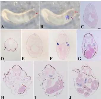
Whole mount in situ hybridization showed that in the tail- bud stage (stage 29/30), CROCC mRNA expression was unambiguously detected in the anterior part of the develop- ing brain (Fig. 4A); however, at the late tailbud stage (stage 35/36), it was expressed in the anterior part of the brain and very weakly expressed in the esophagus and nephrostomes (Fig. 4B). In situ hybridization on sections revealed more detailed expression. At stage 29/30, expression was local- ized to the roof plate of the anterior part of the neural tube (Fig. 4C). CROCC mRNA expression was detected in the olfactory region at stage 35/36 and expression in the anterior part of the developing brain also persisted at this stage (Fig.
4D and E). Interestingly, on this stage, the expression was observed in the nephrostomes of the pronephric kidney (Fig.
4F). At stage 40, along with the nephrostomes, CROCC mRNA expression was detected very weakly in the esophagus (Fig.
4G). At the tadpole stage (stage 45), CROCC mRNA was
first detected in the pharyngeal epithelium that may be con- nected to the gills (Fig. 4H) and subsequently the expression was confined to a ring-like structure of the esophagus (Fig.
4I) that was finally connected to the intestine (Fig. 4J).
Fig. 2. Temporal expression profile of CROCC during Xenopus early development. Expression was analyzed by RT-PCR of RNA extracted from embryos at the indicated developmental stages. EF1α was used as a loading control. Lane M shows a negative control (absence of RNA). In Xenopus laevis, CROCC expression was initiated from stage 1 (maternal) onward (zygotic).
Fig. 3. Developmental expression of CROCC in Xenopus by in situ hybridization. (A) CROCC mRNA was expressed in the animal hemisphere at stage 15. (B) In situ hybridization on sec- tions obtained at stage 15 and (C) at stage 20, CROCC was expressed in the surface ectoderm. Scale bar = 100µm.
Fig. 4. Expression analysis of CROCC gene at different stages of development in Xenopus. (A) Whole mount in situ hybrid- ization showed the expression of CROCC mRNA in the ante- rior brain at stage 29/30. (B) CROCC was expressed in the anterior part of the brain, surface ectoderm at neck region and nephrostomes at stage 35/36. (C) In the sections, CROCC was expressed in the roof plate of the neural tube at stage 29/30. (D) CROCC was expressed in the olfactory placode at stage 35/36.
(E) CROCC expression persisted in the roof plate of the neural tube at stage 35/36. (F) Expression was strongly detected in the nephrostomes. (G) CROCC was expressed in the nephrostomes at stage 40. (H) CROCC also expressed in pharyngeal epithelia at stage 45 that are connected to the gills. (I) Expression was confined to esophagus (stage 45) and (J) Esophageal expression continued to the intestine (stage 45). Red arrow, roof plate of the neural tube; Blue arrow, nephrostomes; Purple arrow, sur- face ectoderm at the pharyngeal region; Black arrow, olfactory placode. Scale bar = 100µm.
184
Md. Mahfujur Rahman, In-Shik Kim, Dong-Choon Ahn, Ho-Seong Cho, Won-Il Kim, Bumseok Kim, Gee-Wook Shin, Jungkee Kwon, Md. Rashedunnabi Akanda, Byung-Yong ParkDiscussion
We reported the CROCC mRNA expression pattern in developing X. laevis embryos. The primary function of the ciliary rootlet is to provide structural support for the cilium [14]. The ciliary rootlet is composed of polymerized rootle- tin bundle fibers into thick filaments [15]. In the present study, the phylogenetic relationship between the Xenopus CROCC and its known homologues from different species including drosophila, rat, chicken, goat, cow, horse and human was analyzed using neighbor joining methods. As a results, CROCC proteins were conserved among all of the species studied. According to RT-PCR results, CROCC is a maternal gene which expression begins in the early cleavage stage and continues onward (zygotic) where expression was persisted up to stage 45, the last stage examined in this study.
Based on in situ hybridization, Xenopus CROCC mRNA was expressed in the ciliated epidermis at the early cleavage stage and in the surface ectoderm at the early neurula stage.
In the tailbud stage, we found that CROCC mRNA was also expressed in the roof plate of the anterior neural tube and the olfactory organ. Mouse CROCC was localized in discrete structures consistent with dendritic knobs of the olfactory sensory neurons [7]. These findings suggest that CROCC may have a role in the development of neural tissue and sen- sory organs. In Xenopus, CROCC may also participate in the development of the pharynx and esophagus as indicated by its expression in the pharyngeal epithelium and esophagus.
Whole mount in situ hybridization revealed surface ecto- dermal expression of CROCC in the pharyngeal region that is connected to the gills. The gills are the respiratory organs of Xenopus comprising comb-like filaments, the gill lamel- lae, which increase the surface area for oxygen exchange.
Along with other cilia-related genes, mouse CROCC mRNA is also expressed in the respiratory epithelium [7]. Thus, CROCC may be responsible or play a role in the develop- ment of the respiratory organs in both vertebrates and mammals.
In the present study, CROCC mRNA was exclusively expressed in nephrostomes of Xenopus that are specific to the amphibian pronephric kidney. Nephrostomes are funnel- shaped ciliated openings of the excretory tubules into the coelom that propel water, metabolic waste, unnecessary hor- mones and other substances into the metanephridium [1]. In Xenopus, CROCC may thus have a vital role in regulation of the disposal function of the pronephric kidney by activating the cilium of the nephrostomes.
The overall conclusion of the present study is that cilia- related Xenopus CROCC is a maternally enriched gene that is expressed in the roof plate of the anterior part of the devel- oping brain, the pharyngeal epithelium connected to the gills, and is exclusively expressed in another ciliated parts includ- ing the nephrostomes, through which Xenopus disposes of waste materials. Functional analysis will, however, be neces- sary to reveal the specific location-dependent functions of CROCC.
Acknowledgments
This study was financially supported by grants from the Korea Research Foundation Grant (2011-0014454).
References
1. Aliaev IuG, Grigor’ev NA. The first experience of percutaneous nephrolithotripsy without nephrostome. Urologiia 2012, 102-104.
2. Basu B, Brueckner M. Cilia multifunctional organelles at the center of vertebrate left-right asymmetry. Curr Top Dev Biol 2008, 85, 151-174.
3. Fariss RN, Molday RS, Fisher SK, Matsumoto B.
Evidence from normal and degenerating photoreceptors that two outer segment integral membrane proteins have separate transport pathways. J Comp Neurol 1997, 387, 148-156.
4. Fliegauf M, Benzing T, Omran H. When cilia go bad:
cilia defects and ciliopathies. Nat Rev Mol Cell Biol 2007, 8, 880-893.
5. Jones CM, Smith JC. Mesoderm induction assays.
Methods Mol Biol 2008, 461, 395-404.
6. Nieuwkoop PD. Normal Table of Xenopus laevis (Daudin):
a Systematical and Chronological Survey of the Development from the Fertilized Egg Till the End of Metamorphosis.
North-Holland, Amsterdam, 1967.
7. McClintock TS, Glasser CE, Bose SC, Bergman DA.
Tissue expression patterns identify mouse cilia genes. Physiol Genomics 2008, 32, 198-206.
8. Park TJ, Mitchell BJ, Abitua PB, Kintner C, Wallingford JB. Dishevelled controls apical docking and planar polarization of basal bodies in ciliated epithelial cells. Nat Genet 2008, 40, 871-879.
9. Pearson WR, Robins G, Zhang T. Generalized neighbor- joining: more reliable phylogenetic tree reconstruction. Mol Biol Evol 1999, 16, 806-816.
10. Sive HL, Grainger RM, Harland RM. Early Development of Xenopus laevis: a Laboratory Manual. Cold Spring Harbor Laboratory Press, New York, 2000.
11. Slack JMW, Forman D. An interaction between dorsal and ventral regions of the marginal zone in early amphibian embryos. J Embryol Exp Morphol 1980, 56, 283-299.
12. Thompson JD, Higgins DG, Gibson TJ. CLUSTAL W:
improving the sensitivity of progressive multiple sequence alignment through sequence weighting, position-specific gap penalties and weight matrix choice. Nucleic Acids Res 1994, 22, 4673-4680.
13. Wessely O, Obara T. Fish and frogs: models for vertebrate cilia signaling. Front Biosci 2008, 13, 1866-1880.
14. Yang J, Gao J, Adamian M, Wen XH, Pawlyk B, Zhang L, Sanderson MJ, Zuo J, Makino CL, Li T. The ciliary rootlet maintains long-term stability of sensory cilia.
Mol Cell Biol 2005, 25, 4129-4137.
15. Yang J, Liu X, Yue G, Adamian M, Bulgakov O, Li T.
Rootletin, a novel coiled-coil protein, is a structural component of the ciliary rootlet. J Cell Biol 2002, 159, 431-440.