Fracture of a Dental Needle during Inferior Alveolar Nerve Block in a Young Child:
A Case Report
Hanbyeol Lee
1, Minkeun Kim
2, Howon Park
1, Hyunwoo Seo
1, Juhyun Lee
11
Department of Pediatric Dentistry and Oral Science Research Center, College of Dentistry, Gangneung-Wonju National University
2
Department of Oral and Maxillofacial Surgery and Oral Science Research Center, College of Dentistry, Gangneung-Wonju National University
The fracture of a needle during local anesthesia in dental treatment is rare; however, when it occurs, the nee- dle should be removed without damage to surrounding structures as soon as possible. A fractured needle frag- ment that is buried in soft tissue would be difficult to remove, and a careful surgical procedure under general anesthesia is recommended in such cases.
Children who require dental treatment are often not capable of cooperative behavior, thus unexpected move- ments can increase the risk of needle fracture. Clinicians can reduce the incidence of needle fracture accidents with a few precautions.
In the present case report, we report a case of needle fracture due to abrupt movement during inferior alveolar nerve block anesthesia in a young child, with the purpose of drawing attention to needle fracture incidents. This report describes the possible causes and prevention methods of local anesthetic needle fracture, and the localiza- tion methods and surgical procedure for needle fragment removal.
Key words : Local anesthetic needle fracture, Inferior alveolar nerve block, Child, Dingman mouth gag Abstract
Ⅰ. Introduction
Despite the reduced incidence of dental needle fracture since the development of disposable needles and the use of flexible materials for needle fabrication, needle frac- ture accidents still occur. The majority of needle frac- tures occur during administration of inferior alveolar nerve block, with the most common site for a fractured needle fragment being the pterygomandibular space
1).
A fractured needle fragment should be removed due to the potential danger for migration and detrimental ef- fects to adjacent structures
2). However, removal of a
fractured needle fragment that is not observed with the naked eye is one of the most difficult procedures, and there is a need for a correct understanding of the anatomical structure surrounding the fractured needle fragment.
Children who require dental treatment are often not capable of cooperative behavior, thus unexpected move- ments can increase the risk of needle fracture.
Therefore, a careful approach is needed when adminis- tering local anesthetics to a child, and clinicians should know the technique variations related to the smaller skull and different anatomy of a child.
Corresponding author : Juhyun Lee
Department of Pediatric Dentistry, College of Dentistry, Gangneung-Wonju National University, 7, Jukheon-gil, Gangneung, 25457, Republic of Korea Tel: +82-33-640-2452 / Fax: +82-33-640-3113 / E-mail: [email protected]
Received June 29, 2015 / Revised July 17, 2015 / Accepted July 17, 2015
Previous reports regarding needle fracture focus main- ly on localization techniques of needle fragments in adults
1,3-5). Accordingly, here, we report a case of surgical retrieval of a fractured needle fragment occurring in a young child during inferior alveolar nerve block anesthe- sia, with a view to drawing attention to needle fracture incidents. In addition, we discuss the possible causes, management, and prevention methods of needle frac- ture, as well as the precautions to be taken when per- forming local anesthesia in children.
Ⅱ. Case report
A healthy three-year, five-month-old boy with severe early childhood caries came to Department of Pediatric Dentistry of Gangneung-Wonju National University Dental Hospital in order to receive dental treatment in January 2015. His body weight was 17 kg and he was administered 1100 mg chloral hydrate and 25 mg hy- droxyzine by a dental assistant. Approximately 90 min-
utes after the drugs were administered, he fell asleep.
When a mouth gag was inserted into the mouth of the patient, he moved suddenly. Approximately 10 minutes after inhalation of nitrous oxide/oxygen, the mouth gag was inserted again and he did not seem to move. When a 21-mm 30-gauge needle was inserted in order to deliver the inferior alveolar nerve anesthesia, the patient sud- denly tilted his head in a direction opposite to that of the needle, woke up, and struggled. The needle had fractured at the hub and the needle fragment was not visible.
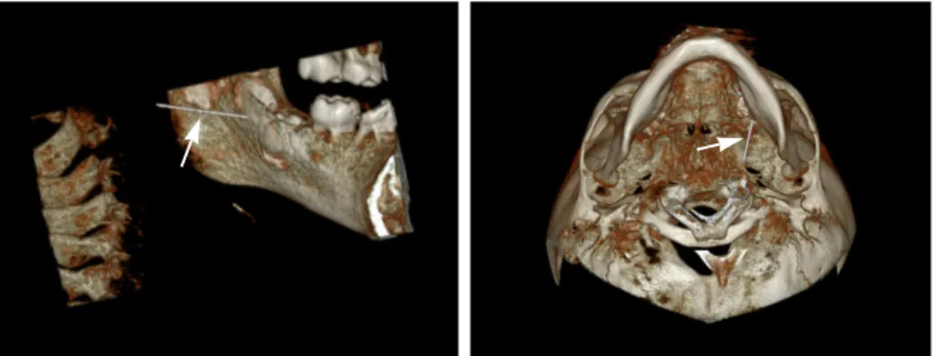
Dental panoramic and computed tomographic (CT) scans with three-dimensional reconstruction were per- formed, which confirmed the presence of the fragment in the left pterygomandibular space adjacent to the para- pharyngeal space (Fig. 1 & 2). Following discussion with the patient’ s caregiver, who was in office at the time of the incident, about possible risks, complications, and available options, the patient’ s caregiver agreed to the removal of the fractured needle fragment under gen- eral anesthesia.
Fig. 1. Panoramic radiograph and axial computed tomographic scans of a patient with a fractured needle fragment in the oral cavity (arrowhead).
Fig. 2. Computed tomographic scans with three-dimensional reconstruction illustrating the fractured needle fragment located at a position medial to the
mandible (arrowhead).
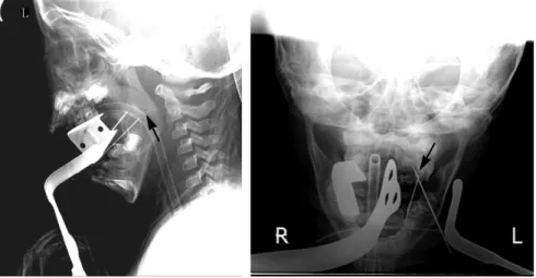
The following day, the patient was admitted to hospi- tal and underwent general anesthesia for removal of the fractured needle fragment. There is always the possibili- ty of migration of the needle fragment since the CT scan was obtained, thus, in the operating room we took a combination of lateral and anteroposterior radiographs with two reference needles inserted based on the previ- ous CT scans, in order to determine the actual location of the fractured needle fragment (Fig. 3). To prevent mi- gration of the needle, we decided to find the middle por- tion of the needle, and a dingman mouth gag was put in place for better visibility and easy surgical access (Fig.
4). Infiltration of local anesthetics (2% lidocaine hy- drochloride with 1:100,000 epinephrine) was carried out in the left ramus area in order to reduce the bleeding.
Intra-oral radiographs taken using two needles of dif- ferent thickness were used for localization of the needle fragment and for determining the incision site. One nee- dle was located in the lingual mucosa of the left mandibular second primary molar, which was used as a
fixed indicator. A second needle was inserted into the soft tissue overlying the medial border of the mandibular ramus, and the intra-oral radiograph was taken (Fig.
5). Subsequently, we made an approximate 4 cm mucos- al incision from the medial side of the left retromolar pad area vertically downwards between the 2 needles in a di- rection almost perpendicular to the needle fragment.
Careful dissection was carried out using a blunt dissec- tion scissor and electrocautery. After moving the position of the second needle, an intra-oral radiograph was taken and the dissection was continued (Fig. 5). Finally, the needle was found and removed in one piece without any complications (Fig. 6). A Penrose drain was inserted and the mucosal incisions were sutured with 4.0 vicryl. The drain was removed after 3 days.
2 weeks later, the patient had progression of caries treatment and endodontic treatment under general anesthesia, the results confirm the surgical site was well healed without infection or complications.
Fig. 3. Lateral and anteroposterior radiographs with two reference needles to determine the position of the fractured needle fragment at the time of operation (arrowhead).
Fig. 4. Dingman mouth gag allowing for better visibility and easy surgical access in surgical procedures of the oral cavity.
Ⅲ. Discussion
In adults and children, there is a difference in the posi- tion of the mandibular foramen and in the injection depth of the needle. In adults, the mandibular foramen exists above the occlusal plan, however, in children, the fora- men can be found from below the occlusal plane to the occlusal plane level
2). Previous studies have reported that the needle migth be inserted approximately 21-mm deep into the soft tissue for inferior alveolar nerve block in adults, while it might be 15-mm deep in a child
6,7). Clinicians should be sure to remember these differences when delivering the inferior alveolar nerve anesthesia.
A previous case study of 31 patients, in which needle fracture during inferior alveolar nerve anesthesia was re-
ported, a 30G needle was used in 23 patients and a 27G needle was used in 8 patients
1). The dimension and length of the needle should be carefully selected depend- ing on the situation, and insertion of the full length of the needle should be avoided. Malamed
8)reported that the abrupt movement of a patient after the needle is already inserted was the major cause of needle fracture. In addi- tion, a change in angulation of a needle that was already inserted into the tissue, would raise the risk of needle fracture. Pre-bending of the needle, particularly the hub area, may be a possible cause of needle fracture. If the needle is bent, the preset breaking point will be activat- ed. In addition, the quality of the needle may affect the needle fracture, however, there have been no studies re- garding needle quality, and further research is needed.
Fig. 5. Intra-oral radiographs for the localization of the fractured needle fragment (arrowhead).
Fig. 6. Clinical photographs and post-operative panoramic radiograph. (A) Intra-oral photograph showing the needle fragment lodged in the soft tissue. (B) The removed needle fragment. (C) Postoperative panoramic radiograph showing no residual needle fragment.
A
B
C
There are some details that need attention in order to prevent needle fracture during local anesthesia. The use of a too-thin or too-short needle should be avoided
1,9). The most common gauges of dental needles are 25, 27, and 30 gauge. The higher the gauge number, the small- er the internal diameter. As the deflection of the needle is increased, the possibility of needle fracture rises. A previous study has reported that thicker needles are less deflected compared with thinner needles, as they pene- trate the soft tissue
10). There are three lengths of dental needle, long (31-mm), short (25-mm), and ultra-short (12-mm). The length of the needle should be decided by the type of injection, size of patient, and thickness of the target tissue. The hub portion of the needle should not be inserted near the mucosa, and there should be at least a 5-mm length of needle outside the tissue
11).
In the majority of pediatric patients, the 27-gauge short needle is recommended, and in most adult pa- tients, 25-27 gauge long needle is recommended for infe- rior alveolar nerve block
12). In addition, the needle should not be bent prior to injection, and insertion into tissue should not change the angulation. The angle should be changed after taking the needle out of the tis- sue. Finally, prevention of sudden movements should be taken by explaining the possible pain to the patient, es- pecially a child, and by controlling the patient’ s head.
1,11)A fractured needle fragment should be removed as soon as possible due to the potential danger for migra- tion to adjacent structures
2). If the fractured needle is observed with the naked eye, it can be removed using a hemostat. However, if the entire length of the fractured needle fragment is embedded in the tissue, the attempt to determine the location of the needle can result in the deeper migration of the needle fragment. Moreover, the possibility of needle fragment migration can be increased by mouth movements such as swallowing and chewing.
Thus, in such cases, it is recommended that at least mouth opening be minimized before removal of the nee- dle fragment, and that removal of the needle fragment is performed under general anesthesia
13).
During removal of a foreign body from the oral cavity, the visibility of the surgical site and the identification of the exact position of the foreign body are essential. In the present case, since the fractured needle fragment was in the pterygomandibular space adjacent to the parapharyngeal space, we used a dingman mouth gag during the operation for ease of access. A dingman mouth gag is designed to retract the cheek while de-
pressing the tongue, and offers good mouth opening and anchorage for sutures. It is a useful instrument for al- lowing easy surgical access during surgical procedures of the mouth and tonsils
14).
Various methods are used to find a needle fragment inserted in soft tissue. CT scans with three-dimensional reconstruction are recommended to localize the needle fragment and the surrounding structures before opera- tion. Here, we conducted the operation after confirming that the exact location of the needle fragment was is in the pterygomandibular space adjacent to the parapha- ryngeal space, through CT scans with three-dimensional reconstruction. However, there is a possibility of migra- tion of the needle fragment since the CT scans were ob- tained, thus imaging techniques with the purpose of lo- cating the needle fragment during the operation is nec- essary. During the surgical procedure, plan film radiog- raphy, intra-oral radiography, magnetic resonance imag- ing, intraoperative fluoroscopic imaging (C-arms), and ultrasonography can be used in order to detect foreign bodies
4). In our case, we used plan film radiography (a combination of lateral and anteroposterior radiographs) and intra-oral radiography using two different thickness of needles as indicators. We could confirm the position of the fractured needle fragment correctly with these intra- operative imaging techniques.
In the present case, a 21-mm 30-gauge needle was used for injection without bending in a three-year, five- month-old boy under conscious sedation. When the nee- dle was inserted into the tissue while the clinician stabi- lized the patient’ s head by supporting the head against the clinician’ s body, there was a sudden movement of the patient. As a result, the needle was inserted up into the hub, which is the most susceptible point for break- age, and needle fracture occurred. Through this, in chil- dren, there is an increased risk of sudden movement, we keep in mind that firm stabilization is very important el- ement during local anesthesia.
If the fracture of a needle during local anesthesia oc-
curs, the management of patient or caregiver is essential
in order to prevent medical disputes. It is important to
explain sufficiently to patient or caregiver and to offer an
expression of sincere regret. In the present case, the
caregiver was in office at the time of the incident and we
were directly involved in all process including dental
treatment, radiography, and surgical removal of the nee-
dle fragment. So we could easily explain the situation
and get agreement on the process.
Ⅳ. Summary
The fracture of a needle during local anesthesia in dental treatment is rare, however when it occurs, the needle should be removed without damage to surround- ing structures as soon as possible. Nevertheless, the re- moval of a needle fragment often requires the patient to endure traumatic surgery under general anesthesia and to be exposed to further ionizing radiation. Therefore, ef- forts are required to prevent needle fracture. In children, there is an increased risk of sudden movement, thus we should pay more attention even during conscious seda- tion, and use a sufficient dimension of needle that is not pre-bent.
References
1. Augello M, von Jackowski J, Gratz KW, Jacobsen C : Needle breakage during local anesthesia in the oral cavity-a retrospective of the last 50 years with guidelines for treatment and prevention. Clin Oral Investig, 15:3-8, 2011.
2. Khalil H : A basic review on the inferior alveolar nerve block techniques. Anesth Essays Res, 8:3-8, 2014.
3. Ethunandan M, Tran AL, Brennan PA. et al. : Needle breakage following inferior alveolar nerve block: implications and management. Br Dent J, 202:395-397, 2007.
4. Holmes PJ, Miller JR, Gutta R, Louis PJ : Intraoperative imaging techniques : a guide to retrieval of foreign bodies. Oral Surg Oral Med Oral Pathol Oral Radiol Endod, 100:614-618, 2005.
5. Thompson M, Wright S, Cheng LH, Starr D :
Locating broken dental needles. Int J Oral Maxillofac Surg, 32:642-644, 2003.
6. Kronman JH, el-Bermani AW, Wongwatana S, Kumar A : Preferred needle lengths for inferior alve- olar anesthesia. Gen Dent, 42:74-76, 1994.
7. Korean academy of Pediatric Dentistry : Textbook of Pediatric Dentistry. 5th ed. Yenang INC, 326-328, 2014.
8. Malamed SF : Handbook of Local Anesthesia. 5th ed. Mosby, 275-277, 2004.
9. Pogrel MA : Broken local anesthetic needles: a case series of 16 patients, with recommendations. J Am Dent Assoc, 140:1517-1522, 2009.
10. BR Chrcanovic, DC Menezes Jr, AL Custodio : Complication of local dental anesthesia - a broken needle in the pterygomandibular space. Braz J Oral Sci, 8:159-162, 2009.
11. Ram D, Peretz B : Administering local anaesthesia to paediatric dental patients -- current status and prospects for the future. Int J Paediatr Dent, 12:80- 89, 2002.
12. Zeltser R, Cohen C, Casap N : The implications of a broken needle in the the bandit needle prior to injec- tion should be avoided.pterygomandibular space:
clinical guidelines for prevention and retrieval.
Pediatr Dent, 24:153-156, 2002.
13. Shah A, Mehta N, Von Arx DP : Fracture of a den- tal needle during administration of an inferior alveo- lar nerve block. Dent Update, 36:20-22, 25, 2009.
14. Rao LP, Peter S : Modification of the dingman
mouth gag for better visibility and access in the
management of cleft palate. Cleft Palate Craniofac J,
52:250-253, 2015.
어린 아동의 하치조신경 전달마취 시 발생한 주사바늘 파절 : 증례보고 이한별
1∙김민근
2∙박호원
1∙서현우
1∙이주현
11
강릉원주대학교 치과대학 소아치과학교실 및 구강과학연구소
2