Effect Effect Effect
Effect of of of Mandibular of Mandibular Mandibular Repositioning Mandibular Repositioning Repositioning Repositioning Device Device Device on Device on on on Airway Airway Airway Airway Size Size Size and Size and and and Airway Airway Airway Airway Collapsibility
Collapsibility Collapsibility
Collapsibility in in in Obstructive in Obstructive Obstructive Obstructive Sleep Sleep Sleep Apnea Sleep Apnea Apnea Syndrome Apnea Syndrome Syndrome: Syndrome : :Cine : Cine Cine Cine CT CT CT CT during during during during Sleep Sleep Sleep Sleep
Seung Bong Hong,1 Seung-Hyun Kyung,2 Hyun Jung Han,5 Dong Kyu Na,3 Young Ik Son4 and Young-Chel Park6
ABSTRACT
Objectives: To investigate the effect of mandibular repasitioning device on airway sige and airway collapsibility in patients with obstructive sleep apnea syndrome(OSAS).
Methods: Cine CT with polysomnographic monitoring was performed during sleep in nine(OSAS) patients before and after man- ibular repositioning device(MRD) application. Axial CT images were obtained in five upper airway levels(retropalatal-high, retro- alatal-low, retroglossal, epiglottis, and hypopharynx levels). In each airway level, one axial CT image was obtained during sleep apnea period and 10 serial axial CT images were scanned every 1 second during normal sleep breathing. After wearing MRD, all CT images were obtained by the same method. The cross-sectional areas of airway were measured by automatic tracing method.
The changes of minimum airway size and maximum airway size after MRD were evaluated. The airway collapsibility was calcula- ed before and after MRD.
Results: During sleep apnea, the airway of retropalatal-low level was the most frequently narrowest site. During normal sleep brea- hing the minimum airway size was increased significantly after MRD at retropalatal-low level(p=0.011). The mean airway collapsi- bility was the highest at retropalatal-low level. MRD decreased the airway collapsibility significantly at retropalatal-low level(p=
0.021) and epiglottis level(p=0.038).
Conclusions: The enlargement of the minimum airway size and decreased airway collapsibility may be the therapeutic mechanism of MRD in obstructive sleep apnea. Sleep Medicine and Psychophysiology 1999;;;6((((2)))):; ::110-115 :
Key words: Obstructive sleep apnea syndrome·Mandibular repositioning device·Cine CT·Airway size·Airway collapsibility.
Since early 1980s, nasal continuous positive airway pres- sure(CPAP) has been successfully used in treating obstru- ctive sleep apnea syndrome(OSAS). Although nasal CPAP was very effective to treat obstructive sleep apnea, many patients, especially younger and less severe, showed non- compliance because of nasal congestion, pressure sensation, airleak, mask intolerance, anxiety of chronic use and other
problems(1,2). One of alternatives to CPAP treatment is oral mandibular repositioning device(MRD)(3,4). Our MRD consists of upper and lower parts that attach to the ma- xillary teeth and the mandibular teeth, and advances the mandible. MRD treats OSAS in some patients and are not successful in others. In our sleep laboratory, the patient with OSAS has another overnight polysomnography with wea- ring preliminary MRD to decide whether or not MRD treats OSAS. If the patient was cured and tolerable to MRD with intolerability to CPAP, permanent MRD was made. The changes of airway opening size during respiration have fundamental importance in the pathogenesis of OSAS. Com- puter-based upper airway imaging techniques(5) provide methods to evaluate these airway dimensional changes. Dyn- amic upper airway imaging using ultrafast CT has demon- strated that the upper airways of snorers with and without OSA are more collapsible than the upper airways of non- snorers, even during awake respiration(6,7). To investigate
1성균관대학교 의과대학 삼성서울병원 신경과학교실, 2교정과학교실,
3영상의학과학교실, 4이비인후과학교실
1Departments of Neurology, 2Orthodontics, Institute of Oral Health,
3Radiology, and 4Otorhinolaryngology, Samsung Medical Center, Sungkyunkwan University School of Medicine, Seoul, Korea
5안양병원 신경과
5Department of Neurology, Anyang General Hospital, Anyang, Korea
6연세대학교 치과대학 교정과학교실
6Department of Orthodontics, College of Dentistry, Yonsei University, Seoul, Korea
Corresponding author: Seung Bong Hong, Department of Neurology, Samsung Medical Center, Sungkyunkwan University School of Medicine 50 ILWON-Dong, Kangnam-Ku, Seoul 135-710, Korea Tel: 02) 3410-3592, Fax: 02) 3410-0052
E-mail: [email protected]
how the MRD acts on upper airway opening sizes and colla- psibility in OSAS patients, we performed cine CT of upper airway with recording polysomnography during sleep.
METHODS
Fourty-six OSAS patients had two overnight polysom- nography(PSG) without and with MRD. Fifteen patients were cured with MRD(32.6%)(Fig. 1). Cure was defined as a 50% or more reduction in apnea-hypopnea index(AHI) and post-MRD AHI of 10 or less. Nine patients of 15 cured with RMD had upper airway CT scanning during sleep before and after MRD. There were 7 men and 2 women.
Their ages were from 16 to 73 years old(mean=48 years).
The mean body mass index was 24.7(20.1-29). All patients had overnight PSG in one night. Next day another over- night PSG was performed with wearing MRD which was made by orthodontist. In daytime, cine CT of upper airway was done with polysomnographic monitoring during sleep.
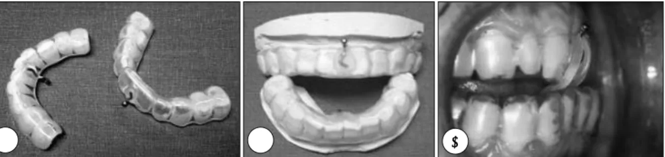
1. Mandibular repositioning device((((MRD))))(Fig. 2) All subjects had a thermoplastic MRD, which was made from compound material by co-extrusion. It consists of a
hard polycarbonate base material and a soft polyurethane material. MRD has two parts, upper and lower ones. Both of them have a metal open ring for hooking interarch elastic bands for mandibular advancement. The metal ring was attached with resin on the labial side of maxillary incisor at the level of apical middle one thirds in upper part and on the lingual side of mandibular incisor at the level of apical middle one thirds in the lower one. The elastic band was Hippo(ORMCO, extraoral elastics). In this study, the ver- tical separation of upper and lower teeth was minimized with contacting upper and lower parts of the MRD. To me- asure the amount of mandibular movement, two lateral ce- phalograms were taken at a centric occlusion state without MRD and with wearing MRD of which upper and lower parts were connected with an elastic band. To calculate the amount of mandibular movement, the incisor movement was measured from the Frankfort-Horizontal plane without and with MRD. The amount of mandibular movement was 4.6±1.7 mm(mean±SD) horizontally and 8.5±0.6 mm vertically.
2. CT scanning at five upper airway levels with poly- somnographic monitoring(Fig. 3)
Unenhanced CT scans were obtained by using a helical CT scanner(Hispeed Advantage;GE Medical Systems, Mil- waukee, Wis) with the parameters of 120 kVp and 200 mA.
Cine CT was performed at five levels from hard palate to hypopharynx parallel to the Frankfort-Horizontal plane.
Five levels of upper airway were selected on scout CT film(retropalatal-high, retropalatal-low, retroglossal, epig- lottis and hypopharynx levels). In each level, one axial CT image was obtained during sleep apnea and 10 axial images were obtained every 1 second during normal sleep breathing before MRD trial. After wearing MRD, another 10 serial axial CT images were obtained every 1 second at five airway levels during normal sleep breathing. The thickness of each CT image was 7 mm.
Fig. 1. The change of apnea-hypopnea index(AHI) before and after MRD trial in 46 OSAS patients. X means the average of AHI. P value was calculated by paired t-test.
Fig. 2. A:Upper(right) and low(left) parts of MRD, B:MRD worn on teeth molding, C:MRD is worn on patient's teeth (mandibular teeth were advanced by elastic band hooked over a metal ring of upper part of MRD).
AAA
A BBBB CC CC
For polysomnographic monitoring during CT scanning, we used a Sanei EE1121 21-channel recorder. We recorded four channels of EEG(C3-A2, C4-A1, O1-A2, O2-A1), two channels for eye movement, one channel for chin EMG, one channel for nasal airflow, and two channels for chest and abdominal movements. All patients had CT scanning during sleep apnea period and while normal sleep breathing after at least 10 seconds from the end of sleep apnea.
3. Analysis
Night PSG was interpreted for sleep staging and res- piratory scoring. AHI was obtained in PSG without MRD and PSG with MRD. The cross-sectional area of the airway was measured with an automatic tracing on GE Vantage PACS system. During sleep apnea, cross-sectional areas of upper airway CT images at five anatomical levels were measured and plotted on Fig. 4. During normal sleep brea- thing, 10 axial CT images were obtained every 1 second at five airway levels. This total 10 second cine CT included at least one full cycle of respiration. The cross-sectional areas of airways in 10 axial CT images were measured by auto- matic tracing method. The minimum and maximum sizes of airway opening were determined at each airway level before and after MRD application. The changes of airway sizes produced by MRD were tested statistically at five air- way levels.
During normal sleep breathing, airway collapsibility was calculated at five airway levels before and after MRD ap- plication by the following formula:
Airway Collapsibility=
Maximum airway size-Minimum airway size
Minimum airway size × 100%
The changes of airway collapsibility after MRD applica- tion were evaluated by Wilcoxon signed ranks test at five airway levels.
RESULTS
In nine good responders to MRD, AHI was reduced from 47.1(mean) to 6.2(mean)(p<0.05). During night PSG with MRD, all 9 patients were tolerable with MRD.
1. The level of narrowest airway size during sleep apnea(Fig. 4)
During sleep apnea five patients showed the narrowest airway size(AW1) at retropalatal-low level(55.6%), two (22.2%) at retroglossal level and two(22.2%) at epiglottis level. The locations of secondly narrowest airway size (AW2) were retropalatal-high level in three, retropalatal- low level in two, retroglossal level in two, and epiglottis
Fig. 3. Axial CT images in retropalatal-low level. Image No. 23 was scanned during obstructive sleep apnea. Image No. 24-33 were scanned every 1 second during normal sleep breathing. The cross-sectional areas of airway opening were measured by automatic tracing method in all CT images.
Fig. 4. The cross-sectional area of airway opening at five upper airway levels during obstructive sleep apnea period in 9 patients with OSAS.
level in two patients. Inter-site ratios were less than 1.0 in four patients. Inter-site ratios were calculated by(Area of AW2-Area of AW1)/Area of AW1. Inter-site ratios of nine patients were 0.68, 3.91, 0.53, 1.11, 0.99, 2.49, 94.66, 0.79 and 0.66. Inter-site ratio less than 1.0 means that the area of secondly narrowest airway size is less than two times of the narrowest airway size.
2. The changes of minimum and maximum airway sizes before and after MRD during normal sleep breathing
After MRD, the cross-sectional area of minimum airway size was increased significantly at retropalatal-low level(p=
0.011)(Table 1). The minimum airway sizes of retroglo- ssal and hypopharynx levels appeared to increase consid-
erably with MRD, but showed no statistically significant changes. The maximum airway sizes showed no significant changes after MRD application(Table 2). They rather app- eared to become smaller after MRD.
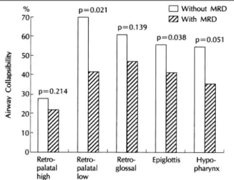
3. The changes of airway collapsibility at five airway levels by MRD(Fig. 5)
The airway collapsibility was the highest at retropalatal- low level and then retroglossal, epiglottis, hypopharynx and retropalatal-high levels in the order of decreasing collap- sibility. The airway collapsibilities of the other four airway levels were significantly higher than that of retropalatal- high level(retropalatal-low, p=0.008;retroglossal, p=0.
008;epiglottis, p=0.011;hypopharynx, p=0.015). No statistically significant difference of collapsibility was obse- rved among retropalatal-low, retroglossal, epiglottis and hy- popharynx levels. After MRD, the airway collapsibility was reduced significantly at retropalatal-low level(p=0.021) and epiglottis level(p=0.038). The airway collapsibilities of retropalatal-high, retroglossal and hypopharynx levels also decreased non-significantly after MRD application.
DISCUSSION
During obstructive sleep apnea, the narrowest anatomic airway level was retropalatal-low level in five patients, retro-glossal level in two and epiglottis level in two. In four out of nine patients, the secondly narrowest airway levels were also narrow and the changes of airway sizes during respiratory disturbance through five airway levels were not steep in half of subjects. The airway collapsibility was the
Table 1. The changes of minimal airway opening size after wearing MRD at five airway levels during normal sleep breathing Retropalatal-high Retropalatal-low Retroglossal Epiglottis Hypopharynx Mm2
MRD- MRD+ MRD- MRD+ MRD- MRD+ MRD- MRD+ MRD- MRD+
Mean 299.19 239.82 33.01 112.94 112.81 160.23 173.14 188.15 183.34 241.48 SD 297.74 161.57 24.13 118.26 79.88 114.45 97.2 106.21 132.03 108.87
p value 0.95 0.011 0.173 0.26 0.314
Mean:mean size of airway opening before and after MRD application, MRD-:without MRD, MRD+:with MRD, SD:standard deviation, p value:significance of Wilcoxon signed ranks test
Table 2. The changes of maximum airway opening size after wearing MRD at five airway levels during normal sleep breathing Retropalatal-high Retropalatal-low Retroglossal Epiglottis Hypopharynx mm2
MRD- MRD+ MRD- MRD+ MRD- MRD+ MRD- MRD+ MRD- MRD+
Mean 367.82 308.42 216.81 170.96 282.62 278.02 418.46 304.21 482.61 352.92 SD 288.30 223.08 279.39 159.18 88.02 154.55 289.57 146.37 319.28 116.19
P value 0.26 0.86 0.86 0.17 0.31
Mean:mean size of airway opening before and after MRD application, MRD-:without MRD, MRD+:with MRD, SD:standard deviation, p value:significance of Wilcoxon signed ranks test
Fig. 5. The changes of airway collapsibility at five airway levels after MRD application. p value:Wilcoxon Signed Ranks test(2-tailed).
highest at retropalatal-low level and the airway collapsi- bilities of retroglossal, epiglottis and hypopharynx were not significantly different from that of retropalatal-low level.
These results suggest that OSAS frequently involves multi- segment airway levels. Pharyngeal narrowing may contri- bute to snoring and OSA(8,9). In good responders to MRD, the narrowest airway sizes during normal sleep breathing of OSAS became larger significantly at retropalatal-low level and mildly at retroglossal and hypopharynx levels. Mandi- bular advancement by MRD appeared to enlarge a small air- way size mechanically and by stretching floppy pharyngeal muscles. The maximum airway size was not affected signi- ficantly by MRD, but appeared to decrease during normal sleep breathing. We think the reduction of maximum airway size may be secondary to tonal changes of upper airway dilator muscles produced by MRD. Langin et al.(10) per- formed upper airway CT scanning in 20 OSAS patients before and after uvulopalatophayngoplasty(UPPP). The change of minimal cross-sectional area of oropharyngeal level after UPPP was significantly correlated with the change in AHI(r=-0.54, p<0.02). The changes of smallest cross- sectional area at upper airway may be an important thera- peutic mechanism in both UPPP and MRD.
The results of our study showed that not only the upper airway size but also the upper airway collapsibility during sleep was changed by MRD in OSAS. We demonstrated the use of cine CT scanning to calculate airway collapsibility during sleep. MRD reduced significantly airway collapsi- bility at retropalatal-low and epiglottis levels. Earlier studies showed the velopharynx is the area where upper airway collapse most often begins(11-16) and velopharyngeal coll- apsibility seemed to be critical to determine the upper air- way obstruction during sleep(9). Oropharyngeal obstruction may also occur, usually promoted by increased negative inspiratory pressure after primary velopharyngeal obstru- ction. In this study, MRD appeared to prevent obstructive sleep apnea by decreasing airway collapsibility in both velo- pharyngeal and oropharyngeal levels. Tsushima et al.(17) reported the velopharynx of the good responders after laser UPPP became less collapsible than that of the poor resp- onders. Our study and their results indicate that the dec- reased airway collapsibility of the upper airway after UPPP or MRD may be an essential factor in the treatment of OSAS.
We confirmed that MRD can treat OSAS by increasing the smallest airway size and decreasing the airway colla- psibility during sleep breathing in some OSAS patients.
Night polysomnography with disposable MRD is recom- mended to assess the therapeutic effect of MRD in OSAS patients who fail to tolerate nasal CPAP or want simple and handy alternative therapeutic device.
Acknowledgement
We are grateful to Gi Bong Kim, Min Sung Kim and other EEG technicians of our EEG-sleep laboratory for recording the polysomnography and the CT technicians for perform-ing cine CT of OSAS patients asleep. We thank Woo Suk Tae for measuring the cross-sectional areas of airways.
REFERENCES
1. Meurice JC, Dore P, Paquereau J, et al. Predictive factors of long- term nasal continuous positive airway pressure treatment in sleep apnea syndrome. Chest 1994;105:429-433
2. Sanders MH, Stiller RA. Positive airway pressure in the treatment of sleep-related breathing disorders. In: Chokoverty P, ed. Sleep disor- ders medicine. Boston. MA: Butterworth-Heinemann;1994. p.455-469 3. Riley RW, Powell NB, Guilleminault C. Obstructive sleep apnea:
Trends in therapy. West J Med 1995;162:143-148
4. Schmidt-Nowara WW, Meade TE, Hays MB. Treatment of snoring and obstructive sleep apnea with a dental orthosis. Chest 1991;99:
1378-1385
5. Schwab RJ, Gefter WB, Pack AI, Hoffman EA. Dynamic imaging of the upper airway during respiration in normal subjects. J Appl Physiol 1993;74(4):1504-1514
6. Schwab RJ, Gefter WB, Hoffman EA, Gupta KB, Pack AI. Dynamic upper airway imaging during awake respiration in normal subjects and patients with sleep-disordered breathing. Am Rev Respir Dis 1993;148:1385-1400
7. Galvin JR, Rooholamini SA, Stanford W. Obstructive sleep apnea:
Diagnosis with ultrafast CT. Radiology 1989;171:775-778 8. Haponik EF, Smith PL, Bohlman ME, Allen RP, Goldman SM, Ble-
ecker ER. Computerized tomography in obstructive sleep apnea:
Correlation of airway size with physiology during sleep and wake- fulness. Am Rev Respir Dis 1983;127:221-226
9. Tsushima Y, Antila J, Svedstrom E, Vetrio A, Laurikanine E, Polo O.
Upper airway size and collapsibility in snorers: Evaluation with di- gital fluoroscopy. Eur Respir J 1996;9:1611-1618
10. Langin T, Pepin JL, Pendlebury S, et al. Upper airway changes in snorers and mild sleep apnea sufferers after uvulopalatopharyngo- plasty. Chest 1998;113(6):1595-1603
11. Shepard JW, Gefter WB, Guilleminault C, et al. Evaluation of the upper airway in patients with obstructive sleep apnea. Sleep 1991;
14(4):361-371
12. Suratt PM, Dee P, Atkinson L, Armstrong P, Wilhoit SC. Fluorosco- pic and computed tomographic features of the pharyngeal airway in obstructive sleep apnea. Am Rev Respir Dis 1983;127:487-492 13. Crumley RL, Stein M, Gamsu G, Golden J. Determination of ob-
structive site in obstructive sleep apnea. Laryngoscope 1987;97:
301-308
14. Stein MG, Gamsu G, deGeer G, Golden JA, Crumley RL, Webb WR.
Cine-CT in obstructive sleep apnea. AJR 1987;148:1069-1084 15. Hudgel DW, Hendricks C. Palate and hypopharynx: Sites of inspir-
atory narrowing of the upper airway during sleep. Am Rev Respir
Dis 1988;138:1542-1547
16. Katsantonis GP, Walsh JK. Somnofluoroscopy: Its role in the sele- ction of candidates for uvulopalatopharyngoplasty. Otolaryngol Head Neck Surg 1986;94:56-60
17. Tsushima Y, Antila J, Laurikainen E, Svedstrom E, Polo O, Kormano M. Digital fluoroscopy before and after laser uvulopalato-pharyn- goplasty in obstructive sleep apnea. Acta Radiologica 1997;38(2):
214-221
수면무호흡증 환자에서 Mandibular Repositioning Device가 Airway size와 Airway Collapsibility에 미치는 효과
홍승봉·경승현·한현정·나동규·손영익·박영철
목 적:수면무호흡증 환자들에서 수면 중 구강내 기구의 착용이 수면 중 기도의 크기와 airway collapsibility에 미치는 영향을 알아보기 위하여.
방 법:9명의 폐쇄성 수면무호흡(obstructive sleep apnea) 환자들에서 구강내 기구를 착용하기 전과 후에 수면다원검 사와 axial airway CT를 시행하였다. 상기도의 다섯 곳(retropalatal-high, retropalatal-low, retroglossal, epiglottis, hypopharynx levels)에서 axial CT를 시행하였는데, 각 위치에서 수면무호흡 상태에서 하나의 axial CT image를 얻고, 정 상적인 수면호흡 중에 1초 간격으로 10장의 axial CT images를 연속적으로 얻었다. 이와 같은 방법으로 구강내 기구를 착용하 기 전과 후에 airway CT를 각각 2회 실시하였다. 각 CT 영상에서 기도의 횡단면적(cross-sectional area)을 측정하여서 구강내 기구의 착용 전과 후에서 기도 횡단면적의 최소 크기, 최대 크기 및 airway collapsibility의 측정치를 비교하여 유의한 변화가 있는지 알아보았다.
결 과:수면무호흡 중에 가장 흔하게 좁아지는 부위는 retropalatal-low level이었다. 정상 수면호흡 중에 retropalatal- low level의 최소 기도면적이 구강내 기구에 의하여 유의하게 넓어졌다(p=0.011). 평균 airway collapsibility는 retropal- atal-low level에서 가장 높았다. 구강내 기구는 retropalatal-low level(p=0.021) 뿐만아니라 epiglottis level(p=0.038)에 서도 유의하게 airway collapsibility를 감소시켰다.
결 론:이상의 결과를 종합하여 보면 구강내 기구는 일부 환자에서 수면 중 기도의 최소면적을 넓히고, airway collap- sibility를 낮추어서 수면무호흡을 예방할 수 있음을 시사한다.
중심 단어:폐쇄성 수면무호흡증・구강내 기구・기도면적・기도 collapsibility.
초 록