• 검색 결과가 없습니다.

Application of a Split-thickness Skin Graft after the Removal of Huge Cutaneous Squamous Cell Carcinoma on the Right Lower Posterior Neck and Right Shoulder: Case Report

N/A
N/A
Protected

Academic year: 2021

Share "Application of a Split-thickness Skin Graft after the Removal of Huge Cutaneous Squamous Cell Carcinoma on the Right Lower Posterior Neck and Right Shoulder: Case Report"

Copied!
4
0
0

로드 중.... (전체 텍스트 보기)

전체 글

(1)

- 45 -

Introduction

Cutaneous squamous cell carcinoma (cSCC) is a common cutaneous malignancy that often presents as an elevated, indurated lesion with varying degrees of ulceration and crusting. cSCC accounts for 20% of all cutaneous malig- nancies and is the second most common malignancy, with

an incidence that continues to increase.

2)

The diagnosis of cSCC is primarily based on clinical features. A biopsy or excision and histologic confirmation should be performed in all clinically suspicious lesions in order to facilitate the prognostic classification and correct management of cSCC.

The first line treatment of cSCC is complete surgical ex- cision with histopathological control of excision margins.

6)

When lesions are larger than 2 cm in diameter, SCCs can be associated with a greater risk for disfigurement, local recurrence, and metastasis.

1)

They tend to be more invasive when the lesion is larger or when the cellular differentiation is worse. While squamous cell carcinoma is a common cuta- neous malignancy, few cases of squamous cell carcinoma larger than 5 cm in diameter have been reported. We report here our successful treatment of huge SCC using a

대한두경부종양학회지, 제32권 제1호, 2016. pp.45-48

Korean Journal of Head & Neck Oncology, Vol.32, No.1

http://dx.doi.org/10.21593/kjhno/2016.32.1.45 ISSN 1229-5183(Print)

Application of a Split-thickness Skin Graft after the Removal of Huge Cutaneous Squamous Cell Carcinoma on the Right

Lower Posterior Neck and Right Shoulder: Case Report

Jong Chan Kim, MD

1

, In Pyo Hong, MD, PhD

2+

Department of Plastic and Reconstructive Surgery

1

, National Medical Center, Seoul, Korea Department of Plastic and Reconstructive Surgery

2

, Eulji University Hospital, Eulji University School

of Medicine, Daejeon

목과 오른쪽 어깨 부위에 발생한 거대 편평세포암에 대해 피부이식술로 치험한 증례보고

김종찬

1

·홍인표

2+

국립중앙의료원 성형외과학교실,

1

을지대학교 의과대학 대전 을지대학교 병원 성형외과학교실

2

= Abstract =

거대한 피부편평세포암은 드문 피부 종양이다. 편평세포암은 모든 피부 악성 종양 중 20%을 차지한다.

피부편평세포암은 주로 햇빛의 노출되는 것이 주요 원인으로 알려져 있으며, 때때로 피하층을 침범을 하는

경향이 있다 . 편평세포암이 흔한 종양인 것과는 다르게, 5cm 이상의 거대 편평세포암은 보고된 바가 드물다.

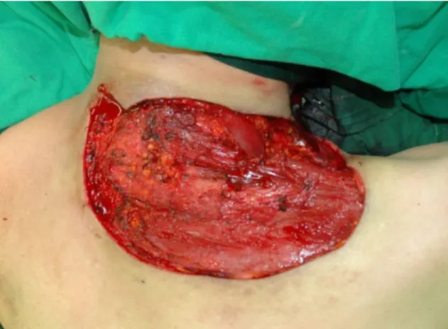
이에 저자는 경부에 발생한 14.5cm × 11.5cm × 9.5cm 크기의 거대한 피부편평세포암을 피부이식술을 통해 성공적으로 치료하여 이에 대해서 문헌 고찰과 함께 보고하는 바이다.

중심 단어:편평세포암ㆍ피부이식술ㆍ피부 종양ㆍ피부편평세포암ㆍ거대피부편평세포암.

Received Re v i se d Accepted

: May 6, 2016 : May 16, 2016 : May 24, 2016

+

Corresponding author: In Pyo Hong, Department of Plastic and Reconstructive Surgery, Eulji University Hospital, Eulji University School of Medicine, 95 Dunsanseo-ro, Seo-gu, Daejeon 302-799, Korea

Tel: (042) 611-3031 Fax: (042) 259-1111

E-mail: [email protected]

(2)

- 46 - split-thickness skin graft (STSG) in the right lower posterior neck and right shoulder.

Case report

A 58-year-old female patient was admitted to our hospital with a gradually growing mass on her right lower posterior neck and right shoulder. According to the patient, a smaller solid mass had appeared on her right lower posterior neck and right shoulder 15 years previously, but she never had analyzed or treated the mass. Upon physical examination, the mass of 14.5 cm × 11.5 cm × 9.5 cm was found accom- panied by an ulcer that bled easily (Fig. 1). The patient did not complain of pain or tenderness, and no hypertrophy of the lymph node was found in the physical examination.

The mass was biopsied and diagnosed as cSCC (Fig. 2).

The fact was revealed that the Deepest of mass had invaded the superficial layer of the right trapezius muscle with the computed tomography, but without evidence of local meta- stasis in the neck lymph nodes or distant metastasis in any other organs (Fig. 3). According to clinical stage based on American Joint Committee on Cancer (AJCC), the diameter of the cSCC was bigger than 2cm and there was the deep infiltration to skeletal muscle. It was confirmed as T3.

Additionally, because of no regional lymph node metastasis and distant metastasis, the stage of the cSCC was T3N0M0 and stage 3.

Because the cSSC could be curative by the complete ex- cision, a wide resection of the mass was performed that included the resection of at least 2 cm of the surrounding tissue horizontally and 6 mm thickness of a portion of the right trapezius muscle with the unaided eye using numerous frozen section biopsies evaluation (Fig. 4, Fig. 5). The fro- zen biopsy was a crucial method for complete excision us- ing numerous repeated frozen biopsies confirmed negative margin (Fig. 6). Biopsy of the lymph nodes and the margin of excised mass were revealed no evidence of metastasis and local invasion. Since the depth of the resected area was not deep, a STSG was used to complete the operation. The wound healed without any problems or complications (Fig.

7). She showed no sign of local recurrence or metastasis during the three years follow-up period. Although she had some secondary contracture and itching sensation in the late clinic follow-up, she had no problems with neck or shoulder

movement.

Fig. 1. A 58-year-old woman with a 14.5 cm x 11.5 cm x 9.5 cm mushroom-shaped mass on the right shoulder

Fig. 2. Histologic findings (H&E, 200×). Tumor cells transform into keratinized squames and form round nodules with con- centric, laminated layers, called "cell nests" or "epithelial/

keratinous pearls"

Fig. 3. Computed tomography indicated that the mass had

invaded the superficial portion of the right trapezius muscle

(3)

- 47 -

Discussion

cSCC has relatively high cure rate, but sometimes invades the dermis or the subcutaneous layer of the skin.

3)

Poor

prognostic factors include cSCC larger than 2 cm in diame- ter, located in the periorificial zones, or occurring in an injured area (e.g., on burn scars, chronic ulcers, or areas of radiodermatitis); mucoepidermoid, acantholytic, and des- moplastic histologic subtypes; perineural invasion; and poorly differentiated or undifferentiated tumors.

4)

Moreover, immunosuppression, incomplete resection, lymph node in- vasion, or metastasis all lead to a poor prognosis.

4)

The 5-year cure rate for huge cSCC is 70%, which is sub- stantially worse than that for smaller lesions, at 98.4%.

Moreover, cSCC greater than 4 cm in diameter is associated with a much higher mortality.

2)

Huge cSCC has been known to present in patients without a family history of skin cancer and occur from chronic le- sions located in areas that are difficult for a patient to notice.

For example, approximately 66.7% of huge SCCs are thought to occur on the back.

1)

On our case, it was similar that the patient had suffered from right lower posterior neck and right shoulder. Surgical resection with safety margins is the best treatment for most cSCC cases including those with very large lesions.

4)

When initial removal is in- complete, cSCC is more likely to recur, mostly locally or less frequently in regional lymph nodes.

6)

The more massive the cSCC, the larger the margins recommended for a safe resection.

5)

Thus, complete negative margin was very important. A 4 mm margin is sufficient to remove 95%

of clinically well-defined low risk tumors measuring less than 2 cm in diameter. For cSCC of more than 2 cm in clinical diameter, or for tumors with more than 6 mm thick- ness, or tumors with other high risk prognostic character- istics, a margin of at least 6 mm is considered necessary

Fig. 7. At two months postoperatively, some itching sensa- tion occurred in the surgical area, but no signs of any wound complications or secondary contracture appeared Fig. 4. Wide excision of the mass including the superficial

layer of the trapezius muscle

Fig. 5. Excised tumor, gross view. The mass was completely excised with ligation of the neovascularized vessels and weighed 580g.

Fig. 6. Histologic findings (H&E, 100×). cSSCs on the right up-

per side and trapezius muscle cell on the left down side

were seen

(4)

- 48 - to obtain the same result.

6)

In our case, we decided a hori- zontally 2cm margin and a vertically 6 mm margin with the unaided eye. Using numerous intraoperative frozen bi- opsies, we performed the negative margin excision.

Suitable reconstruction methods should be determined af- ter tumor resection according to the size and location of the defect.

5)

In our case, we chose the STSG as coverage for two reasons. First, the resected region was not deep but wide (18 cm × 15 cm). Due to the large size of huge cSCC, if surgical resection with safety margins is con- ducted, the defect size will be enlarged, which can make coverage very difficult. Secondly, though at that time no impression of lymph node invasion or distant metastasis was evident, huge cSCC in itself has a high level of relapse.

Therefore, a secondary operation using a flap is necessary in case of later recurrence. Taking these reasons into ac- count, we concluded that when the invasion depth is not deep, it would be more appropriate to implement STSG than to use a flap for primary reconstruction of the defect region and then observe the progress.

Adjuvant or post-operative RT should be considered in the following situations: (i) cSCC with substantial named perineural involvement, and (ii) when tissue margins are not tumor free after surgical excision and further surgery is not possible or unlikely to completely eradicate the tumor.

6)

Additionally, adjuvant RT should be also consid- ered in all patients with regional disease of the head and neck, trunk or extremities who have undergone lymph node dissection, particularly if multiple nodes are affected.

6)

In our case, there were no lymph nodes infiltration and named perineural involvement. Additionally, tissue margins were negative resulting from successful excision. Thus, we did not apply adjuvant or post-operative RT.

The longer that treatment for a cSCC is delayed, the more likely it is that the cSCC will increase to a size greater than 2 cm in diameter. Therefore, early detection and treat- ment of SCCs can have a positive impact on morbidity and mortality rates as well as costs.

1)

Nevertheless, in case of already enlarged SCCs, if the invasion depth is not deep, our case study suggests that STSG application, considering the characteristics of huge SCCs, could be a good primary surgery option.

References

1) Renzi C, Mastroeni S, Passarelli F, et al. Factors associated with large cutaneous squamous cell carcinomas [published online ahead of print July 2, 2010]. J Am Acad Dermatol.

2010;63:404-411.

2) Weinberg AS, Ogle CA, Shim EK. Metastatic cutaneous squ- amous cell carcinoma: an update. Dermatol Surg. 2007;33:

885-899.

3) Madan V, Lear JT, Szeimies RM. Non-melanoma skin cancer. Lancet. 2010;375:673-685.

4) French Society of Dermatology. Guidelines for the diagnosis and treatment of cutaneous squamous cell carcinoma and precursor lesions. arguments

May 2009 [in French]. Ann Dermatol Venereol. 2009;136(suppl 5):S189-S242.

5) Papadopoulos O, Frantzoglou M, Chrisostomidis C, et al.

Neglected squamous cell carcinoma of the frontal area: a clinical report. J Craniofac Surg. 2006;17:1015-1020.

6) Stratigos A, Garbe C, Lebbe C, Malvehy J, del Marmol V,

Pehamberger H, Peris K, Becker JC, Zalaudek I, Saiag P,

Middleton MR, Bastholt L, Testori A, Grob JJ; European

Dermatology Forum; European Association of Dermato-

Oncology; European Organization for Research and

Treatment of Cancer. Diagnosis and treatment of invasive

squamous cell carcinoma of the skin: European con-

sensus-based interdisciplinary guideline. Eur J Cancer. 2015

Sep;51(14):1989-2007.

수치

Fig. 1. A 58-year-old woman with a 14.5 cm x 11.5 cm x 9.5  cm mushroom-shaped mass on the right shoulder
Fig. 5. Excised tumor, gross view. The mass was completely  excised with ligation of the neovascularized vessels and  weighed 580g.

참조

관련 문서

Concentration-Dependent inhibition of cell viability by adenosine in human oral fibroblast and FaDu human head and neck squamous cell carcinoma · · ·

Differential Expression of Desmoglein1, Desmoglein3, Epithelial Membrane Antigen, Ber-EP4 and CD10 in Basal Cell Carcinoma and Squamous Cell Carcinoma

In 4-week group, the group filled with bone graft with decortication revealed larger new bone formation area than shown in the group that had a defect area

Objective: The purpose of this study was to analyze recent trend in incidence of basal cell carcinoma and squamous cell carcinoma in patients from the Gwangju City

The purpose of the achievement standard in the Chinese letters area was more focused on understanding and appreciation of the actual work than on the form

The index is calculated with the latest 5-year auction data of 400 selected Classic, Modern, and Contemporary Chinese painting artists from major auction houses..

1 John Owen, Justification by Faith Alone, in The Works of John Owen, ed. John Bolt, trans. Scott Clark, "Do This and Live: Christ's Active Obedience as the

Select: Skin surface (Target of intersection, white border on upper figure) Click: [GO] (in the lower right side of modeling window). Select: original and duplicated curves