Archives of Craniofacial Surgery
Copyright © 2013 The Korean Cleft Palate-Craniofacial Association
This is an Open Access article distributed under the terms of the Creative Commons Attribution Non-Commercial License (http://creativecommons.org/
licenses/by-nc/3.0/) which permits unrestricted non-commercial use, distribution, and reproduction in any medium, provided the original work is properly cited.
www.e-acfs.org pISSN 2287-1152 eISSN 2287-5603
65 Archives of Craniofacial Surgery
유방암, 위암, 대장암과 동반된 피지선암
윤민지·민경원
서울대학교 의과대학 서울대학병원 성형외과학교실
Muir-Torre syndrome is defined by concurrent or sequential development of internal malignancy and sebaceous neoplasm or multiple keratoacanthomas. Muir-Torre syndrome is very rare, with only 205 cases reported in the literature. We reported a patient with Muir-Torre syndrome with three internal malignancies. A 64-year-old patient with a history of breast cancer, stomach cancer and colon cancer visited our department for treatment of the skin lesion that occurred five years before on the left cheek. The lesion was excised completely with a resection margin of 1 cm, followed by full-thickness skin graft from left postauricular area for reconstruction. Histopathology revealed a 0.2 × 0.2 × 0.1 cm sized sebaceous carcinoma with 4 mm safety margin. The skin graft was well taken within 7 days after surgery and the patient was discharged to outpatient follow-up. There was no complication related with surgery. Muir-Torre syndrome is very rare, as are sebaceous gland tumors. So if a cancer of the sebaceous gland is diagnosed, screening workup for internal malignancy is recommended. Because of its good prognosis, surgical removal of primary or metastatic cancers may be curative and should be attempted where possible.
Keywords: Sebaceous carcinoma / Muir-Torre syndrome
Sebaceous Carcinoma Associated with Breast Cancer, Stomach Cancer, and Colon Cancer: Muir-Torre Syndrome
Min Ji Yun, Kyung Won Minn
Department of Plastic and Reconstructive Surgery, Seoul National University Hospital, Seoul National University College of Medicine, Seoul, Korea
Introduction
Muir-Torre syndrome is defined as concurrent or sequential de- velopment of an internal malignancy−most commonly colorectal cancer-and sebaceous neoplasm or multiple keratoacanthomas in the absence of other precipitating factors such as radiotherapy or AIDS [1].
It is rare for more than two internal malignancies to occur in Muir-Torre syndrome. We herein report a rare case of Muir-Torre syndrome with breast cancer, gastric cancer, colon cancer and ex- tra-ocular sebaceous carcinoma in a 64-year-old female.
Case Report
A 64-year-old patient visited our department for treatment of the skin lesion that occurred on the left cheek five years before. The patient has been treated with laser therapy to the mass three times since 2007, and the last therapy was performed in 2009. It recurred in January of 2011, when a punch biopsy was performed and seba- ceous carcinoma confirmed.
The patient presented in 1988 with breast cancer, and was treat- ed by surgical resection without adjuvant therapy. Adenocarcino- ma of the stomach was diagnosed at 1989, and radical total gastrec- tomy was performed. A right hemicolectomy was performed at 2006 with adjuvant chemotherapy, and histology confirmed a mu- cinous adenocarcinoma with no lymph node metastasis. Screen- ing for further malignancy including chest radiography, abdomen sonography, tumor marker screening (carcinoembryonic antigen)
Arch Craniofac Surg Vol.14 No.1, 65-68 http://dx.doi.org/10.7181/acfs.2013.14.1.65
Correspondence: Kyung Won Minn
Department of Plastic and Reconstructive Surgery, Seoul National University College of Medicine, 101 Daehak-ro, Jongno-gu, Seoul 110-744, Korea Tel: +82-2-2072-3642 / Fax: +82-2-2072-3680 / E-mail: minnkw@snu.ac.kr Received February 1, 2013 / Revised March 4, 2013 / Accepted March 5, 2013
Case Report
Archives of Craniofacial Surgery Vol. 14, No. 1, 2013
www.e-acfs.org
66
were all negative.
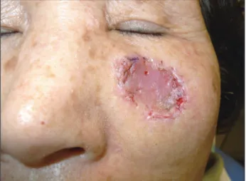
On physical examination, an asymptomatic 0.5 × 0.2 cm-sized pink-colored cyst was present on the left cheek (Fig. 1). The tumor was freely movable, firm on palpation. The patient had no symptoms of tenderness, edema, local heating, discharge, and no palpable lymph node was found on physical examination. The patient had no significant family history. The results of laboratory examination were nonspecific. Further evaluation with chest computed tomography (CT) neck CT were confirmed that there was no metastatic lesion.
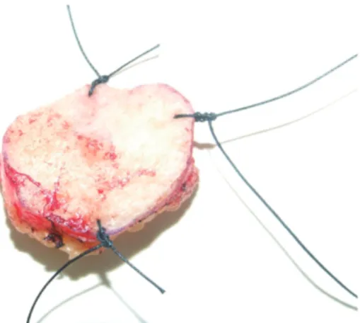
The lesion was excised completely with a resection margin of 1 cm including subcutaneous tissue (Fig. 2), followed by full-thick- ness skin graft from left postauricular area for reconstruction. Fro- zen biopsy was requested intraoperatively, and no malignant cells were found at the resection margin. Histopathology revealed a
0.2 × 0.2 × 0.1 cm sized sebaceous carcinoma with 4 mm safety mar- gin (Fig. 3). The skin graft took well within 7 days after the opera- tion and the patient was discharged to outpatient follow-up (Fig. 4).
There were no complications related with surgery of graft loss, in- fection, or hematoma.
Discussion
Muir-Torre syndrome was first described independently by Muir in 1967 and Torre in 1968, and it has since been recognized as a subtype (1% to 3%) of Lynch type II hereditary nonpolyposis colon cancer [2]. It is caused by an autosomal dominant inherited germline mutations in DNA mismatch repair genes, MSH2, MLH1, PMS2, MSH6 resulting in microsatellite instability (MSI)
Fig. 1. A 64-year-old patient with an asymptomatic 0.5×0.2 cm-sized
pink waxy cyst that occurred five years before on the left cheek. Fig. 2. The skin lesion was excised completely with a resection margin of 1 cm.
Fig. 3. (A) Section of colon shows adenocarcinoma with well differentiated and forming glandular structures (H&E, × 100). (B) Section of the skin shows well-differentiated circumscribed proliferation of sebaceous and undifferentiated cells showing marked cytologic atypia, nuclear pleomor- phism and mitotic activity (H&E, × 100).
A B
67
www.e-acfs.org
Min Ji Yun, et al. Muir-Torre syndrome and sebaceous carcinoma
[1]. Testing of sebaceous lesions for MSI is one method of evaluat- ing a patient for Muir-Torre syndrome. In our case, MSI was de- tected during an immunohistochemical study of colon cancer at 2006. Therefore, we did not perform a repeat immunohistochemi- cal study for the skin lesion. However, immunohistochemical study for a sebaceous neoplasm that precedes an internal malig- nancy is a valid tool to identify patients who are at risk for Muir- Torre syndrome.
Criteria for defining the syndrome have been proposed as con- current or sequential diagnosis of a sebaceous neoplasm (adenoma, epithelioma, seboacanthoma, or carcinoma), and a minimum of one internal malignancy or a family history of Muir-Torre syn- drome with a personal history of multiple keratoacanthoma and visceral malignancies [1]. In this case, the patient was pathologically diagnosed with sebaceous carcinoma associated with breast can- cer, gastric cancer, colon cancer, and fulfilled the criteria of Muir- Torre syndrome (Table 1) [3] according to Cohen et al. [4]. The se- baceous tumors can precede the internal malignancy by as much as 25 years and follow the diagnosis of the initial cancer by a maxi- mum of 37 years. In our case, the sebaceous carcinoma followed the diagnosis of breast cancer by 23 years.
The most common visceral malignancies in Muir-Torre syn- drome in one study were colorectal carcinoma (47%), genitouri- nary tumors (21%), breast carcinoma (12%), and hematologic disor- ders (9%) [4]. Other types of internal malignancies that have been found include those of the parotid gland, larynx, biliary, paragan- glioma, and chondrosarcoma.
Although the internal malignancies associated with Muir-Torre syndrome generally metastasize more often, median survival ap-
pears to be significantly longer than for those not associated with Muir-Torre syndrome [1]. It has been suggested that because of their relatively good prognosis, surgical removal of primary or metastatic cancers may be curative and should be attempted where possible.
The most recent review identified 205 reported cases in the world literature [5]. In Korea, five cases of Muir-Torre syndrome have been reported. Four cases were associated with only one visceral malig- nancy, and one case was associated with multiple visceral malignan- cy, B-cell lymphoma, esophageal cancer and gastric cancer [3,6-8].
Typical skin tumors associated with this syndrome include seba- ceous adenomas, epitheliomas and carcinomas, keratoacanthomas.
All of these sebaceous gland tumors are rare in the general popula- tion. Sebaceous neoplasms may occur without internal malignancy, but are rare, and Muir-Torre syndrome should be considered when they are found. So if a cancer of the sebaceous gland is diagnosed, screening workup for internal malignancy is recommended.
Sebaceous neoplasm is a rare and aggressive malignant cutane- ous tumor derived from the adnexal epithelium of sebaceous glands. Therefore, this neoplasm can originate anywhere in the body where these glands are found. Because the periocular region is rich in this type of gland, head and neck region is a common site of origin. The diagnosis of a cutaneous sebaceous neoplasm pres- ents the opportunity for early diagnosis of a visceral malignancy for the head and neck surgeon [1]. The identification of any seba- ceous neoplasm should prompt consideration for screening for Muir-Torre syndrome. Once a diagnosis of sebaceous neoplasm is considered, a detailed history and physical examination should be performed, with an emphasis on a thorough internal malignancy screening workup and genetic counseling.
Fig. 4. At postoperative 14 days, full-thickness skin graft was well taken.
Table 1. Diagnostic criteria for Muir-Torre syndrome [3]
Group A
Sebaceous adenoma Sebaceous epithelioma Sebaceous carcinoma
Keratoacanthoma with sebaceous differentiation Group B
Visceral malignancy Group C
Multiple keratoacanthomas Multiple visceral malignancies Family history of Muir-Torre syndrome
In order to achieve a diagnosis of Muir-Torre syndrome, the patient must fulfill one criterion each from groups A and B or fulfill all three criteria from group C.
Archives of Craniofacial Surgery Vol. 14, No. 1, 2013
www.e-acfs.org
68
Conflict of Interest
No potential conflict of interest relevant to this article was reported.
References
1. Eisen DB, Michael DJ. Sebaceous lesions and their associated syn- dromes: part II. J Am Acad Dermatol 2009;61:563-78.
2. Schwartz RA, Torre DP. The Muir-Torre syndrome: a 25-year retro- spect. J Am Acad Dermatol 1995;33:90-104.
3. Kim MS, Park OJ, Won CH, Chang SE, Lee MW, Choi JH, Moon KC.
A case of Muir-Torre syndrome: extra-ocular sebaceous carcinoma in a patient with breast cancer. Korean J Dermatol 2010;48:696-9.
4. Cohen PR, Kohn SR, Kurzrock R. Association of sebaceous gland tu-
mors and internal malignancy: the Muir-Torre syndrome. Am J Med 1991;90:606-13.
5. Akhtar S, Oza KK, Khan SA, Wright J. Muir-Torre syndrome: case re- p o r t o f a patient with concurrent jejunal and ureteral cancer and a review of the literature. J Am Acad Dermatol 1999;41:681-6.
6. Lee JH, Yun SJ, Kim SJ, Lee SC, Won YH, Lee JB. A case of sebaceous carcinoma on the extraocular area associated with B-cell lymphoma, esophageal cancer and gastric cancer: a case of Muir-Torre syndrome.
Korean J Dermatol 2007;45:702-5.
7. Lee JE, Kim J, Chung YL, Lee SH. A case of Muir-Torre syndrome. Ko- rean J Dermatol 2004;42:1484-7.
8. Chang DS, Seo SJ, Hong CK. Two cases of sebaceous carcinoma devel- oped on the unusual site: a case of Muir-Torre syndrome. Korean J Dermatol 2001;39:587-91.
Archives of Craniofacial Surgery Vol. 14, No. 1, 2013
www.e-acfs.org
68
No potential conflict of interest relevant to this article was reported.
1. Eisen DB, Michael DJ. Sebaceous lesions and their associated syn- dromes: part II. J Am Acad Dermatol 2009;61:563-78.
2. Schwartz RA, Torre DP. The Muir-Torre syndrome: a 25-year retro- spect. J Am Acad Dermatol 1995;33:90-104.
3. Kim MS, Park OJ, Won CH, Chang SE, Lee MW, Choi JH, Moon KC.
A case of Muir-Torre syndrome: extra-ocular sebaceous carcinoma in a patient with breast cancer. Korean J Dermatol 2010;48:696-9.
4. Cohen PR, Kohn SR, Kurzrock R. Association of sebaceous gland tu-
mors and internal malignancy: the Muir-Torre syndrome. Am J Med 1991;90:606-13.
5. Akhtar S, Oza KK, Khan SA, Wright J. Muir-Torre syndrome: case re- port of a patient with concurrent jejunal and ureteral cancer and a re- view of the literature. J Am Acad Dermatol 1999;41:681-6.
6. Lee JH, Yun SJ, Kim SJ, Lee SC, Won YH, Lee JB. A case of sebaceous carcinoma on the extraocular area associated with B-cell lymphoma, esophageal cancer and gastric cancer: a case of Muir-Torre syndrome.
Korean J Dermatol 2007;45:702-5.
7. Lee JE, Kim J, Chung YL, Lee SH. A case of Muir-Torre syndrome. Ko- rean J Dermatol 2004;42:1484-7.
8. Chang DS, Seo SJ, Hong CK. Two cases of sebaceous carcinoma devel- oped on the unusual site: a case of Muir-Torre syndrome. Korean J Dermatol 2001;39:587-91.