Background: During lumbar epidural injection (LEI) using a midline approach, we might encounter failure of identifying the epidural space owing to an equivocal or absent loss of resistance (LOR) sensation. The reason for such absence of LOR sensation has been suggested as paucity of midline ligamentum flavum, paravertebral muscle, and cyst in the interspinous ligament of the lumbar spine. Despite its low specificity, LOR is the most commonly used method to identify the epidural space.
Objectives: The purpose of this study was to analyze lumbar epidural pressure decrease patterns and identify factors contributing to this pressure decrease.
Study Design: Prospective randomized trial.
Setting: An interventional pain management practice in South Korea.
Methods: This prospective study included 104 patients receiving LEI due to lumbar radiculopathy.
A midline or paramedian approach of LEI was determined with randomization. Among various factors, gender, age, body mass index (BMI), and diagnosis were analyzed using a subgroup that included 60 cases of only a paramedian approach.
Results: Grades I, II (abrupt decrease), and III (gradual decrease) were found as patterns of epidural pressure decrease. Abrupt pressure decrease was more frequently observed in the paramedian group (P < 0.001). Age, gender, BMI, and diagnosis did not show any significant difference in frequencies between abrupt and gradual pressure decrease.
Limitations: We could not match LOR sensation with epidural pressure decrease shown in the monitor.
Conclusions: This study demonstrates that abrupt pressure decrease occurs more frequently with the paramedian approach. However, age, gender, BMI, or diagnosis did not affect the incidence of epidural pressure decrease.
Key words: Epidural, paramedian, midline, pressure decrease Pain Physician 2020: 23:E203-E210
Randomized Trial
Comparison of Epidural Pressure Decrease
Pattern According to Different Lumbar Epidural Approaches
From: 1Department of Anesthesiology and Pain Medicine, Keimyung University School of Medicine, Republic of Korea; 2Department of Psychiarty, Keimyung University School of Medicine, Republic of Korea
Address Correspondence:
Jihee Hong, MD, PhD Department of Anesthesiology and Pain Medicine, Keimyung University School of Medicine 1095 Dalgubeol-daero, Dalseo-gu, Daegu 42601 Republic of Korea E-mail: [email protected]
Disclaimer: This work was supported by the National Research Foundation of Korea (NRF) Grant funded by the Korea Government (MSIP) No.2014R1A5A2010008).
Conflict of interest: Each author certifies that he or she, or a member of his or her immediate family, has no commercial association (i.e., consultancies, stock ownership, equity interest, patent/licensing arrangements, etc.) that might pose a conflict of interest in connection with the submitted manuscript.
Manuscript received: 06-04-2019 Revised manuscript received:
07-17-2019 Accepted for publication:
08-20-2019 Free full manuscript:
www.painphysicianjournal.com
Jiseob Kim, MD1, Sungwon Jung, MD, PhD2, Eunyoung Cho, MD1, and JiHee Hong, MD, PhD1
L
Lumbar epidural injection (LEI) is frequently used for the treatment of lumbar radiculopathy (1-3). Loss of resistance (LOR) technique is the most commonly used method to identify the lumbar epidural space. If the needle is placed within the dense
ligamentum flavum, a vital structure to create a highly sustained intraneedle pressure, one can feel LOR during entry to the epidural space owing to an abrupt decrease of pressure. However, gaps in ligamentum flavum, paravertebral muscle, cyst or cavity formation due to
efits and risks before they provided informed consent.
This trial was registered prior to patient enrollment at clinicaltrials.gov (NCT03245294, date of registration:
August 3, 2017).
A total of 107 patients receiving LEI from August 2017 to April 2018 were enrolled. Inclusion criteria were patients with lumbar radicular pain and back pain due to spinal stenosis, herniated nucleus pulposus, and internal disc disruption. Patient age was between 20 and 80 years. Patients showed pain intensity (Numeric Rating Scale) and disability (Oswestry Disability Index) levels of more than 5 and more than 20, respectively.
They all failed to improve with conservative treatment.
Patients with contraindication to epidural anesthe- sia, including allergy to contrast media, coagulopathy, and infection at the needle insertion site were excluded.
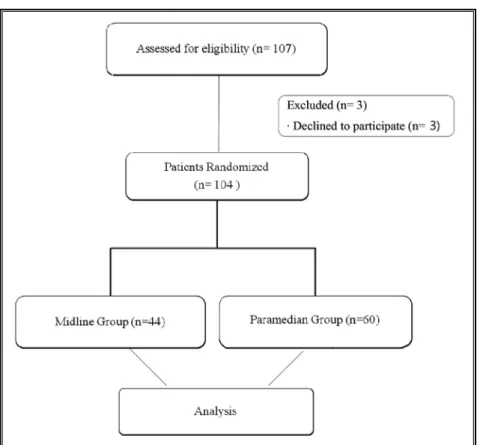
Patients with previous lumbar spine surgery, pregnancy, absence of lumbar magnetic resonance imaging (MRI), and neurologic symptoms requiring immediate reevalu- ation were also excluded. During enrollment, 3 patients were excluded owing to refusal to participate in this study. Finally, 104 patients were included in this study for the analysis (Fig. 1).
All cases of LEI were performed by one pain expert (J.H.) with more than 15 years of experience via either the midline or paramedian approach at the L3-4, L4-5, or L5-S1 level. A midline or paramedian approach of LEI was determined with randomization. The randomiza- tion list was computer generated by the department of research support of our hospital using SPSS version 11.0 (SPSS Inc., Chicago, IL). Decision concerning the side of injection (right or left) for the paramedian approach and the level of both approaches were made based on the presenting symptoms and the level of discogenic lesion or spinal stenosis confirmed by MRI.
Under fluoroscopic guidance and after aseptic preparation of the lower back, a 21-gauge Tuohy needle (Tae Chang Industrial Co., Kongju, Republic of Korea) was inserted via a midline or paramedian ap- proach following local skin infiltration. The midline approach was performed with Tuohy needles located within the midline trajectory and entering the ligamen- tum flavum at the midline in true antero-posterior (AP) images. Skin entry was done at the center of the lumbar interlaminar space in cases of a midline approach. The midline approach was defined when the final needle tip position was located within the width of the up- per or lower spinous process. The skin entry of the paramedian approach was done at 1.5 cm lateral and 2 cm caudal to the standard midline needle insertion degeneration process in the interspinous ligament
can modify the resistance during injection, leading to subsequent failure to identify the epidural space (4-6). Although more study is needed, different types of needles might be one of reasons for compromised resistance during LEI. If the failure rate is high, repeated attempts of LEI are required, causing additional discomfort or pain to the patient.
During LEI using a midline approach, we could en- counter failure of identifying the epidural space owing to an equivocal or absent LOR sensation. The reason for such absence of LOR sensation has been suggested as the paucity of midline ligamentum flavum in the lumbar spine. Reported incidence rates of ligamentum flavum midline gaps at L1-2 and C7-T1 levels are as high as 22.2% and 68%, respectively (5,6). Therefore highly elastic pressure generated by the ligamentum flavum might be attenuated or even absent if LOR technique is used via a midline approach.
Cervical epidural pressure shows a highly dynamic changing pattern depending on the body position. In addition, cervical epidural injection using a parame- dian approach has demonstrated more frequent abrupt pressure decrease in epidural pressure wave compared with a midline approach (7,8). This result implies that if we use a paramedian approach rather than a midline approach, we can improve the accuracy of identifying the epidural space.
Lumbar epidural pressure in patients with spinal stenosis shows dynamic change depending on body position, with higher epidural pressure compared with normal individuals (9,10). In addition, it remains con- troversial whether epidural pressure is truly positive or negative (11-13).
Epidural pressure waveform can be visualized in the monitor using a closed measurement system, and this method can be an alternative way to identify the epidural space (14-16).
The primary endpoint of this study was to compare the incidence of lumbar epidural pressure decrease pat- terns during needle advancement between the para- median and midline groups. The secondary endpoint was to identify factors other than approach method contributing to such pressure decrease patterns.
Methods
This prospective and randomized study was ap- proved by the institutional review board (IRB #05-039) of our institution. All patients were given written and verbal information about the trial and of potential ben-
point. We adjusted all needle tips to stay away from the midline at least 10 mm in an AP image (Fig.
2A-C). The final locations of the needle of each group were saved to the hard disc of C-arm. They were analyzed to assess the prop- erty of final needle position.
When the needle was firmly located with a lateral image showing at least more than 5 to 10 mm distance left to reach the epidural space, the pressure was started to be measured in a closed measurement system using a pressure transducer (Ed- wards Lifesciences, Irvine, CA). At the same time, video recording of pressure changes shown in the monitor was started using a mo- bile phone. Under the guidance of lateral image of C-arm (Ziehm 8000, Ziehm, Germany) and observing the pressure pattern shown in the monitor, the epidur- al needle was slowly advanced while keeping the same trajecto- ry in the lateral plane. Before ad- vancement of the needle, 2 mL of normal saline solution was inject- ed through the epidural needle, and a saline solution–filled sterile extension tubing was connected
to one side to the epidural needle hub while the other side was connected to the pressure transducer. The level of pressure transducer was adjusted to heart level. The pressure scale was set in a range of 0 to 30 mmHg on a portable monitor. When a highly sustained intraneedle pressure decreased abruptly, we assumed that the needle had entered the epidural space. The waveform appeared in the monitor soon after the abrupt pressure decrease showed a characteristic pulsatile waveform superimposed on respiratory oscillation.
Fig. 1. Flow chart showing patients who participated in this study.
Fig. 2. Locations of the final needle tip in the midline and paramedian groups. In the midline group (A, B), needle tip was located within the width of spinous process (dotted line). In the paramedian group (C), needle tips were adjusted to stay away from the midline at least 10 mm in an AP image.
After confirming the abrupt or gradual pressure decrease and subsequent pulsatile wave- form, video recording using a mobile phone was stopped.
A 3 mL of contrast medium was injected subsequently after identifying characteristic pulsa- tile waveform. On completion of LEI, fluoroscopic images of AP and lateral views were saved to the hard disk of C-arm, and were transmitted to a picture archiving and communication system.
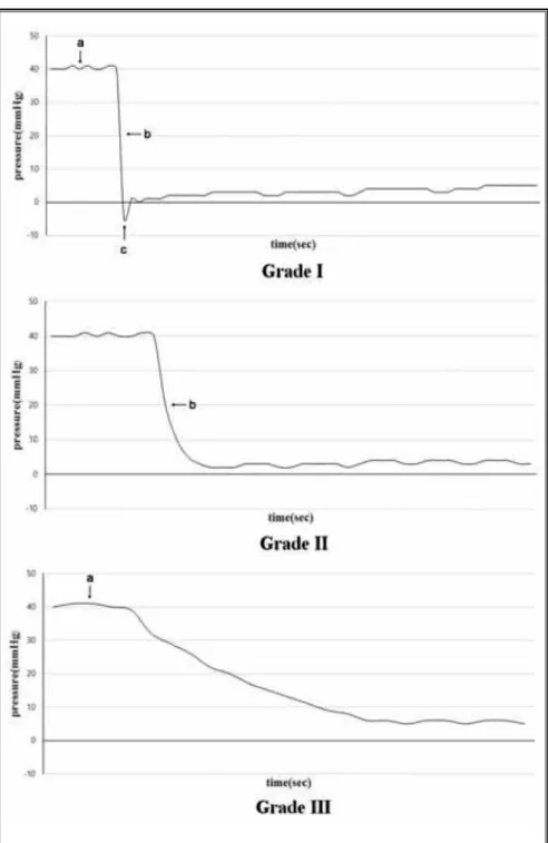
An investigator who was blinded to the fluoroscopic im- age, approach method of LEI, and demographic data (gen- der, body mass index [BMI], and diagnosis) of the patient assessed the epidural wave- form using a video recorded at the time of LEI. The pattern of pressure decrease changes was classified into grades I, II, and III. Grade I showed the following characteristic compo- nents in sequence: (1) a highly sustained intraneedle positive pressure before entering the epidural space, (2) an abrupt pressure decrease at the mo- ment of entering the epidural space with a tactile sensation of popping, and (3) a negative peak pressure before lumbar epidural pressure equilibration.
Grade II was defined as a high- positive pressure (1) followed by an abrupt pressure drop with popping (2). Grade III was defined as a high-positive pres- sure (1) followed by continuous pressure drop without initial negative pressure (Fig. 3). Age, gender, BMI, and diagnosis (stenosis vs. discogenic pain) were identified prior to LEI.
Fig. 3. Grade I showed the following characteristic components in sequence: (A) a highly sustained positive pressure before entering the epidural space, (B) an abrupt pressure decrease at the moment of entering the epidural space with a tactile sensation of popping, and (C) a negative peak pressure before lumbar epidural pressure equilibration. Grade II was defined as a high-positive pressure (A) followed by an abrupt pressure drop with popping (B). Grade III was defined as a high-positive pressure (A) followed by continuous pressure drop without initial negative pressure.
They were used to analyze factors contributing to pres- sure decrease changes of LEI.
Patients with spinal stenosis were defined as hav- ing prominent claudication clinically demonstrating walking distance of < 200 m and showing MRI findings compatible to spinal stenosis. MRI evaluation was per- formed by one pain physician who was not involved in this study. Dural sac cross sectional area (DSCSA) and stenotic levels were evaluated. For the diagnosis of spinal stenosis, critical size of < 100 mm2 was used as an objective diagnostic criterion for lumbar spinal stenosis.
DSCSA (mm2) was measured at the central part of the disc level on axial T1 images using a region of interest curve on a diagnostic workstation (Maro view version 5.4.10.57, INFINITT, Seoul, Korea). In cases of multilevel spinal stenosis, the most stenotic level was chosen to measure the DSCSA.
Patients with discogenic pain were defined as hav- ing a low back pain with or without a radicular leg pain and having MRI findings of intervertebral disc extrusion or protrusion.
Our primary outcome, compared between the midline and paramedian groups, was the difference in incidence of epidural pressure decrease pattern dur- ing needle advancement from the ligamentum flavum to the epidural space. Our secondary outcome was to identify factors other than approach method contribut- ing to such pressure decrease patterns.
Statistical Analysis
This study was powered to detect differences in oc- currence of pressure decrease patterns between abrupt pressure decrease (grade I and II) and gradual pressure decrease (grade III) according to results of a previous study (8). On the basis of an α error level of 0.05, a β error level of 0.2, and odds ratio of 4, 34 injections were required to obtain a power of 80%. All statisti- cal evaluations were performed using SPSS version 11.0 (SPSS Inc.). The Fisher exact test or the chi-square test was used to compare frequency differences of various factors, such as gender, diagnosis, and type of approach method. Mean values of age and BMI were compared using an independent t test. Odds ratio of various fac- tors for identifying an abrupt pressure decrease was calculated with 95% confidence intervals (CI). Differ- ences were considered statistically significant when P values were < 0.05.
Results
A total of 107 patients were assessed for eligibility,
and 3 patients were excluded owing to refusal to par- ticipate in this study. Thus 104 patients were randomly allocated to the midline or paramedian groups (Fig. 1).
Demographic data were similar between the 2 groups. Regarding the level of injection, L5-S1 was the most frequent level in both groups. We observed 3 types of epidural pressure decrease patterns, and they were classified as grades I, II, and III (Fig. 3). Frequencies of each grade were compared between the 2 groups.
In the midline group, grade III, which demonstrated a gradual pressure decrease, was observed the most commonly (n = 29), followed by grade II (n =10). In the paramedian group, grade II was the most commonly observed waveform (n = 36), followed by grade III (n = 16) (Table 1).
Grades I and II were considered as an abrupt pres- sure decrease, whereas grade III was considered as a gradual pressure decrease. The odds ratio of approach method (midline vs. paramedian) was then identified.
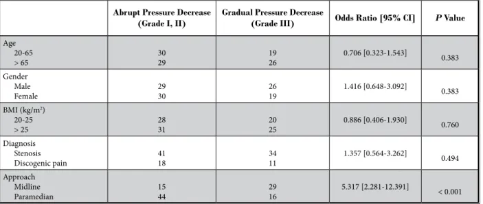
Abrupt pressure decrease was more frequently observed in the paramedian group (P < 0.001). The odds ratio of the paramedian approach with an abrupt pressure decrease at the moment of epidural space entry was 5.317 (95% CI, 2.281-12.391). Age, gender, BMI, and diagnosis were also analyzed. However, we could not find any significant differences in frequencies between abrupt and gradual pressure decrease groups (Table 2).
discussion
Tactile sensation of LOR is still an advocated method to confirm epidural space despite its lack of specificity (17). Therefore more distinct LOR sensation during needle advancement is very important. It can be associated with easier and successful epidural injection.
We observed 3 patterns of epidural pressure de- crease, which means grades I, II, and III. The difference between grade I and II is the presence of a negative peak pressure before lumbar epidural pressure equili- bration. Only group I showed such negative pressure.
A previous study, which investigated the epidural pres- sure decrease pattern in the cervical area, demonstrat- ed grade IV in the midline group in addition to grades I through III (8). Grade IV was defined as having no pressure change before and after entering the epidural space, whereas high-positive pressure was not attained before entering the epidural space. The high-positive pressure is generated when the needle passes through the dense ligamentous structure, such as supraspinous or interspinous ligament. In the midline group studied
Table 1. Demographic data and frequency of each grade of study patients.
Midline Group (n = 44) Paramedian Group (n = 60) P value
Age 62.2 ± 2.6 61.5 ± 2.7 0.705
Male/Female 21/23 33/27 0.552
BMI (kg/m2) 24.6 ± 2.8 24.7 ± 3.7 0.809
Stenosis/discogenic pain 34/10 41/19 0.379
Level of injection
L3-4 5 (11.4) 4 (6.7)
L4-5 17 (38.6) 11 (18.3) 0.030
L5-S1 22 (50.0) 45 (75)
Epidural pressure after entering the epidural space (mmHg) 8.5 ± 3.2 7.8 ± 3.4 0.661 Frequencies of grades
Grade I 5 (11.4) 8 (13.3)
Grade II 10 (22.7) 36 (60.0) < 0.001
Grade III 29 (65.9) 16 (26.7)
Results are presented as mean ± standard deviation for quantitative variables, and n (%) for qualitative variables.
Grade I showed the following characteristic components in sequence: (1) a highly sustained positive pressure before entering the epidural space, (2) an abrupt pressure decrease at the moment of entering the epidural space with a tactile sensation of popping, and (3) a negative peak pressure before lumbar epidural pressure equilibration. Grade II was defined as having the same components with grade I, but not showing component 3. Grade III showed a highly sustained positive pressure before entering the epidural space followed by gradual pressure decrease without initial negative pressure.
Table 2. Odds ratio for variables associated with abrupt pressure decrease during lumbar interlaminar epidural injections.
Abrupt Pressure Decrease
(Grade I, II) Gradual Pressure Decrease
(Grade III) Odds Ratio [95% CI] P Value Age 20-65
> 65 30
29 19
26 0.706 [0.323-1.543] 0.383
Gender Male
Female 29
30 26
19 1.416 [0.648-3.092] 0.383
BMI (kg/m2) 20-25
> 25 28
31 20
25 0.886 [0.406-1.930] 0.760
Diagnosis Stenosis
Discogenic pain 41
18 34
11 1.357 [0.564-3.262] 0.494
Approach Midline
Paramedian 15
44 29
16 5.317 [2.281-12.391] < 0.001 Results are presented as n (%), and odds ratio with [95% CI].
Grade I showed the following characteristic components in sequence: (1) a highly sustained positive pressure before entering the epidural space, (2) an abrupt pressure decrease at the moment of entering the epidural space with a tactile sensation of popping, and (3) a negative peak pressure before lumbar epidural pressure equilibration. Grade II was defined as having the same components with grade I, but not showing component 3. Grade III showed a highly sustained positive pressure before entering the epidural space followed by gradual pressure decrease without initial negative pressure.
Abrupt pressure decrease was more frequently observed in the paramedian group (P < 0.001). The odds ratio of the paramedian approach with an abrupt pressure decrease at the moment of epidural space entry was 5.317 [95% CI, 2.281-12.391].
by Joo et al, (8) the needle might have been deviated to adjacent paravertebral muscle layers or encountered with cyst in the interspinous ligament. However, we could not find any case of grade IV in either group of our study.
Our study demonstrated that abrupt pressure de- crease occurred 5 times more frequently with the para- median approach compared with that of the midline approach. When performing epidural injection with the midline approach, we should consider the possibil- ity of midline gaps of ligamentum flavum, although it is not frequent in the lower lumbar level (6). Defect or midline gaps in the ligamentum flavum may imply that the LOR sensation is dependent on supraspinous and interspinous ligaments before entering the epi- dural space. Compared with the ligamentum flavum of elastic fiber, which produces distinct and high elastic resistance, supraspinous and interspinous ligaments are composed of collagen fibers (4-6). Therefore the distinct LOR sensation may be attenuated or even absent com- pared with the sensation generated by the ligamentum flavum. If we correlate the distinct LOR sensation and the existence of abrupt pressure decrease measured using a closed measurement system, the paramedian approach provides a higher chance of feeling a distinct LOR sensation.
Regarding the presence of initial negative epidural pressure observed in grade I, there has been many con- troversies regarding the mechanism. Previous studies have suggested artifacts of dura tenting or retraction of ligamentum flavum, and difference in the shape of needle when entering the epidural space (11,12,18).
Our results showed similar incidence of grade I between the midline and paramedian groups. Further study is required to clarify this mechanism.
As distinct from the benefit of clear LOR sensation, the paramedian approach also has clinical effectiveness for patients with lumbar radiculopathy. Paramedian LEI has shown equivalent or superior efficacy with shorter procedure time and radiation exposure compared with transforaminal or midline LEI (19-22). If the paramedian approach is performed in the cervical area, one should consider the increased risk of dural puncture because the epidural space becomes thinner from the midline to the paramedian. However, epidural space of the lumbar area becomes less thin compared with the cervical area (7,8,17). No patient in either group had any side effect, such as dural puncture, in the present study.
We performed subgroup analysis to identify fac- tors affecting the pressure decrease pattern other than the approach method. Lumbar epidural pressure in patients with spinal stenosis showed dynamic change depending on body position and higher epidural pres- sure compared with normal individuals (9,10). Higher epidural pressure found in patients with spinal stenosis might have influenced LOR sensation. However, we could not find any significant difference in frequencies of epidural pressure decrease patterns between pa- tients with spinal stenosis and those without stenosis.
We presume that the generation of epidural pressure decrease is more dependent on ligamentous struc- ture before entering the epidural space rather than increased epidural pressure caused by spinal stenosis.
High elastic resistance created by ligamentous structure could be changed with the aging process. Patients aged from 61 to 79 years have shown interspinous cyst in the lumbar region up to 85% when autopsy was conducted (5-7). In the elderly, the degeneration of the interspi- nous ligament with cavity formation has been observed more frequently (23). However, age, gender, and BMI did not affect the pattern of epidural pressure in the present study.
Our study includes several limitations. First, to correlate distinct LOR sensation and the existence of abrupt pressure decrease, one should match LOR sen- sation with pressure decrease shown in the monitor.
However, we could not use the LOR technique during needle advancement because the needle was con- nected to a closed measurement system. In addition, a LOR sensation is very subjective depending on the phy- sician, and various tissue structures encountered during needle advancement.
Second, the level of LEI was not unified. It was performed from L3-4 to L5-S1 in accordance with the stenotic or disc protrusion level.
conclusions
Our study demonstrates that abrupt pressure de- crease occurs more frequently with the paramedian approach. However, age, gender, BMI, and diagnosis did not affect the pressure decrease pattern. This study suggests that the accuracy of identifying the epidural space can be improved with the paramedian approach if we suppose that LOR sensation occurs owing to an abrupt pressure decrease during needle advancement from the ligamentum flavum to the epidural space.
RefeRences
1. Manchikanti L, Buenaventura RM, Manchikanti KN, et al. Effectiveness of therapeutic lumbar transforaminal epidural steroid injections in managing lumbar spinal pain. Pain Physician 2012;
15:E199-E245.
2. Cohen SP, Bicket MC, Jamison D, Wilkinson I, Rathmell JP. Epidural steroids: A comprehensive, evidence- based review. Reg Anesth Pain Med 2013;
38:175-200.
3. Manchikanti L, Cash KA, Pampati V, Falco FJ. Transforaminal epidural injections in chronic lumbar disc herniation: A randomized, double- blind, active-control trial. Pain Physician 2014; 17:E489-E501.
4. Lirk P, Colvin J, Steger B, et al. Incidence of lower thoracic ligamentum flavum midline gaps. Br J Anaesth 2005;
94:852-855.
5. Lirk P, Kolbitsch C, Putz G, et al. Cervical and high thoracic ligamentum flavum frequently fails to fuse in the midline.
Anesthesiology 2003; 99:1387-1390.
6. Lirk P, Moriggl B, Colvin J, et al. The incidence of lumbar ligamentum flavum midline gaps. Anesth Analg 2004;
98:1178-1180.
7. Moon JY, Lee PB, Nahm FS, Kim YC, Choi JB. Cervical epidural pressure measurement: Comparison in the prone and sitting positions. Anesthesiology 2010; 113:666-671.
8. Joo Y, Moon JY, Kim YC, Lee SC, Kim HY, Park SY. A pressure comparison between midline and paramedian approaches to the cervical epidural space. Pain Physician 2014; 17:155-162.
9. Takahashi K, Miyazaki T, Takino T, Matsui T, Tomita K. Epidural pressure measurements. Relationship between
epidural pressure and posture in patients with lumbar spinal stenosis. Spine (Phila Pa 1976) 1995; 20:650-653.
10. Takahashi K, Kagechika K, Takino T, Matsui T, Miyazaki T, Shima I. Changes in epidural pressure during walking in patients with lumbar spinal stenosis.
Spine (Phila Pa 1976) 1995; 20:2746-2749.
11. Okutomi T, Watanabe S, Goto F. Time course in thoracic epidural pressure measurement. Can J Anaesth 1993;
40:1044-1048.
12. Zarzur E. Genesis of the ‘true’ negative pressure in the lumbar epidural space.
A new hypothesis. Anaesthesia 1984;
39:1101-1104.
13. Visser WA, Gielen MJ, Giele JL, Scheffer GJ. A comparison of epidural pressures and incidence of true subatmospheric epidural pressure between the mid- thoracic and low-thoracic epidural space.
Anesth Analg 2006; 103:1318-1321.
14. Lennox PH, Umedaly HS, Grant RP, White SA, Fitzmaurice BG, Evans KG. A pulsatile pressure waveform is a sensitive marker for confirming the location of the thoracic epidural space. J Cardiothorac Vasc Anesth 2006; 20:659-663.
15. de Medicis E, Tetrault JP, Martin R, Robichaud R, Laroche L. A prospective comparative study of two indirect methods for confirming the localization of an epidural catheter for postoperative analgesia. Anesth Analg 2005;
101:1830-1833.
16. Arnuntasupakul V, Van Zundert TC, Vijitpavan A, et al. A randomized comparison between conventional and waveform-confirmed loss of resistance for thoracic epidural blocks. Reg Anesth Pain Med 2016; 41:368-373.
17. Tran DQ, Gonzalez AP, Bernucci F, Finlayson RJ. Confirmation of loss-of- resistance for epidural analgesia. Reg Anesth Pain Med 2015; 40:166-173.
18. Gil NS, Lee JH, Yoon SZ, Jeon Y, Lim YJ, Bahk JH. Comparison of thoracic epidural pressure in the sitting and lateral decubitus positions.
Anesthesiology 2008; 109:67-71.
19. Gharibo CG, Varlotta GP, Rhame EE, Liu EC, Bendo JA, Perloff MD. Interlaminar versus transforaminal epidural steroids for the treatment of subacute lumbar radicular pain: A randomized, blinded, prospective outcome study. Pain Physician 2011; 14:499-511.
20. Ghai B, Bansal D, Kay JP, Vadaje KS, Wig J. Transforaminal versus parasagittal interlaminar epidural steroid injection in low back pain with radicular pain:
A randomized, double-blind, active- control trial. Pain Physician 2014;
17:277-290.
21. Ghai B, Vadaje KS, Wig J, Dhillon MS.
Lateral parasagittal versus midline interlaminar lumbar epidural steroid injection for management of low back pain with lumbosacral radicular pain: A double-blind, randomized study. Anesth Analg 2013; 117:219-227.
22. Hong JH, Park EK, Park KB, Park JH, Jung SW. Comparison of clinical efficacy in epidural steroid injections through transforaminal or parasagittal approaches. Korean J Pain 2017;
30:220-228.
23. Sharrock NE. Recordings of, and an anatomical explanation for, false positive loss of resistance during lumbar extradural analgesia. Br J Anaesth 1979;
51:253-258.