Journal of Trauma and Injury Vol. 26, No. 3, September, 2013
[ J Trauma Inj 2013;26:131-138 ]
� Address for Correspondence : Hyung Sik Hwang, M.D.
Department of Neurosurgery, Dongtan Sacred Heart Hospital, College of Medicine, Hallym University, 40 Sukwoo-dong, Hwaseong, 445-170, Korea
Tel : 82-31-8086-2810, Fax : 82-31-8086-2809, E-mail : [email protected] Submitted : August 13, 2013 Revised : August 24, 2013 Accepted : August 28, 2013
측두골 골절후 발생한 안면마비 환자의 안면신경감압술:
25명 환자들의 증례분석
한림대학교 동탄성심병원 신경외과학교실, 1순천향대학교 부천병원 신경외과학교실
남한가위, 황형식, 문승명, 신일영, 신승훈, 정제훈1
- Abstract -
Facial Nerve Decompression for Facial Nerve Palsy with Temporal Bone Fracture: Analysis of 25 Cases
Han Ga Wi Nam, M.D., Hyung Sik Hwang, M.D., Seung-Myung Moon, M.D., Il Young Shin, M.D., Seung Hun Sheen, M.D., Je Hoon Jeong, M.D.
1Department of Neurosurgery, Dongtan Sacred Heart Hospital, College of Medicine, Hallym University, Hwaseong, Korea
1Department of Neurosurgery, Bucheon Hospital, College of Medicine, Soonchunhyang University, Bucheon, Korea
Purpose: The aim of this study is to present a retrospective review of patients who had a sudden onset of facial palsy after trauma and who underwent facial nerve decompression.
Methods: The cases of 25 patients who had traumatic facial palsy were reviewed. Facial nerve function was graded according to the House-Brackmann grading scale. According to facial nerve decompression, patients were categorized into the surgical (decompression) group, with 7 patients in the early decompression subgroup and 2 patients in the late decompression subgroup, and the conservative group(16 patients).
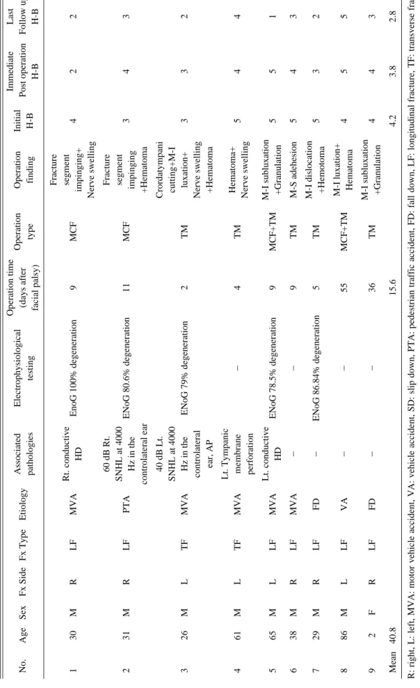
Results: The facial nerve decompression group included 8 males and 1 female, aged 2 to 86 years old, with a mean age of 40.8. In early facial nerve decompression subgroup, facial palsy was H-B grade I to III in 6 cases (66.7%); H-B grade IV was observed in 1 case(11.1%). In late facial nerve decompression subgroup, 1 patient (11.1%) had no improvement, and the other patient(11.1%) improved to H-B grade III from H-B grade V. A com- parison of patients who underwent surgery within 2 weeks to those who underwent surgery 2 weeks later did not show any significant difference in improvement of H-B grades (p>0.05). The conservative management group included 15 males and 1 female, aged 6 to 66 years old, with a mean age of 36. At the last follow up, 15 patients showed H-B grades of I to III(93.7%), and only 1 patient had an H-B grade of IV(6.3%).
Conclusion: Generally, we assume that early facial nerve decompression can lead to some recovery from trau- matic facial palsy, but a prospective controlled study should and will be prepared to compare of conservative treatment to late decompression.
Key Words: Facial paralysis, Temporal bone, Facial nerve injuries, Decompression, Facial nerve
I.
Introduction
Facial nerve palsy (FNP) after temporal bone frac- tures occurs in 7% to 10% of patients.(1,2) FNP that is associated with temporal bone fractures is classi- fied into immediate palsy or delayed palsy, depend- ing on the timing of the onset of the FNP after the head trauma.(2) When complete FNP occurs immedi- ately after head trauma, surgical intervention is indicated. Conventionally, facial nerve decompres- sion (FD) is considered for the following conditions:
1) severe or complete paralysis of House-Brackmann (H-B) grades of 4 to 6,(3) 2) immediate onset of FNP, and 3) degree of denervation exceeding 90%
according to electroneuronography (ENoG).
Some studies have reported beneficial effects of late FD in patients who had facial paralysis caused by temporal bone fractures and who could not be operated on earlier.(4,5) Others have suggested that surgical decompression of the facial nerve is indi- cated at any time and may be beneficial, even in very old injuries.(6) However, most surgeons think that surgical intervention should be performed within 2 weeks of the onset of total FNP for it to be effective and favor surgical decompression as soon as electrical studies indicate severe injury and pre- dict a poor prognosis.(7-10) Otherwise, a patient’s unstable condition may complicate decisions about the optimal timing of surgical decompression. The best timing of FD remains controversial.(11,12)
The aim of this study was to report surgical results for patients with FNP after temporal bone fractures according to the timing of surgical explo- ration. We investigated the outcomes in facial func- tion according to the location of the temporal bone fracture, the severity, and the type of injury.
II.
Materials and Methods
The study design was a retrospective review of 25 patients who had FNP due to temporal bone frac- tures from because of head injury that occurred between 2003 and 2011. We determined patient age, and sex, type of temporal bone fracture, severity of FNP, type of operation, operation timing, follow-up period, and recovery of FNP. Evaluation of facial
nerve function was graded according to the H-B grading scale.(3) According to the FD performed, 25 patients were divided into the FD group or the con- servative management group. The FD group con- sisted of 9 patients, and the conservative manage- ment group consisted of 16 patients. The FD group was subdivided into 2 groups according to early decompression(7 patients) or late decompression(2 patients, with late defined as the surgical decom- pression was conducted 2 weeks after the onset of FNP). Electromyography or ENoG, if possible, was conducted on the patients with facial paralysis. In total 12 patients (FD group, 6 patients; conservative management group, 6 patients) underwent electro- physiological assessments. For radiological evalua- tions, high-resolution computerized tomography was performed in all cases. Magnetic resonance imaging (MRI) was not routinely performed in this series but was performed in cases in which MRI was indicated. Thus, MRI played a complementary role in the diagnosis of concomitant intracranial complica- tions.
The outcomes after the decompression surgery were also assessed with the H-B grading scale. In this study, an H-B grade of I was defined as com- plete recovery, and grades I-III with slight sequelae were defined as good recovery.
Statistical analyses were performed with the Statistical Package for the Social Sciences (SPSS) for Windows (version 18, IBM Corporation, Amonk, NY, USA). The improvement in H-B grades between the early FD group and the late FD group was analyzed with Crosstab. p values less than 0.05 were consid- ered statistically significant.
III.
Results
The FD group had 8 male and 1 female patients who were aged 2 to 86 years, with a mean age of 40.8 years. The mean operation time after the onset of FP was 15.6 days (ranging from 2 to 55 days). The mean follow-up period was 22 months (ranging from 2 months to 8 years). FP involved the left side in 4 cases and the right side in 5 cases. Seven patients had temporal bone fractures of the longitu- dinal type(77.7%), and 2 patients(22.3%) had tempo-
Table 1.The Demography of Facial Nerve Decompression Group AssociatedElectrophysiologicalOperation time OperationOperationInitialImmediateLast No.AgeSexFx SideFx TypeEtiology pathologiestesting(days after typefindingH-BPost operation Follow up facial palsy)H-BH-B Fracture 130MRLFMVARt. conductive EnoG 100% degeneration9MCFsegment 422 HDimpinging+ Nerve swelling 60 dB Rt.Fracture 231MRLFPTASNHL at 4000 ENoG 80.6% degeneration11MCFsegment 343 Hz in theimpinging controlateral ear+Hematoma 40 dB Lt.Crordatympani SNHL at 4000cutting+M-I 326MLTFMVAHz in theENoG 79% degeneration2TMluxation+332 controlateralNerve swelling ear, AP+Hematoma Lt. Tympanic Hematoma+ 461MLTFMVAmembrane-4TM Nerve swelling544 perforation 565MLLFMVALt. conductive ENoG 78.5% degeneration9MCF+TMM-I subluxation 551 HD+Granulation 638MRLFMVA--9TMM-S adehesion543 729MRLFFD-ENoG 86.84% degeneration5TMM-I dislocation 532 +Hemotoma 886MLLFVA--55MCF+TMM-I luxation+ 455 Hematoma 92FRLFFD--36TMM-I subluxation 443 +Granulation Mean40.815.64.23.82.8 R: right, L: left, MVA: motor vehicle accident, VA: vehicle accident, SD: slip down, PTA: pedestrian traffic accident, FD: fall down, LF: longitudinal fracture, TF: transverse frac- ture, SNHL: sensory neural hearing loss, HD: hearing disturbance, MCF: middle cranial fossa approach, TM: transmastoid approach, M-S: malleus-stapes, M-I: malleus-incus, I-S: incus-stapes
Fig. 1. Change of Mean H-B Grade Between Decompression Group and Conservative Group
Table 2. The Demography of Conservative Management Group
Initial Last
No. Age Sex Fx Side Fx Type Etiology Associated pathologies
H-B Electrophysiological testing Follow up H-B
1 14 M R LF VA 40 dB Lt. SNHL 3 - 3
at 4000 Hz in the controlateral ear
2 22 M R LF MVA Rt. Conductive HD 3 - 3
3 41 M R LF FD AP 4 - 3
4 51 M L LF FD AP 2 - 2
5 49 M R LF VA Rt. Conductive HD 3 ENoG 23.1% degeneration 3
6 30 M R LF VA Rt. Conductive HD 2 - 2
7 65 M R LF VA Rt. Conductive HD 4 - 3
8 23 M B LF TA Both Conductive HD 2 ENoG 0% degeneration 2
9 19 M L LF MVA Both Conductive HD 2 ENoG 82.1% degeneration 2
10 66 F R LF PTA Rt. Conductive HD 2 - 2
11 53 M R LF MVA Rt. Conductive HD 2 ENoG 35.7% degeneration 2
12 6 M R TF PTA - 2 - 1
13 50 M B TF MVA Rt. Conductive HD 3 - 2
14 14 M L LF MVA Lt. Temporal Pseudo- 4 - 4
aneurysm
15 45 M L LF FD Lt. Optic nerve injury 3 - 2
16 28 M L LF FD Lt. Conductive HD 3 ENoG 69.7% degeneration 1
Mean 36 2.8 2.3
R: right, L: left, MVA: motor vehicle accident, VA: vehicle accident, SD: slip down, PTA: pedestrian traffic accident, FD: fall down, LF: longitudinal fracture, TF: transverse fracture, AP: abducens palsy, SNHL: sensory neural hearing loss, HD: hearing dis- turbance
ral bone fractures of the transverse type. One case of sixth nerve palsy was evaluated. There were 6 road traffic accidents(66.7%), 2 falls(22.2%), and 1 vehicle accident(11.1%).
For the preoperative H-B grades of the FD group (9 patients), 2 patients were normal to moderate (grades I-III), 3 patients were moderately severe (grade IV), and 4 patients were poor (grades V-VI).
The immediate postoperative H-B grades of 3 patients were normal to moderate (grades I-III), of 4 patients were moderately severe (grade IV), and of 2 patients were poor (grade V-VI). At the last follow up, facial nerve function was grade I to III in 7 cases. H-B grade IV was observed in 1 case(11.1%), and grade V was found in 1 case(11.1%). Grade VI was not found in any of the cases. The mean improvement in the H-B grade was a grade of 1.4.
The rate of goof recovery was 85.7%(6 of 7 patients) if the surgery was performed within 2 weeks. Of the 2 patients who underwent FD after 2 weeks, 1 patient had no improvement, and the other patient had an improved H-B grade of III from grade V (Table 1) (Fig. 1).
The conservative management group included 15 males and 1 female who were aged 6 to 66 years old and with a mean age of 36. The mean follow-up period was 14.7 months (ranging from 2 months to 9 years). FNP involved the left side in 5 cases and the
right side in 11 cases. One case of a left temporal traumatic pseudoaneurysm was identified, and 2 cases of sixth nerve palsy were evaluated. Fourteen patients had temporal bone fractures of the longitu- dinal type(87.5%), and 2 patients(12.5%) had them of the transverse type. Eight FPs(50.0%) were caused by road traffic accidents, 4(25.0%) were due to falls, and 4(25.0%) were from vehicle accidents. For the initial H-B grades of the conservative management group, 13 patients were normal to moderate (grades I-III)(81.2%), 3 patients were moderately severe (grade IV)(18.8%), and no cases were poor (grades V- VI). At the last follow up, 15 patients had H-B grades of I to III(93.7%), and only 1 patient had an H-B grade of IV(6.3%). The mean improvement in the H-B grade was a grade of 0.6. The demograph- ics of the conservative management group are shown in Table 2.
1. Facial nerve decompression
Of the 25 FP cases managed, 9 were surgically treated(36%), and 16 were medically treated(64%).
For the 9 surgically treated patients, surgery was performed any time between 2 days and 55 days after the onset of FNP, depending on the severity of the patient’s general condition and intracranial injury. Most patients were operated on within 2
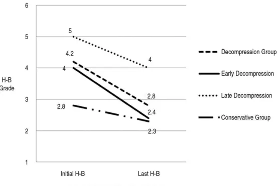
Fig. 2. Preoperative (A, B) and postoperative (C, D) HRCT images of patient no. 1, longitudinal fracture line (white arrow) of the temporal bone in axial view of the right temporal bone (A), Traumatic fluid collection of both mastoids and right middle ear cavity (B), Middle cranial fossa approach of axial view (C) and coronal view (D)
A B
C D
weeks (mean, 7 days; ranging from 2 to 11 days) after the onset of FNP(7 cases, 77.8%), and 2 patients (patients 8 and 9, 22.2%) underwent the decompression 2 weeks later (mean, 45.5 days;
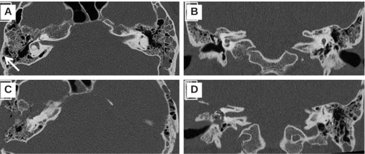
ranging from 36 to 55 days). Patients who under- went surgery within 2 weeks and those who under- went surgery 2 weeks later did not have any signifi- cant difference in improvement of H-B grades (p>0.05). A combined middle cranial fossa (MCF) and transmastoid (TM) approach was chosen in 2 cases (22.2%). A pure MCF route was used in 2 cases (22.2%) (Fig. 2). A pure TM route was used in 5 cases (55.6%) (Fig. 3). During the operation, it was observed that 5 cases had massive hematomas sur- rounding the facial nerve, 4 cases had malleus- incus subluxation, 3 cases had severe nerve swelling, 2 cases had granulation with the facial nerve, 2 cases had a fracture segment impinging on the facial nerve, 1 case had chorda tympani cutting, and 1 case had malleus-stapes subluxation.
IV.
Discussion
There is still controversy concerning the efficacy, timing, and choice of approach in the management of traumatic FNP. First, Nash et al. reported the results of a systemic review of 612 patients and con- cluded that the efficacy of FD was debatable because
the rate of complete recovery was only 23% in the surgery group, whereas it was 66% in patients who were followed without treatment. Dorrouzet et al.(13) reported that, among their 115 patients, 65 patients received FD, and 94% of them recovered to H-B grades of I to III, 2 years later. They suggested that FD was effective. These different results may be explained by differences in the severity of the FNP between the patients receiving conservative therapy and those treated with FD. In our study, 7(77.7%) of the 9 patients who underwent FD recov- ered to H-B grades of I to III.
Second, FD has been shown to provides beneficial effects if it is performed within 14 days of injury. In addition, later FD should be done if the facial nerve function does not show any recovery in order to ascertain acute or delayed FP in an unconscious patient.(1) In many studies, it has been emphasized that early FD is more effective than late FD. Hato et al.(2) found that 14 of 66 patients who underwent FD within the first 2 weeks after trauma had an excellent therapeutic outcome with a rate of com- plete recovery of 85.7% and a rate of good recovery of 92.9%. However, recent studies have reported that late FD had sufficient effects for traumatic FNP. Lieberherr et al.(14) reported on 14 patients with acute FP, with more than 90% nerve degenera- tion shown on ENoG, 1 to 3 months following the
Fig. 3. Preoperative (A, B) and postoperative (C, D) HRCT images of patient no. 3, transverse fracture line (white arrow) of the tem- poral bone in axial view of the left temporal bone (A) and coronal view (B), Transmastoid approach of axial view (C) and coronal view (D)
A B
C D
trauma and found that 53~100% of the facial nerve function returned to normal. In addition, Quaranta et al.(5) found good recovery in patients who under- went FD even 3 months after the temporal bone fracture and with >95% denervation on ENoG. They found that 77.7% of the patients showed H-B grades of I or II recovery followed for at least 1 year. Ulug and Ulubil(15) reported that 11 patients with FNP after traumatic temporal bone fracture underwent FD within the first 3 months after the onset of paralysis. Five patients showed H-B grade of I, 4 patients had an H-B grade of II, and 2 patients had an H-B grade of III facial recovery.
In our study, the rate of goof recovery was 85.7%
(6 of 7 patients) if the surgery was performed within 2 weeks. This result supported the beneficial effects of early FD. Among the 2 patients who underwent FD after 2 weeks, 1 patient had no improvement, and the other patient had an improved H-B grade of III from a grade of V.
Third, the choice of the FD approach in traumatic FP considerably depends on the presence of service- able hearing following the trauma and the types of temporal bone fractures. According to Fisch,(16) the facial nerve is injured distal to the geniculum in the majority of the cases with longitudinal fractures.
In general, for longitudinal fractures, the MCF approach is the preferred technique. For mixed and transverse fractures, when serviceable hearing is present, a middle fossa craniotomy combined with a TM exploration is used.(13,17,18) We used the MCF approach and combined it with TM exploration in 2 such affected patients, and total decompression of the facial nerve was achieved. A pure MCF approach was performed in 2 patients. We agree that the MCF approach is adequate for direct exposure of the internal auditory meatus and the cisternal, meatal, labyrinthine, and tympanic segments of the facial nerve, and, even more so, the perigeniculate area.
Last, when the FP is moderate and the patient’s condition is unstable, medical treatment is consider- able. In this study, 16 patients were treated with prednisolone for 2~3 weeks. At the last follow up, 15 patients had H-B grades of I to III(93.7%), and only 1 patient had an H-B grade of IV(6.3%). Thus, med- ical treatment was effective in moderate FP patients
and when some aspect of iatrogenic injury was con- sidered during the decompression surgery.
A retrospective study to examine the usefulness of FD is a readily available method.(2,19) However, our study had some limitations. The 2 groups (FD group and conservative management group) had differ- ences in their severity of facial paralysis, and there was a small number of patients. Therefore, further studies with prospective randomization, and a suffi- cient number of patients are needed.
V.
Conclusion
We think that the early decompression for trau- matic FP provides beneficial effects to patients. If the patients are delayed and have an unstable gen- eral condition, late surgery might be considered bet- ter when the patient’s condition is stable. Medical treatment is effective in moderate FP patients, especially when considering with some aspect of iatrogenic injury during the decompression surgery.
Finally, a prospective randomized and blinded study to examine the usefulness of FD is needed.
REFERENCES
01) Chang CY, Cass SP. Management of facial nerve injury due to temporal bone trauma. Am J Otol. 1999; 20(1): 96-114.
02) Hato N, Nota J, Hakuba N, Gyo K, Yanagihara N. Facial nerve decompression surgery in patients with temporal bone trauma: analysis of 66 cases. J Trauma. 2011; 71(6): 1789-92;
discussion 92-3.
03) House JW, Brackmann DE. Facial nerve grading system.
Otolaryngol Head Neck Surg. 1985; 93(2): 146-7.
04) Sanus GZ, Tanriover N, Tanriverdi T, Uzan M, Akar Z. Late decompression in patients with acute facial nerve paralysis after temporal bone fracture. Turk Neurosurg. 2007; 17(1): 7- 12.
05) Quaranta A, Campobasso G, Piazza F, Quaranta N, Salonna I.
Facial nerve paralysis in temporal bone fractures: outcomes after late decompression surgery. Acta Otolaryngol. 2001;
121(5): 652-5.
06) Brodsky L, Eviatar A, Daniller A. Post-traumatic facial nerve paralysis: three cases of delayed temporal bone exploration with recovery. Laryngoscope. 1983; 93(12): 1560-5.
07) Gantz BJ, Rubinstein JT, Gidley P, Woodworth GG. Surgical management of Bell’s palsy. Laryngoscope. 1999; 109(8):
1177-88.
08) U F, editor. Lacrimation in Facial Nerve Surgery.
Birmingham, AL; 1977.
09) May M. Total facial nerve exploration: transmastoid, extral- abyrinthine, and subtemporal indications and results.
Laryngoscope. 1979; 89(6 Pt 1): 906-17.
10) Kim J, Moon IS, Shim DB, Lee WS. The effect of surgical timing on functional outcomes of traumatic facial nerve paral- ysis. The Journal of trauma. 2010; 68(4): 924-9.
11) Adour KK, Boyajian JA, Kahn ZM, Schneider GS. Surgical and nonsurgical management of facial paralysis following closed head injury. Laryngoscope. 1977; 87(3): 380-90.
12) Yanagihara N. Transmastoid decompression of the facial nerve in temporal bone fracture. Otolaryngol Head Neck Surg.
1982; 90(5): 616-21.
13) Darrouzet V, Duclos JY, Liguoro D, Truilhe Y, De Bonfils C, Bebear JP. Management of facial paralysis resulting from temporal bone fractures: Our experience in 115 cases.
Otolaryngol Head Neck Surg. 2001; 125(1): 77-84.
14) Lieberherr U SD, Fisch U. Management of severe facial nerve paralysis in the temporal bone-a review of 82 cases. In: Castro D, ed. Proceedings of the Sixth International Symposium on the facial nerve. 1990: 285-9.
15) Ulug T, Arif Ulubil S. Management of facial paralysis in tem- poral bone fractures: a prospective study analyzing 11 operat- ed fractures. Am J Otolaryngol. 2005; 26(4): 230-8.
16) Fisch U. Management of intratemporal facial nerve injuries. J Laryngol Otol. 1980; 94(1): 129-34.
17) Angeli SI, Chiossone E. Surgical treatment of the facial nerve in facial paralysis. Otolaryngol Clin North Am. 1997; 30(5):
683-700.
18) U F. Facial paralysis in fractures of the petrous bone.
Laryngoscope. 1974; 84: 2141-54.
19) Lathrop FD. Lesions of the Facial Nerve Due to War Injuries and Their Repair. Proc R Soc Med. 1945; 38(11): 629-34.