DOI 10.17480/psk.2020.64.5.377
Dimethylnitrosamine으로 유발된 생쥐 간섬유화에 대한 풋귤 추출물의 억제 효과
한상철*·윤지현**·윤원종***·윤선화***·강희경*·유은숙*,#
*제주대학교 의학전문대학원 의학과, **제주관광대학 창업보육센터 농업회사법인(주)삼원네이처
***제주테크노파크 생물종다양성연구소
Suppressive Effects of Premature Citrus unshiu Extract on Dimethylnitrosamine-induced Hepatic Fibrosis in Mice
Sang-Chul Han*, Ji-Hyun Yun**, Weon-Jong Yoon***, Seon-A Yoon***, Hee-Kyoung Kang*, and Eun-Sook Yoo*,#
*Department of Medicine, School of Medicine, Jeju National University
**SamWon Nature Co., Ltd., Business Incubation Center, Jeju Tourism University
***Biodiversity Research Institute, Jeju Technopark
(Received June 30, 2020; Revised August 6, 2020; Accepted August 15, 2020)
Abstract Hepatic fibrosis is a wound healing process of the liver, which is characterized by overproduction and accumulation of extracellular matrix. Hepatic fibrosis occurs when chronic inflammation causes a deposition of scar tissue.
Dimethylnitrosamine (DMN) is a potent liver toxin that can lead to fibrosis of the liver. DMN causes massive hepatocyte damage, cell death by necrosis or apoptosis and liver tissue distribution. Citrus unshiu, which is known to contain several flavonoids, has pharmacological effects on anti-oxidative, anti-cancer, anti-microviral and anti-inflammation activity. Therefore, we investigated the effect of premature C. unshiu and fermented premature C. unshiu extract on DMN-induced hepatic fibrosis mouse model. Here, we show that administration of premature C. unshiu and fermented premature C. unshiu reduces the generation of triglyceride, alanine aminotransferase (ALT), aspartate aminotransferase (AST), malondialdehyde (MDA) and cytochrome P450 2E1 (CYP2E1) in mouse serum. Also, the extract reduced the mRNA expression of glutathione S-transferases (GSTs), Heme oxygenase-1 (HO-1), NAD(P)H quinone oxidoreductase 1 (NQO1), and increased inflammatory factors such as IL-6, COX-2 and iNOS. These results suggest that premature C. unshiu has anti-hepatic fibrosis activity by regulating metabolic detoxification pathways such as cytochrome P450s and GSTs.
Keywords hepatic fibrosis, dimethylnitrosamine, Citrus unshiu, cytochrome P450s, glutathione S-transferases
서 론(Introduction)
간섬유화는 다양한 종류의 간 손상(만성 B형 간염, C형 간염, 자가면역성 질환, 알코올성 지방 간염 및 비알코올성 지방간질 환 등)에 의한 지속적인 염증, 상처재생 과정 및 콜라겐 축적에 의해 발생되는 질환으로 만성 간섬유화로 인해 간경변이 유발 되며, 간경변증은 간암이나 간부전으로 발전하게 된다.
1)간섬유 화의 다양한 원인 중 지방간의 발생은 간에 과도하게 축적된 중
성지방(triglyceride)과 지속적으로 염증을 유발하는 다양한 인자 들(interlukin-1β, -6, -8, COX-2, iNOS 등)이 지방형성 성분의 발 생을 유도하여 나타난다.
2,3)간성상세포(hepatic stellate cell, HSC) 는 간섬유화의 중요한 세포로서 사이토카인 및 산소유도 자유 라디칼의 산물인 MDA에 의해서 활성화고 이는 콜라겐 등 세 포외기질(extracellular matrix, ECM) 구성 단백질을 합성한다.
4,5)화학 발암물질 중 간섬유증을 유발하는 물질인 dimethylnitro- samine (DMN) 은 간의 mixed function oxidase system에 의해 전 자친화력이 있는 대사물질로 전환되고 높은 반응성을 나타내어 DNA 및 단백질 등에 작용하여 methyl 유도체를 형성하며 ECM 단백질을 간에 축적시키고 간의 괴사를 일으킴으로써 최종적으 로 간, 신장, 폐에 암을 발생시키는 물질이다.
6,7)DMN 은 저용량 으로 장기간 투여 시 간암 발생을 유도하지만 고용량으로 단기 간 투여 시 간 조직 괴사와 이차적 섬유화증을 유발한다.
8)DMN
#
Corresponding author
Eun-sook Yoo, Department of Medicine, School of Medicine, Jeju National University, 102 Jejudaehak-ro, Jeju-si, Jeju Special Self- Governing Province 63243, Korea
Tel: +82-64-754-3847, Fax: +82-64-702-2687
E-mail: [email protected]
은 주로 cytochrome P450s에 의해 간에서 대사되며 대사과정에 서 발생되는 물질인 hydroxyl radicals, reactive oxygen intermedi- ates등은 간 중심 정맥 주위에 간성상세포 손상 및 괴사의 원인 이 되고, 이후 섬유화를 발생시킨다.
9)인체에 영향을 미칠 수 있는 독성물질은 환경적 요인에 의한 독성물질(외독소, exotoxins)과 체내에서 생산되는 독성물질(내독 소, endotoxins)로 분류할 수 있다. 인간은 현대사회에서 음식, 대 기, 페인트, 카페트, 염색제 등의 독성물질에 쉽게 노출되고 있 으며, 이러한 독성물질들은 체내의 detoxification pathway에 의 해 대부분 해독되어지나 오랜 기간 동안 노출 시 체내에 조금 씩 축적된다.
10)독성물질은 수용성이나 지용성 물질로 간에 유 입되며, 수용성 독성물질은 비교적 쉽게 대사되어 신장, 장, 피 부, 폐 등에 의해 배출되지만 지용성 독성물질은 지방세포에 저 장되기 때문에 인체 내 DNA, 세포막, 단백질 등의 결합을 방해 하여 정상적인 대사활동에 부정적인 영향을 미칠 수 있다.
11)독 성물질에 의한 반복적인 노출은 단기적으로 두통, 메스꺼움, 피 로감 등을 유발할 수 있으며, 장기적으로는 체중증가 및 치매, 섬유근육통, 심혈관 질환, 암, 백혈병 등의 각종 만성질환을 유 발할 수 있다고 알려져 있다.
12)마리아엉겅퀴(Silybum marianun)의 열매로부터 추출한 물질인 실리마린은 민간에서 간질환의 치료를 목적으로 이용되던 약용 식물로써 간세포보호, 간세포 재생작용, 항산화작용 및 글루타 치온 보존작용을 통한 간장의 해독능을 증가시키는 효능이 있 고 현재 다양한 간질환에 대해 치료보조제로 사용되고 있다.
13-16)고기능성 성분을 함유하고 있는 감귤에는 flavonoid 화합물인 naringin, naringenin, hesperidin, hesperetin, tangeretin, nobiletin 등이 함유되어 있고 다양한 질환에 대한 효능 연구가 진행되고
있다.
17,18)최근 연구에 의하면 감귤 내 다양한 flavone 중 poly-
methoxyflavone 는 암 예방에 있어 중요한 활성 성분으로 밝혀졌 고, carotenoid는 인체 내 흡수 후 vitamin A로 전환됨으로써 면 역기능 증진 및 발암물질 해독에 효능이 있다고 알려져 있다.
19-21)
또한, flavonoid류가 감귤 과육보다 박에 많이 함유되어 있어 항산화 작용을 하고 휘발성 물질인 pinene, linalool 등이 존재하 고 있어 항균작용이 있으며, 염증 및 암에 carotenoids, biofla- vonoids 와 같은 물질이 효과를 나타낸다고 보고되고 있다.
22-25)감 귤 미숙과(풋귤)는 완숙과와 비교하여 organic acid, polyphenol 및 다양한 flavonoid가 많이 함유되어 있고, 특히 미숙과의 감귤 박에 이와 같은 생리활성 물질이 다량 함유되어 있어 항염과 항 산화에 높은 활성을 나타내는 것으로 보고된다.
26)미생물을 이용한 발효공정은 최종 영양성분의 변화 또는 생 리활성 물질의 효능을 극대화시킬 수 있으며, 미생물의 다양한 가수분해효소와 천연물의 세포 내 조직에 결합되어 있던 생리 활성 물질이 유리됨으로써 발효된 천연물은 인체 내 흡수율이 보다 높아진다.
27-29)또한, 천연물을 발효하면 발효 전과 비교하 여 항산화, 항균, 항비만, 항염 등의 효과가 더욱더 극대화된다 고 알려져 있다.
30-33)따라서 본 연구에서는 풋귤 주정추출물과 발효풋귤 주정추출물의 간 기능 개선 및 detoxification 효능을
검증하기 위해 DMN으로 유도된 간 섬유화 동물 모델에 추출 물을 투여하여 체중 변화, 혈청 중 지질대사 및 간 손상 지표 분석, 간 조직의 조직학적 분석, 디톡스 관련 유전자 발현 등을 분석하여 추출물의 효능을 확인하고자 하였다.
실험 방법(Experimental Methods)
시약 및 기기
Dimethylnitrosamine (DMN) 은 Wako (Junyaku, OSaka, Japan) 에서 구입하였고 cytochrome P450 2E1 (CYP2E1) ELISA kit는 Mybiosource (San Diego, CA, USA)에서 구입하여 사용하였다.
또한, triglyceride, alanine aminotransferase (ALT), aspartate ami- notransferase (AST) ELISA kit는 Biovision (Milpitas, CA, USA) 에서 구입하였고, EZ-lipid peroxidation (TBARS) ELISA kit 는 Dogenbio (Seoul, Korea)에서 구입하여 사용하였다.
추출물 제조
풋귤은 세척 후 파쇄하여 건조하였으며, 풋귤 건조물과 효모 (Saccharomyces cerevisiae) 를 접종한 10% 설탕수용액을 1:9 비율 로 넣고 20
oC 저온에서 15일간 발효 후 건조하였다. 발효된 풋 귤 및 풋귤 건조물은 각각 50% 주정으로 24시간 추출하여 사 용하였다.
실험동물
수컷 5주령의 ICR 생쥐는 중앙실험동물(Central Lab. Ani. Inc, Korea)에서 공급받았다. 동물은 실험 당일까지 고형사료와 물을 충분히 공급하고, 온도 22±2
oC, 습도 55±15% 조건하에서 12시 간 light-dark cycle의 환경을 유지하며 1주간 적응시킨 후 실험 에 사용하였다. 실험동물은 그룹별 10마리로 나누었고 정상대조 군(NC), 유발대조군(DMN), 양성대조군(Silymarin, Sily; 200 mg/
kg), 50% 풋귤 에탄올추출물 투여군(PCU; 100 and 300 mg/kg), 발효풋귤 50% 에탄올추출물 투여군(FPCU; 100 and 300 mg/kg) 으로 총 7그룹이며, 식이와 식수는 자유롭게 섭취하도록 하였다.
동물실험의 윤리적, 과학적 타당성 검토 및 효율적인 관리를 위 하여 제주대학교 동물실험윤리위원회(Jeju National University Animal Care and Use Committee)의 승인을 받았다.
DMN 을 이용한 간 섬유화 동물모델 제작 및 시험물질 투여
생쥐를 1주간 기본사료로 적응시킨 후 DMN (10 mg/kg)을 주
3 회 8주간 복강으로 반복투여 하였고 양성대조군과 추출물은 최
초 DMN 투여 일과 동시에 경구투여용 금속제 존대(zonde)를
이용하여 위 내로 강제 투여 하였다. 최종 실험 종료 후 체중
측정 및 심장천자법으로 채혈하여 혈청을 분리하고 blood chemistry
와 ELISA 분석을 실시하였다. 또한, 간 조직 적출 후 10% 포
르말린 용액에 보관하여 haemotoxylin and eosin (H&E) 염색에
사용하였고 간 조직을 분쇄하여 RNA 추출 후 디톡스 관련 유
전자를 real-time PCR을 이용하여 분석하였다.
혈중 지질생화학적 분석
추출물을 8주간 투여 후 각 실험동물로부터 분리한 혈액 내 혈청에서 중성지방(triglyceride)의 함량을 ELISA kit를 이용하여 측정하였다. 각 well에 희석한 혈청 50 μL와 Lipase를 분주하고, 20분 동안 실온에서 반응시킨 후 triglyceride reaction mix를 분 주하여 30분 동안 암소에서 반응시켰다. 이후, ELISA 판독기를 이용하여 570 nm 파장에서 흡광도를 측정하고 분석하였다.
혈중 ALT, AST, CYP2E1 및 MDA 분석
실험 종료 후 각 실험동물로부터 분리한 혈액 내 혈청에서 ALT, AST, CYP2E1, MDA 의 함량을 제조사에서 제시한 실험방 법을 이용하여 ELISA kit로 분석하였다.
간 조직 내 다양한 유전자 발현 분석
실험 종료 후 각 실험동물로부터 적출한 간 조직의 유전자 발 현 양상을 real-time PCR 증폭법을 사용하여 분석하였다. RNAsol
B(Tel-Test) 용액을 사용하여 각 조직으로부터 RNA를 추출한 뒤 KAPA SYBR
®FAST qPCR kit (Kapa Biosystem, Woburn, MA, USA) 를 이용하여 cDNA 합성 및 real-time PCR 분석을 하 였다. 조직에 RNAzol
B를 넣고 homogenizer를 이용하여 조직을 분쇄 후 chloroform을 첨가하여 혼합하였다. 이후 원심 분리를 시행한 후 상층액을 회수하여 2-propanol과 혼합 후 RNA를 추 출하고 cDNA를 합성하였다. Real time quantitative PCR은 iQ
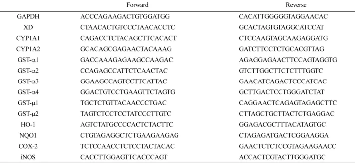
TM5 Multicolor Real-Time PCR Detection System (Bio-Rad Labora- tories, Inc.) 를 이용하여 수행하였고 실험에 사용된 primer sequence 들은 Table 1에 나타내었다.
간 섬유화 동물모델의 간 조직 병리학적 분석
실험 종류 후 간 조직을 절취하여 10% neutral buffered for-
malin 에 24시간 동안 고정 시킨 후 graded alcohol로 탈수시키고 파라핀으로 포매하여 block을 제작하였다. Microtome으로 4 μm 두께의 조직절편을 제작하여 H&E 염색을 시행한 후 조직의 특 이 병변의 유무를 관찰하였다.
통계 처리
본 연구의 통계처리는 Student’s t-test 분석을 이용하여 검정하 였고 값은 평균과 표준편차로 표현했다.
결과 및 고찰(Results and Discussion)
생쥐 체중변화 및 조직학적 분석
생쥐에 DMN (10 mg/kg)을 주 3회 8주간 복강으로 반복투여 하였고 양성대조군과 추출물은 최초 DMN 투여 일과 동시에 경 구 투여하였다. 실험 종료 후 체중 측정 및 간 조직 적출 후 10% 포르말린 용액에 고정하여 H&E 염색을 실시하였다. 생쥐 체중 변화는 풋귤(PCU) 및 발효풋귤 추출물(FPCU) 투여로 인 해 DMN군과 비교하여 유의적으로 증가되었고(p<0.05 또는 p<0.01), 양성대조군과도 큰 차이를 보이지 않았다(Fig. 1A). 간 조직의 H&E 염색에서 DMN 투여군은 출혈성 괴사소가 관찰되 었고 섬유성 띠, 섬유성 망 형성, 격막상 붕괴 및 섬유증이 유 발되었다. 반면에 다른 모든 군에서는 위와 같은 증상들이 많이 완화된 것을 관찰할 수 있다(Fig. 1B).
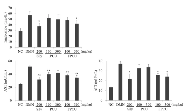
간 섬유화 동물모델에서 혈중 중성지방 함량 및 ALT, AST 생 성 변화
혈중 중성지방의 양이 증가하면 관상 동맥질환이 발생되고 이 는 말초조직을 중심으로 중성지방을 축적시켜 인슐린 저항성을 유도한다.
34)또한, 중성지방은 간세포에 축적되어 지방간을 발생
Table 1. Primer sequence for qRT-PCR
Forward Reverse
GAPDH ACCCAGAAGACTGTGGATGG CACATTGGGGGTAGGAACAC
XD CTAACACTGTCCCTAACACCTC GCACTAGTGTAGGCATCCAT
CYP1A1 CAGACCTCTACAGCTTCACACT CTCCAAGTAGCAAGAGGATG
CYP1A2 GCACAGCGAGAACTACAAAG GATCTTCCTCTGCACGTTAG
GST- α1 GACCAAAGAGAAGCCAAGAC AGAGGAGAACTTCCAGTAGGTG
GST- α2 CCAGAGCCATTCTCAACTAC GTCTTGGCTTCTCTTTGGTC
GST- α3 GGAAGCCAGTCCTTCATTAC GAACATCAGACTCCCATCAC
GST- α4 GGACTGTCCTGAAGTTCTAGTG GCTTGACTCCTGGGATCTAT
GST- μ1 TGCTCTGTTACAACCCTGAC CAGGAACTCAGAGTAGAGCTTC
GST- μ2 TAGTCTCCTCCTATCCCTTGTC CTTAGCTGCTTACTCTGAGGAC
HO-1 AGTCTATGCCCCACTCTACTTC GGAGACGCTTTACATAGTGC
NQO1 CTGTAGAGGCTCTGAAGAAGAG CTAGAGATGACTCGGAAGGA
COX-2 TCTCCAACCTCTCCTACTACAC GAACTCTCTCCGTAGAAGAACC
iNOS CACCTTGGAGTTCACCCAGT ACCACTCGTACTTGGGATGC
시키고 지속적인 지방간은 간 손상을 의미하는 ALT와 AST의 생성을 증가시킨다.
35)따라서 풋귤 주정추출물이 혈중 중성지방 함량 및 ALT와 AST 생성을 억제하는지 ELISA를 이용하여 평 가하였다. 그 결과, 중성지방, ALT 및 AST의 함량은 DMN 자 극에 의해서 증가하였고 추출물 투여로 인해 DMN군과 비교하 여 중성지방 및 ALT의 함량은 양성대조군(중성지방, ALT; P<0.05) 과 발효풋귤 추출물 투여군(중성지방, ALT; p<0.05)에서 유의적 으로 억제되었다. 또한, AST의 함량은 풋귤 및 발효풋귤 추출 물 투여군(p<0.01) 모두 유의적으로 감소하였다(Fig. 2). 이러한 결과들은 추출물이 중성지방 생성 억제를 통해 지방간의 발생 및 간 손상을 완화시켰다는 것임을 의미한다.
생쥐 혈중 Malonedialdehyde 분석
지질과산화의 생산은 여러 가지 독성화합물이나 약물에 의한 간 손상 발생의 가장 대표적인 물질이다.
36)이러한 생성의 원인
으로는 세포 내 산화적 스트레스의 증가 즉, free radical 생성의 증가 및 항산화적 방어력의 감소로 인해 야기된다.
37)과산화 지 질의 대표적인 marker인 MDA의 측정은 TBARS의 측정을 이용 한다. Lipid peroxidation screen 방법으로, MDA가 thiobarbituric acid (TBA)와 반응하여 형성하는 MDA-TBA Adduct를 측정함으 로써 lipid peroxidation 정도를 알 수 있다. 결과적으로 생쥐 혈중 MDA 함량은 저농도의 청귤 추출물 투여로 인해 DMN군과 비 교하여 유의적으로 억제되었다(p<0.05). 반면에, 다른군에서는 MDA의 함량이 감소되었으나 유의적인 결과는 관찰되지 않았다 (Fig. 3). 이러한 결과들은 추출물이 DMN 자극으로 발생된 간 손 상을 지질과산화 생성 억제를 통해 완화시켰다는 것을 의미한다.
생쥐 혈중 및 간 조직 내 Cytochrome P450 family 측정
간의 detoxification pathways의 가장 중요한 과정은 3단계로 분
류할 수 있다. 그 중 1단계(phase I)에서는 주로 cytochrome
Fig. 1. Changes in body weight in DMN-stimulated mice. (A) Hepatic fibrosis was induced in mice by intraperitoneal administration of
DMN. Each mouse received DMN (10 mg/kg) for the first three consecutive days of the week for 8 weeks. Mice in the control groups (NC)
received the intraperitoneal injection of the same volume of 0.9% saline. The positive control (sily), premature C. unshiu (PCU) and fermented
premature C. unshiu (FPCU) extract were forcibly administered into the stomach at the same time as the initial DMN administration day. Mouse
weights were measured after the end of the experiment. (B) Paraffin-embeded sections of liver tissue stained with hematoxylin and eosin. (n=10
mice per group). Scale bar=0.1mm. Values represent the mean±SD. *p<0.05 and **p<0.01 compared with mice in the DMN group.
P450s가 관여하며, detoxification 동안 수용성 독성물질을 더 많 이 만들고, 독성이 적은 분자로 변환시켜 이에 따라 쉽게 혈액 내로 유입하게 하며 신장을 통해 소변으로 배출되게 한다.
38-40)따라서 생쥐 혈중 또는 간 조직 내 cytochrome P450 family의 함량 및 발현정도를 ELISA와 real-time PCR을 이용하여 분석함 으로써 간의 디톡스 기능이 원활하게 일어나고 있는지를 확인 하였다. 그 결과, 생쥐 혈중 CYP2E1 함량은 풋귤 및 발효풋귤 추출물 투여로 인해 DMN군과 비교하여 유의적으로 감소되었 다(p<0.05 또는 p<0.01; Fig. 4A). 반면에 간 조직 내 CYP1A1, CYP1A2 mRNA 발현은 양성대조군 포함 모든 추출물 투여군에
서 DMN군과 비교하여 억제되었으나 유의적인 결과는 관찰되 지 않았다(Fig. 4B). 이러한 결과들은 추출물이 간의 detoxification pathways 중 phase I에 관여하는 인자는 CYP2E1 생성을 억제함 으로써 독성물질의 해독을 용이하게 하고 체외로 배출을 유도 한다는 것을 의미한다.
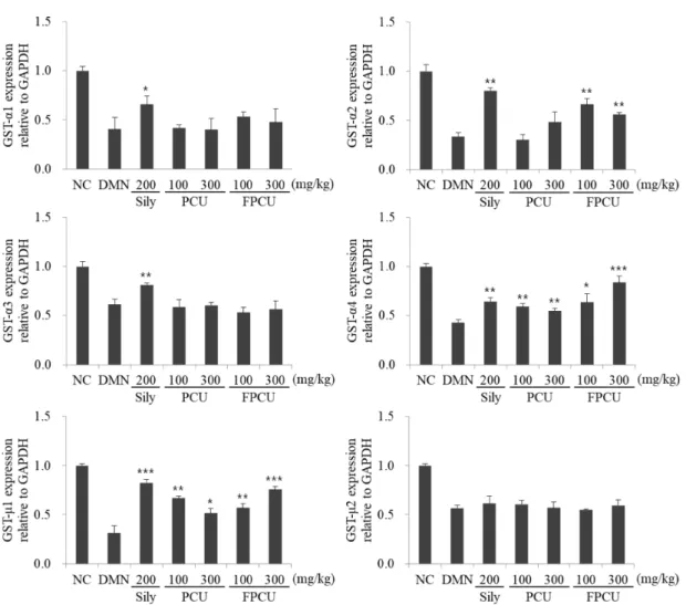
생쥐 간 조직 내 glutathione S-transferases family 분석 간의 detoxification pathways 중 2단계(phase II)에서는 포합반응 (conjugation) 등의 해독기전(detoxifying mechanism)을 통해 phase I 과정에서 생성된 중간대사산물을 수용성으로 최종 변환시켜 체 외로 배설될 수 있도록 한다.
41)Phase II 의 해독 작용에 관여하는 대표적인 효소로는 글루타치온-S-전달효소(glutathione S-transferases;
GST)가 존재하고 GST는 조직마다 분포하는 종류가 다르다. 세 포질에 존재하는 GST는 4가지 종류가 있다(α, μ, π, θ 타입). 특 히, 간에는 GST-α와 GST-μ가 존재하는데, GST-α가 75% 이상을 차지한다.
42,43)따라서 생쥐 간 조직 내 GST family의 발현정도를 real-time PCR을 이용하여 분석함으로써 간의 디톡스 기능이 원 활하게 일어나고 있는지를 확인하였다. 그 결과, 생쥐 간 조직 내 GST-α1, GST-α3, GST-μ2 mRNA 발현은 모든 추출물 투여군에 서 DMN군과 비교하여 증가되었으나 유의적인 결과는 관찰되지 않았다. 반면에, DMN군과 비교하여 풋귤 및 발효풋귤 추출물 투 여로 인해 GST-α2 (p<0.01), GST-α4 (p<0.05; p<0.01 또는 p<0.001), GST-μ1 (p<0.05; p<0.01 또는 p<0.001) mRNA 발현이 유의적으로 증가되었다(Fig. 5). 이러한 결과들은 추출물이 간의 detoxification pathways 중 phase II에 관여하는 인자는 GST 발현 Fig. 2. The effects of premature Citrus unshiu extract on blood biochemical markers in DMN-stimulated mice. The triglyceride, AST and ALT levels in serum were measured by ELISA. Values represent the mean±SD. Data are representative of three independent experiments.
*p<0.05 and **p<0.01 compared with mice in the DMN group.
Fig. 3. The effects of premature Citrus unshiu extract on MDA
level in DMN-stimulated mice. The MDA level in serum was
measured by ELISA. Values represent the mean±SD. Data are
representative of three independent experiments. *p<0.05 compared
with mice in the DMN group.
을 억제함으로써 phase I에서 제거되지 못한 독성물질을 포합하 여 물질의 해독 및 배설을 유도한다는 것을 의미한다.
생쥐 간 조직 내 항산화 효소 발현 분석
세포 내 항산화 효소로 알려진 heme oxygenase-1 (HO-1)은 세 포 내에 존재하는 heme을 분해하는 효소로서 분해 시 생산되는 부산물들이 세포 내 여러 가지 역할을 수행한다.
44)HO-1 에 의 한 heme의 분해로 Fe(II), bilirubin 및 CO가 생성되는데 Fe(II)는 cytoprotection 효과가 있고 bilirubin은 항산화 효과가 있으며, CO 는 염증반응 및 세포 사멸을 억제하는 것으로 알려져 있다.
45-47)NAD(P)H:quinone oxidoreductase 1 (NQOl)은 detoxification pathways 중 Phase II의 대사 활성화에 의해서 생성되는 활성 quinone체로 활성 산소를 제거하여 무독화 시키는 효소로 알려 져 있다.
48)NQO1은 산화적 스트레스에 의해서 발현이 증가하 며 세포 내 산화적 손상에 대한 세포 방어 기능을 수행한다.
49)따라서 생쥐 간 조직 내 항산화 효소계인 HO-1과 NQO1의 발 현정도를 real-time PCR을 이용하여 분석하였다. 그 결과, 생쥐 간 조직 내 HO-1, NQOl mRNA 발현은 DMN군과 비교하여 풋 귤 및 발효풋귤 추출물 투여로 인해 HO-1 (p<0.01 또는 p<0.001), NQOl (p<0.05 또는 p<0.001) mRNA 발현이 유의적으로 증가되 었다(Fig. 6). 결과적으로 DMN 자극으로 생성된 활성 산소는 간 손상의 원인이 되는데 추출물이 항상화 효소인 HO-1과 NQO1 의 발현을 증가시킴으로써 활성 산소 생성 및 세포 내 산화적 손상을 억제시켰다는 것을 의미한다.
생쥐 간 조직 내 염증성 인자 발현 분석
간 손상의 원인으로 초기 단계에는 virus, 술, 약물, 독성물질 등이 작용한다. 초기 간 독성에서 나타나는 염증반응이나 세포 독성을 매개하는 것은 virus 또는 간 독성물질의 대사에 의하여 생성되는 free radical이나 IL-6와 같은 cytokines, 염증관련 인자 인 cyclooxygenase-2 (COX-2)나 nitric oxide synthase (iNOS) 등이 주가 되어 간독성을 유발하는 것으로 알려져 있다.
50,51)따 라서 생쥐 간 조직 내 IL-6, COX-2, iNOS의 발현을 real-time PCR을 이용하여 분석하였다. 그 결과, 생쥐 간 조직 내 IL-6, COX-2, iNOS mRNA 발현은 DMN군과 비교하여 풋귤 또는 발 효풋귤 추출물 투여로 인해 IL-6 (p<0.01 또는 p<0.001), COX- 2 (p<0.05), iNOS (p<0.01 또는 p<0.001) mRNA 발현이 유의적 으로 억제되었다(Fig. 7). 결과적으로 DMN 자극으로 발생된 염 증은 간 손상의 원인이 되는데 추출물이 염증성 인자인 IL-6, COX-2, iNOS의 발현을 억제시킴으로서 항염증 효능을 나타내 어 간 손상을 완화시켰다는 것을 의미한다.
결 론(Conclusion)
간섬유화는 중성지방, MDA에 의해서 활성화된 간성상세포, detoxification pathways의 오작동 및 만성 염증 등에 의해서 발 생되는 질환으로 간경병 또는 간암으로 발전하게 된다. 본 연구 에서 사용된 DMN은 단기투여 시 간섬유화를 유도하고 장기투 여 시 간암을 유발하는 독성물질로써 DMN에 의한 독성은 DMN Fig. 4. The effects of premature Citrus unshiu extract on the expression of cytochrome P450s in DMN-stimulated mice. (A) CYP2E1 level in serum was measured by ELISA. (B) The expression of mRNA for CYP1A1 and CYP1A2 in liver tissues was measured by real-time PCR.
Values represent the mean±SD. Data are representative of three independent experiments. *p<0.05 and **p<0.01 compared with mice in the
DMN group.
자체보다는 대사과정에서 활성화된 N-methylformamide (NMF) 나 N-hydroxymethl-N-methylformamide (HMMF)와 같은 중간체 에 의해서 간이 손상되는 것으로 알려져 있다.
52)CYP2E1은 소 수성적인 여러 가지 물질을 대사시키는 기능을 하는데 DMN의 대사는 CYP2E1에 의해 촉진된다.
53)따라서 본 연구에서는 DMN
으로 유도된 생쥐 간섬유화 동물모델에서 풋귤 및 발효풋귤 추 출물의 효능을 연구하였다. 추출물은 DMN 자극으로 유도된 간 손상을 중성지방 및 지질과산화 생성 억제를 통하여 간 손상 지 표인 ALT와 AST의 발현을 억제하였고 CYP2E1과 GST family 를 억제 또는 증가시킴으로써 detoxification pathways 중 phase Fig. 5. Premature Citrus unshiu extract upregulates the expression of glutathione S-transferases in DMN-stimulated mice. The expression of mRNA for GST family in liver tissues was measured by real-time PCR. Values represent the mean±SD. Data are representative of three independent experiments. *p<0.05, **p<0.01 and ***p<0.001 compared with mice in the DMN group.
Fig. 6. Premature Citrus unshiu extract upregulates the expression of antioxidant enzymes in DMN-stimulated mice. The expression of
mRNA for HO-1 and NQO1 in liver tissues was measured by real-time PCR. Values represent the mean±SD. Data are representative of three
independent experiments. *p<0.05, **p<0.01 and ***p<0.001 compared with mice in the DMN group.
I 과 phase II를 활성화하여 간 손상을 완화시켰다. 또한, 추출물 은 항산화 효소의 발현 유도와 염증성 인자들의 생성 억제를 통 하여 항산화 및 항염 효능을 나타내었다. 본 연구의 또다른 목 적은 간섬유화에 대한 풋귤 추출물과 발효풋귤 추출물의 효능 을 비교하는 것으로 몇몇 인자들은 두 군에 효능 차이가 있었 지만 크게 다르지는 않았고 전반적으로 비슷한 효능을 나타내 었다. 이러한 모든 결과는 풋귤 추출물이 간 손상 완화 효과를 나타내며 향후 천연물을 이용한 간섬유화 억제 연구에 대한 기 초 자료로 사용될 수 있을 것이라 사료된다.
감사의 말씀(Acknowledgment)
본 연구는 중소벤처기업부와 한국산업기술진흥원의 “지역기업 개방형혁신 바우처(R&D, P0010788)” 사업의 지원을 받아 수행 된 연구결과임.
Conflict of Interest
모든 저자는 이해 상충을 가지고 있지 않음을 선언한다.
References