Oxidative stress and hepatic injury induced in mice fed a cyst extract JVS
5
0
0
전체 글
(2) Toxicity of Sarcocystis-infected meat 501. (approval No. 2B-1472). Cyst extract Macroscopic Sarcocystis cysts in muscle were separated from cattle carcasses in the slaughterhouse. Histopathological examination based on cyst wall diameter confirmed the presence of S. hirsuta. Cysts were detached by using a surgical blade under sterile conditions. Ten percent suspensions of the collected cysts were prepared by homogenizing one gram of the collected cysts in 9 mL of a saline solution. The suspension was centrifuged at 1,500 × g for 15 min at room temperature and then sterilized under UV-light for 3 h with rocking. The supernatant was decanted into 10 mL tubes and brought to a boil in a conical flask at 100°C for 7 h to create the heat-treated cyst extract. Non-infected meat extracts were prepared by using a method described previously [20]. Non infected meat was provided from an industrial Slaughterhouse center. The histopathological examination was done in order to observe any Sarcocystis infection. Final suspensions were stored in a refrigerator at 4oC until needed. Prior to conducting the experiments, a 1:10 dilution of the suspension in normal saline was prepared. Adult male mice (25–32 g), Mus musculus, were housed with free access to sterile tap water and a standard pellet diet. Mice were randomly divided into five experimental groups (n = 8): Group 1 (control group) received normal saline via an oral gavage technique. Group 2 received 0.5 mL/day of the non-infected meat extract by gavage. Group 3 received 0.5 mL/day of freshly prepared S. hirsuta cyst extract by gavage. Group 4 received 0.5 mL/day of a previously frozen S. hirsuta cyst extract. Group 5 mice were given the extract of heat-treated meat infected with S. hirsuta cysts via gavage. All groups were gavaged once per day for 28 days. Extracts were administered by using a metal gavage needle (22-gauge, 2.54– 3.81 cm) fitted to a 2 mL syringe. At the end of the experiment, serum samples were collected by using a conventional method o and were immediately frozen at −80 C until use. The daily dose levels were based on our pilot experiments and previous studies [14,18]. Serum CAT activity was determined by using the Zell-Bio kit (Zell-Bio, Germany) according to manufacturer’s protocol and is expressed as U/mL. Serum SOD activity was measured by using a colorimetric commercial kit (Zell-Bio) according to the manufacturer’s instructions. Liver and brain lipid peroxidation levels were measured based on the reaction between malondialdehyde (MDA) and 2-thiobarbituric acid (TBA) by using a spectrophotometer (UNICO UV/VIS-2100 Spectrophotometer; United Products and Instruments, USA) [17]. ALT and AST liver enzymes were measured by using the Pars Azmoon reagent kits (Pars Azmoon, Iran), according to the instructions.. Histopathological examination After euthanasia, liver specimens were sliced, preserved in formalin, and then processed for histopathological staining. After paraffin embedding and block making, serial sections were stained with H&E and examined under a light microscope at 40× magnification. Statistical analysis The collected data were analyzed by using IBM SPSS Statistics software (ver. 20.0; IBM, USA). Multiple comparisons were performed by performing ANOVA and Tukey tests. The significant difference level was set at p < 0.05.. Results Four weeks of oral gavage feeding of freshly prepared S. hirsuta extract significantly reduced serum CAT activity compared to that in the control group (p < 0.001) (panel B in Fig. 1). There was also a decrease in CAT activity in mice treated with the frozen S. hirsuta extract compared to that in the control group (p < 0.001). Furthermore, serum SOD activities in mice receiving fresh or frozen extracts were significantly lower compared to that in the control mice (p < 0.001 for both) (panel A in Fig. 1). Gavage administration of heat-treated S. hirsuta extract also resulted in reduced CAT and SOD activities when compared to those in the control group (p < 0.01 and p < 0.05, respectively). As expected, serum CAT and SOD activities were not influenced by oral administration of non-infected meat extract (Fig. 1). Further analysis showed that CAT activity in mice receiving fresh S. hirsuta extract was lower than that in the group receiving heat-treated S. hirsuta extract (p < 0.01); however, serum SOD activity did not significantly differ between the two groups (p > 0.05).. Fig. 1. Serum superoxide dismutase (SOD; A) and catalase (CAT; B) activity levels in mice treated with Sarcocystis hirsuta extracts (mean ± SD, n = 8). *p < 0.05, **p < 0.01, and ***p < 0.001 compared to the control group.. www.vetsci.org.
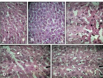
(3) 502 Maryam Sasani et al. Furthermore, there were no significant differences in serum SOD and CAT activities between the groups receiving fresh or frozen extracts (p > 0.05) (Fig. 1). Mice receiving fresh, frozen, and heat-treated S. hirsuta extracts had higher liver MDA levels than that in the control group (p < 0.001, p < 0.001, and p < 0.05, respectively) (panel A in Fig. 2). However, brain MDA levels increased after treatment with fresh and frozen S. hirsuta extracts, but not the heated S. hirsuta extract, compared to that in the control group (p < 0.05 and p < 0.01, respectively) (panel B in Fig. 2). Therefore, oral gavage of heat-treated S. hirsuta extract significantly increased the liver (p < 0.05), but not the brain (p > 0.05), MDA level. Oral administration of the non-infected S. hirsuta extract had no significant effects on the liver and brain MDA levels (p > 0.05). There was also no significant. difference in MDA levels between the group treated with fresh extract and the group treated with frozen extract (all p > 0.05). Our results showed that mice treated with fresh S. hirsuta cyst extract had higher levels of serum ALT and AST compared to those in the control group (p < 0.05 and p < 0.01, respectively) (Fig. 3). There were also significantly higher ALT and AST levels in mice receiving frozen extract compared to those in control mice (p < 0.01). However, there was no significant difference in ALT and AST levels between mice receiving heated extracts and those in the control group (p > 0.05) (Fig. 3). The liver micrographs of the control group and the group receiving the non-infected extract showed normal morphologies with distinct hepatic cells, a central vein, and radiating sinusoids (panels A and B in Fig. 4). Conversely, liver sections of mice treated with fresh or frozen S. hirsuta extracts demonstrated patterns of cell necrosis (panel C in Fig. 4) and disarrangement of hepatic cords (panel D in Fig. 4). In liver micrographs of mice receiving heat-treated extract, histopathological changes were present, but less prominent (panel E in Fig. 4).. Discussion Sarcocystis, one of the most prevalent parasites in muscles of livestock, is responsible for economic and public health burdens worldwide [5], and S. hirsuta is a mildly pathogenic coccidium of cattle [12]. Some studies have reported the presence of Sarcocystis spp. in hamburger meat in Iran;. Fig. 2. Malondialdehyde (MDA) levels in liver (A) and brain (B) of mice treated with Sarcocystis hirsuta extracts (mean ± SD, n = 8). *p < 0.05, **p < 0.01, and ***p < 0.001 compared to the control group.. Fig. 3. Serum levels of aspartate aminotransferase (AST; A) and alanine aminotransferase (ALT; B) in mice treated with Sarcocystis hirsuta extracts (mean ± SD, n = 8). *p < 0.05 and **p < 0.01 compared to the control group. Journal of Veterinary Science. Fig. 4. Photomicrographs of liver sections of the experimental groups. (A) Normal histological appearance of liver tissue of a control mouse. (B) Non-infected extract treated mouse. (C) Fresh extract treated mouse, sinusoidal distention. (D) Liver tissue of a mouse receiving frozen extract, sinusoidal distention. (E) Liver tissue of a mouse received heated extract. H&E stain. 40× (A–E). White arrows, sinusoidal distention; black arrows, the pointer of the microscope..
(4) Toxicity of Sarcocystis-infected meat 503. however, there have been no reports of food poisoning caused by this parasite [7,8,16]. Sarcocystis protozoa can be inactivated by cooking or freezing meat products [11]. Regardless, Sarcocystis spp. are intracellular parasites with several antigens and toxic components that may remain intact after being exposed to extreme (high, low) temperatures; therefore, the aim of this study was to examine the possible toxic effects of frozen and heated Sarcocystis extracts in mice in order to elucidate the toxicity of S. hirsuta cysts when meat products are frozen or heated. Lipid peroxidation is an important mechanism involved in cell membrane destruction and MDA is capable of interacting with amino groups of proteins to form inter-molecular cross-links that inactivate membrane-bound enzymes and receptors. The heat-treated S. hirsuta extract group had increased hepatic MDA levels, distortion of liver architecture, and an insignificant elevation in liver enzyme levels. These results suggest the potential of Sarcocystis cysts to induce lipid peroxidation and liver oxidative damage, probably due to heat-resistant constituents of the cysts. In contrast to the liver lipid peroxidation results, the brain MDA levels were not influenced by administration of the heat-treated extract. A possible explanation for this difference is that the lipid-soluble constituents in the cyst extract, which are capable of penetrating the blood-brain barrier, have been destroyed by heating. Liver damage elevates cytosolic enzyme levels, and it has been shown that elevation of liver enzymes can be a marker of liver toxicity [3]. Mice treated with frozen S. hirsuta extract had higher liver enzymes activities, suggesting that the freezing of Sarcocystis-infected meat does not necessarily reduce or eliminate the toxicity of the parasite. Our results support the results of a previous case study that showed elevation of serum liver enzyme levels in patients with acute muscular sarcocystosis [9]. Our study showed depression of enzymatic antioxidant status in the groups treated with fresh, frozen, and heat-treated S. hirsuta extracts. Superoxide radical anion, peroxy radicals, and hydrogen peroxide are known to induce liver fibrosis through the stimulation of type I procollagen synthesis and the generation of reactive aldehyde end-products [21]. Eukaryotic cells are endowed with a broad array of antioxidant defense mechanisms, including enzymatic antioxidant molecules (e.g., CAT, SOD), glutathione peroxidase, and low-molecular-weight scavengers such as beta-carotene, reduced glutathione (GSH), ascorbic acid, vitamin E, and melatonin [10]. In this investigation, the decreases in SOD and CAT might be due to excessive generation of reactive oxygen species or the decreased availability of NADPH, which is required to maintain antioxidant defense system. These results suggest that the S. hirsuta extracts contain oxidizing agents that inhibit the activity of these endogenous antioxidant enzymes.. Taken together, our study suggests that long-term consumption of Sarcocystis-infected meat induces oxidative stress and liver damage, regardless of whether the infected meat is raw or has been frozen or heat treated. However, the results do indicate that heating can partly diminish some aspects of the toxicity of S. hirsuta-infected meat.. Acknowledgments This study was based on the MS thesis of the first author. We are grateful to the University of Zabol for financial support.. Conflict of Interest The authors declare no conflicts of interest.. References 1. Al-Hyali NS, Khalil LY, Aljawady MA. Sarcotoxin effect on leukocytic finding and phagocytic activity in mice. J Anim Vet Adv 2009, 8, 2395-2398. 2. Bunyaratvej S, Bunyawongwiroj P, Nitiyanant P. Human intestinal sarcosporidiosis: report of six cases. Am J Trop Med Hyg 1982, 31, 36-41. 3. Drotman RB, Lawhorn GT. Serum enzymes as indicators of chemically induced liver damage. Drug Chem Toxicol 1978, 1, 163-171. 4. el-Akkad IN, Mandour AM. On some pharmacological and toxicological effects of a protozoan toxin "sarcocystin". J Egypt Med Assoc 1969, 52, 942-948. 5. Fayer R, Esposito DH, Dubey JP. Human infections with Sarcocystis species. Clin Microbiol Rev 2015, 28, 295-311. 6. Gjerde B. Molecular characterisation of Sarcocystis bovifelis, Sarcocystis bovini n. sp., Sarcocystis hirsuta and Sarcocystis cruzi from cattle (Bos taurus) and Sarcocystis sinensis from water buffaloes (Bubalus bubalis). Parasitol Res 2016, 115, 1473-1492. 7. Hosseini H, Khaksar R, Shemshadi B. Study on infestation of raw hamburgers to Sarcocystis cyst in Tehran. Iranian Nut Sci 2007, 4, 65-70. 8. Jahed-Khaniki GR, Kia EB. Detection of Sarcocystis cysts from meat supplied for hamburger in Iran by histological method. J Med Sci 2006, 6, 18-21. 9. Lau YL, Chang PY, Tan CT, Fong MY, Mahmud R, Wong KT. Sarcocystis nesbitti infection in human skeletal muscle: possible transmission from snakes. Am J Trop Med Hyg 2014, 90, 361-364. 10. Lei XG, Zhu JH, Cheng WH, Bao Y, Ho YS, Reddi AR, Holmgren A, Arnér ES. Paradoxical roles of antioxidant enzymes: basic mechanisms and health implications. Physiol Rev 2016, 96, 307-364. 11. Lhafi KS, Mitzscherling TA, Kühne M. [Parasites in meat: a challenge for veterinarians in meat hygiene]. Dtsch Tierarztl Wochenschr 2004, 111, 277-281. German. 12. Lindsay DS, Blagburn BL, Braund KG. Sarcocystis spp. and sarcocystosis. Br Med J 1995, 5, 249-254. www.vetsci.org.
(5) 504 Maryam Sasani et al. 13. Lunde MN, Jacobs L. Properties of toxoplasma lysates toxic to rabbits on intravenous injection. J Parasitol 1964, 50, 49-51. 14. Mandour AM. Studies on the toxicity of Sarcocystis. J Med Microbiol 1969, 2, 361-363. 15. Mirzaei M, Rezaei H. A survey on Sarcocystis spp. infection in cattle of Tabriz city, Iran. J Parasit Dis 2016, 40, 648-651. 16. Najafiyan HR, Mohebali M, Keshavarz H. Study on frequency of Sarcocystis spp. by macroscopic and microscopic methods in slaughtered cattle in Shahriar district and their public health importance. Paj Saz 2007, 77, 15-19. 17. Ohkawa H, Ohishi N, Yagi K. Assay for lipid peroxides in animal tissues by thiobarbituric acid reaction. Anal Biochem 1979, 95, 351-358.. Journal of Veterinary Science. 18. Saleque A, Bhatia BB, Juyal PD, Rahman H. Toxicity of cyst extract of Sarcocystis fusiformis from buffalo in rabbits and mice. Vet Parasitol 1991, 38, 61-65. 19. Shekarforoush SS, Razavi SM, Abbasvali M. First detection of Sarcocystis hirsuta from cattle in Iran. Iran J Vet Res 2013, 14, 155-157. 20. Sim MK. Cardiovascular actions of chicken-meat extract in normo- and hypertensive rats. Br J Nutr 2001, 86, 97-103. 21. Siwik DA, Pagano PJ, Colucci WS. Oxidative stress regulates collagen synthesis and matrix metalloproteinase activity in cardiac fibroblasts. Am J Physiol Cell Physiol 2001, 280, C53-60..
(6)
수치

관련 문서
In this study, sequential solvent fractions of hot water extract and 70% ethanol extract were prepared from domestic (Imsil region) and foreign (Chile
As a result of performing a compound exercise of spinning and Zumba for 8 weeks, the change in α-amylase showed a significant difference in the exercise group (p<.01), and
Subjects with a smoking period of 1 to 3 years had a high smoking cessation success rate (p=0.024), and the lower the average daily smoking amount, the higher the
The median concentrations of FDP were significantly elevated ( p= 0.02) and the median concentrations of fibronogen were decreased ( p= 0.021) in abnormal bleeding patients
In the control group, lifestyle-related factors were significantly different only in blood glucose (p <.01) and there was no significant difference in
mandibular P-18 inncisor from Nfic-deficient mice (×3,000)... In this study, histological and immunohistochemical studies were carried out to investigate
In other hands, the higher stress levels by parent, academics, appearance and material factors showed more atrophy, physical symptoms,
Defines the failure load and failure moment for a given column for the full range of eccentricities 0 to ∞ g. Compression failure
