Malignant Mesothelioma Diagnosed by Bronchoscopic Biopsy
Yeon-Hee Park, M.D., Jae-Woo Choi, M.D., Sang-Ok Jung, M.D., Min-Ji Cho, M.D., Da-Hyun Kang, M.D., Chae-Uk Chung, M.D., Ph.D., Dong-Il Park, M.D., Jae-Young Moon, M.D., Ph.D., Hee-Sun Park , M.D., Ph.D., Sung-Soo Jung , M.D., Ph.D., Ju-Ock Kim , M.D., Ph.D., Sun-Young Kim, M.D., Ph.D. and Jeong-Eun Lee, M.D., Ph.D.
Department of Internal Medicine, Chungnam National University School of Medicine, Daejeon, Korea
Malignant mesothelioma is a rare malignant neoplasm that arises from mesothelial surfaces of the pleural cavity, peritoneal cavity, tunica vaginalis, or pericardium. Typically, pleural fluid cytology or closed pleural biopsy, surgical intervention (video thoracoscopic biopsy or open thoracotomy) is conducted to obtain pleural tissue specimens.
However, endobronchial lesions are rarely seen and cases diagnosed from bronchoscopic biopsy are also rarely reported.
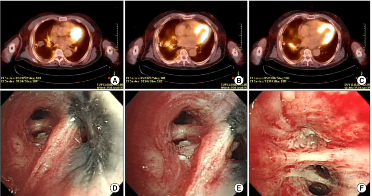
We reported the case of a 77-year-old male who was diagnosed as malignant mesothelioma on bronchoscopic biopsy from obstructing masses of the endobronchial lesion.
Keywords: Mesothelioma, Malignant; Bronchoscopy; Biopsy
and it is rare that originates in other than pleura, especially lung parenchyme or endobronchial mass via direct invasion
7.
Patients with suspected malignant pleural mesothelioma have symptoms such as dyspnea and chest pain and can also have pleural effusion, cough, chest wall mass, weight loss, fever, and sweating
8. The recommended initial evaluation for suspected malignant pleural mesothelioma includes chest computed tomography (CT) or magnetic resonance imag- ing,
18fluoro-2-deoxyglucose positron emission tomography (PET). For tissue diagnosis, thoracentesis for cytology and closed pleural biopsy, thoracoscopic biopsy and open lung biopsy could be done
9-11. Rarely, there are few reports of ma- lignant pleural mesothelioma diagnosed by endobronchial ultrasound-guided transbronchial needle aspiration
12,13.
We reports a case of malignant pleural mesothelioma that was diagnosed by bronchoscopic biopsy of endobronchial mass mimicking lung cancer.
Case Report
A 77-year-old male visited at a secondary hospital with his- tory of dyspnea. He had a previous history of rectal cancer 20 years ago, complete remission state after Mile’s operation and hypertension which was well controlled with losartan. He had Copyright © 2015
The Korean Academy of Tuberculosis and Respiratory Diseases.
All rights reserved.
Introduction
Malignant mesothelioma is a rare cancer associated with asbestos exposure that is estimated to occur in approximately 2,500 people in the United States every year
1,2. In Korea, the in- cidence of malignant mesothelioma is approximately 1–2 per- sons per million
3. Malignant mesothelioma most commonly occurs in pleura, but also in lining of other sites (e.g., perito- neum, pericardium, and tunica vaginalis testis)
4-6. Malignant mesothelioma is a mesodermally derived neoplastic disease
Address for correspondence: Jeong-Eun Lee, M.D., Ph.D.
Department of Internal Medicine, Chungnam National University Hospital, Chungnam National University School of Medicine, 282 Munhwa-ro, Jung-gu, Daejeon 301-721, Korea
Phone: 82-42-280-7147, Fax: 82-42-257-5753 E-mail: [email protected]
Received: Jul. 16, 2014 Revised: Feb. 2, 2015 Accepted: Mar. 23, 2015
cc