• 검색 결과가 없습니다.

Malignant Mesothelioma Presenting as Large Neck Mass

N/A
N/A
Protected

Academic year: 2021

Share "Malignant Mesothelioma Presenting as Large Neck Mass"

Copied!
5
0
0

로드 중.... (전체 텍스트 보기)

전체 글

(1)

Tuberc Respir Dis 2009;67:369-373

CopyrightⒸ2009. The Korean Academy of Tuberculosis and Respiratory Diseases. All rights reserved.

Malignant Mesothelioma Presenting as Large Neck Mass

Departments of

1

Radiology,

2

Pulmonology,

3

Pathology, The Catholic University of Korea College of Medicine, Seoul, Korea Bae Young Lee, M.D.

1

, Hyeon Sook Kim, M.D.

1

, Kyung Sup Song, M.D.

1

, Song Mee Cho, M.D.

1

, Kang Hoon Lee, M.D.

1

, Jung Eun Choi, M.D.

1

, Sang Haak Lee, M.D.

2

, Hwa Sik Moon, M.D.

2

, Ji Young Kang, M.D.

2

, Hyun Hee Kang, M.D.

2

, Ki Ouk Min, M.D.

3

Malignant mesothelioma is the most common primary malignant tumor involving pleura, but its diagnosis is difficult to determine by pathology in addition to the fact that it is rare. We present an unusual case of malignant mesothelioma, which initially presented as large neck mass contrary to the more common presentation of a rind like growth along the pleura demonstrated on imaging and by pathologic findings.

Key Words: Mesothelioma, Neck, Pleura

Address for correspondence: Hyeon Sook Kim, M.D.

Department of Radiology, St. Paul's Hospital, The Catholic University of Korea College of Medicine, 620-56, Jeonnong- dong, Dongdaemoon-gu, Seoul 130-709, Korea

Phone: 82-2-958-2085, Fax: 82-2-960-4568 E-mail: [email protected]

Received: Aug. 21, 2009 Accepted: Sep. 18, 2009

Introduction

Malignant mesothelioma is a rare and aggressive ma- lignant tumor that arises from the pleura or, rarely, peri- toneal and pericardial cavities, and the tunica vaginalis testis. Its incidence is increasing due to long latency pe- riod from asbestos exposure and widespread use of as- bestos before strong relationship of malignant meso- thelioma with asbestos was documented. Immunohisto- chemical staining technique becomes important to con- firm diagnosis

1-3

. We present unusual malignant meso- thelioma presenting as large neck mass.

Case Report

A 61-year-old male admitted for the evaluation of right shoulder pain for several months and acute swel- ling of right lower neck for last 1 week. He had dia- betes mellitus and no history of asbestos exposure. On physical exam, there was diffuse swelling in right lower

neck as compared with left side and decreased breath- ing sound of right lung field. Patient did not complain chest pain or dyspnea. Laboratory findings were within normal range except elevated glucose level.

Chest roentgenogram (Figure 1A) showed large soft tissue mass in right lower neck and apical portion of right hemithorax with minimal pleural reaction at costo- phrenic sulcus. Non-ECG gated 16-row multi-detector computed tomography (MDCT, Light Speed 16; GE, Milwaukee, WI, USA) of chest was performed with 1.25 mm slice thickness. MDCT showed large muscle density mass in right lower neck, connected with apical portion of right hemithorax beyond rib cage, and this mass showed poor contrast enhancement (<HU 10) (Figure 1B). Magnetic resonance imaging (MRI, SIGNA HDx;

GE) was performed, and this mass showed homogenous

iso-signal intensity (SI) on T1 weighted image and ho-

mogenous high SI on T2 weighted image with well de-

fined border compared to the signal of adjacent mus-

cles. This mass showed homogeneous enhancement af-

ter contrast infusion (Figure 1C-E). There was no defi-

nite evidence of necrosis or hemorrhage within the

mass on CT and MR images. Integrated computed to-

mography-positron emission tomography (CT-PET, Bio-

graph LSO; Siemens, Knoxville, TN, USA) was done and

intense uptakes of this large mass and scattered small

pleural plaques were revealed (Figure 1F). There was

(2)

Figure 1. Images of malignant mesothelioma. Chest radiography (A) shows large soft tissue mass (☆) in right lower neck and right apex and small pleural effusion (arrow) in right costophrenic sulcus. There is no definite displacement or destruction of rib and contracture of right hemithorax. Contrast enhanced coronal CT image (B) reveals large muscle density malignant mesothelioma (☆) in right lower neck and right apex through rib cages. Mild pleural thickening (arrow head) and small pleural effusion (arrow) are also seen. The mass (☆) shows iso-signal intensity on T1 weighted axial MR image (C) and high signal intensity on T2 weighted image (D) with good contrast enhancement on coronal T1 weighted image (E). Soft tissue contrast between tumor and adjacent soft tissue is superior in MR than CT. Coronal PET image (F) reveals the intense uptake (SUV=9.6) of large malignant mass (☆) and small pleural nodules (arrow heads).

SUV: standardized uptake valve.

no displacement or destruction of ribs.

Closed pleural biopsy and thoracentesis were done, and poorly differentiated adenocarcinoma was revealed.

Open surgical biopsy of neck mass was performed, and poorly differentiated adenocarcinoma was revealed on hematoxylin-eosin (H&E) stain (Figure 2A). Special im- munohistochemical stains using calretinin and cytoker- atin 5/6 confirmed the epithelial type of malignant mes- othelioma (Figure 2B, C).

Patient has received chemotherapy using pemetrex- ed-cisplatin (9 cycles) and gemcitabine-carboplastin (2 cycles) and been tolerable state for 17 months after di-

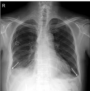
agnosis, and he is still alive with relatively good general status and regression of neck mass (Figure 3).

Discussion

Malignant mesothelioma is originated from the meso- thelial tissues of the pleura, peritoneum, pericardium, and tunica vaginalis testis. Most tumors are originated from pleura (>90%) and followed by the peritoneum (6∼10%)

1-3

. About 2,000∼3,000 new cases are report- ed in the United States every year

4

. Approximately 80%

of malignant mesothelioma is associated with prior as-

(3)

Figure 2. Histology of malignant mesothelioma. The tumor is composed of diffuse asheets of round to polygonal cells (H&E stain, ×200) (A). Immunohistochemical stains by CK 5/6 (B) and calretinin (C) show strong and diffuse cytoplasmic immunoreactivity in the tumor cells.

Figure 3. Follow up chest radiograph after 19 months shows regression of large neck mass and bilateral pleural effusions (arrows).

bestos exposure. Mean age at diagnosis is 60 years be- cause of long latent period between occupational asbes- tos exposure and tumor development and the ratio of male to female is 5:1

1-3,5

. A review about mesotheli- oma revealed that 99% had a latent period of more than 15 years and 96% had at least 20 years latent period.

Median latent period was 32 years

1

.

Frequent symptoms are dyspnea, chest pain, cough, fatigue, and weight loss. Common physical findings are dullness to percussion and decreased breathing sound.

Marked unilateral contraction of affected side of hemi- thorax may be possible in advanced case. Laboratory findings are usually nonspecific

3

. Patient in this case does not complaint dyspnea and chest pain and com- plaint neck mass and shoulder pain. Breathing sound is decreased in right hemithorax.

Common abnormal findings of chest radiograph are

(4)

pleural effusion, pleural plaque, and pleural thickening.

Diffuse pleural thickening is most common finding

3

. CT is the primary modality for tumor staging. Common CT findings are pleural thickening, interlobular fissure thi- ckening, pleural effusion, pleural calcification, and chest wall invasion

5,6

. Pleural thickening is also most common feature on CT

6

. Tumor growth leads to rindlike encase- ment of the lung, and contraction of the affected hemi- thorax is also common. In chest wall involvement, fre- quent manifestations are obliteration of extrapleural fat planes, invasion of intercostal muscles, rib displace- ment, and bone destruction

5

. In our case, malignant mesothelioma showed unusual growth into neck be- yond rib cage and did not show rindlike growth within the hemithorax or rib destruction. MRI permits determi- nation of tumor extent for staging and preoperative evaluation because of its excellent soft tissue contrast resolution. Compared with the SI of adjacent muscle, malignant mesothelioma reveals iso- or slightly high SI on T1-weighted images and moderately high SI on T2- weighted images. With gadolinium-based contrast, this tumor is well enhanced

5

. Our case showed same nature with good enhancement, and tumor was well delineated on MR. MR imaging is problem solving method to eval- uate questionable area of local tumor extension

5,7

. PET in the evaluation of malignant mesothelioma can be useful in the detection of occult distant metastasis and nodal involvement, but the evaluation of local tumor ex- tension is limited due to its poor spatial resolution.

Integrated CT-PET imaging provides anatomic and func- tional information and improves the accuracy of staging.

PET standardized uptake value (SUV) is also used in treatment prognosis. Low SUV and epithelial histology mean the best survival and high SUV and nonepithelial histology mean the worst survival

7

. This case shows high SUV and epithelial histology, and has been toler- able and still alive for 19 months.

Malignant mesothelioma is divided into three histo- logic categories: epithelial, sarcomatous/fibrous, and bi- phasic or mixed. About 50% of pleural and 75% of peri- toneal mesotheliomas is epithelial type. Mixed type is 30% and sarcomatous type is 15∼20%

1

. Sarcomatous

type has poorer prognosis than epithelial or mixed type

8

. Adequate tissue sampling is important to permit accurate diagnosis. Open pleural biopsy, thoracoscopy, and core needle biopsy are recommended to confirm diagnosis but thoracentesis or closed pleural biopsy are not diagnostic in about two thirds of malignant meso- thelioma

3,5

. In our case, open biopsy was performed.

Immunohistochemical stains, especially calretinin and cytokeratin 5/6, are useful to differentiate malignant mesothelioma from adenocarcinoma

3

.

Radiation therapy or chemotherapy alone showed limited response. Combined chemotherapy using peme- trexed with cisplatin is effective in response rate and median survival time

9

. Our case received above men- tioned combined chemotherapy and is well responded.

Surgery alone showed poor survival time less than 1 year, and multimodality therapy composed of surgery, chemotherapy, and radiation therapy showed longer survival time (18.1∼19 months)

5

. So accurate staging is important to select surgical candidate, and CT, MR imaging, and PET are crucial in staging.

We report an unusual case of malignant meso- thelioma appearing as large neck mass with its radio- logical and pathologic findings contrary to usual rindlike growth along the pleura.

References

1. Attanoos RL, Gibbs AR. Pathology of malignant mesothelioma. Histopathology 1997;30:403-18.

2. Gill RR, Gerbaudo VH, Jacobson FL, Trotman-Dicken- son B, Matsuoka S, Hunsaker A, et al. MR imaging of benign and malignant pleural disease. Magn Reson Imaging Clin N Am 2008;16:319-39.

3. Barreiro TJ, Katzman PJ. Malignant mesothelioma: a case presentation and review. J Am Osteopath Assoc 2006;106:699-704.

4. Price B. Analysis of current trends in United States mes- othelioma incidence. Am J Epidermiol 1997;145:211-8.

5. Wang ZJ, Reddy GP, Gotway MB, Higgins CB, Jablons DM, Ramaswamy M, et al. Malignant pleural meso- thelioma: evaluation with CT, MR imaging, and PET.

Radiographics 2004;24:105-19.

6. Kawashima A, Libshitz HI. Malignant pleural meso-

thelioma: CT manifestations in 50 cases. AJR Am J Ro-

(5)

entgenol 1990;155:965-9.

7. Truong MT, Marom EM, Erasmus JJ. Preoperative eval- uation of patients with malignant pleural mesotheli- oma: role of integrated CT-PET imaging. J Thorac Ima- ging 2006;21:146-53.

8. Beer TW, Buchanan R, Matthews AW, Stradling R,

Pullinger N, Pethybridge RJ. Prognosis in malignant mesothelioma related to MIB 1 proliferation index and histological subtype. Hum Pathol 1998;29:246-51.

9. Pistolesi M, Rusthoven J. Malignant pleural mesotheli-

oma: update, current management, and newer ther-

apeutic strategies. Chest 2004;126:1318-29.

수치

Figure  1.  Images  of  malignant  mesothelioma.  Chest  radiography  (A)  shows  large  soft  tissue  mass  (☆)  in  right  lower  neck  and  right  apex  and  small  pleural  effusion  (arrow)  in  right  costophrenic  sulcus
Figure  3.  Follow  up  chest  radiograph  after  19  months  shows regression of large neck mass and bilateral pleural effusions  (arrows).

참조

관련 문서