DOI 10.3349/ymj.2008.49.4.639
Purpose: The incidence of accidentally detected small renal tumors is increasing throughout the world. In this multi- institutional study performed in Korea, histopathological characteristics of contemporarily surgically removed renal tumors were reviewed with emphasis on tumor size. Materials and Methods: Between January 1995 and May 2005, 1,702 patients with a mean age of 55 years underwent surgical treatment at 14 training hospitals in Korea for radiologically suspected malignant renal tumors. Clinicopathological factors and patient survival were analyzed. Results: Of the 1,702 tumors, 91.7% were malignant and 8.3% were benign. The percentage of benign tumors was significantly greater among those 4 cm (13.2%) than those > 4 cm (4.5%) (p < 0.001).
Among renal cell carcinoma patients, the percentage of tumors classed as stage T3 was significantly less among tumors 4 cm (5.2%) than those > 4 cm (26.8%) (p < 0.001). The percentage of tumors classed as Fuhrman's nuclear grades 3 was also significantly less among tumors 4 cm (27.3%) than tumors > 4 cm (50.9%) (p < 0.001). The 5-year cancer- specific survival rate was 82.7%, and T stage (p < 0.001), N stage (p < 0.001), M stage (p = 0.025), and Fuhrman's nuclear (p < 0.001) grade were the only independent predictors of cancer-specific survival. Conclusion: In renal tumors, small tumor size is prognostic for favorable postsurgical histopatho- logies such as benign tumors, low T stages, and low Fuhrman's nuclear grades. Our observations are expected to facilitate
urologists to adopt function-preserving approach in the planning of surgery for small renal tumors with favorable predicted outcomes.
Key Words: Kidney neoplasms, renal cell carcinoma, nephr- ectomy, surgical pathology
INTRODUCTION
Renal cell carcinoma (RCC) accounts for about 3% of all adult cancers. Because of the relative rarity of benign renal tumors, it is a common practice for urologists to consider any renal mass that enhances with intravenous contrast on computed tomography (CT) scan as a malignancy.
If it is localized, they tend to treat such masses radically unless there is definite evidence of a benign pathology. The proportion of incidentally discovered renal tumors is increasing because of the widespread use of abdominal ultrasound and CT scans for reasons unrelated to renal tumors.1,2 Characteristically, these tumors are small and present with low stages and grades.2-4 Thus, a variety of less invasive and function-preserving treatment modalities are being applied to small tumors of 4 cm or less in diameter with oncologi- cal outcomes similar to those of radical nephrec- tomy.5,6 Furthermore, data from several institu- tions indicate that the percentage of benign
A Multi-institutional Study on Histopathological
Characteristics of Surgically Treated Renal Tumors: the Importance of Tumor Size
Sun Il Kim,1 Yeung Deuk Choi,2 Se Joong Kim,1 Byung Ha Chung,2 Do Hwan Seong,3 Chun Il Kim,4 Sang Hyeon Cheon,5 Jin Seon Cho,6 Yun Seob Song,7 Young Sig Kim,8 In Rae Cho,9 Dong Hyeon Lee,10 Ki Hak Song,11 Hong Sup Kim,12 Joong Shik Lee,13 Won Jae Yang,7 and Sung Joon Hong2
Department of Urology, 1Ajou University School of Medicine, Suwon; 2Yonsei University College of Medicine, Seoul; 3Inha University College of Medicine, Incheon; 4Keimyung University School of Medicine, Daegu; 5University of Ulsan College of Medicine, Ulsan; 6Hallym University College of Medicine, Anyang; 7Soonchunhyang University College of Medicine, Seoul;
8National Institute of Health Corporation Ilsan Hospital, Goyang;9Inje University College of Medicine, Goyang;10Ewha Woman's University College of Medicine, Seoul; 11Konyang University College of Medicine, Daejeon; 12Konkuk University School of Medicine, Chungju; 13Sungkyunkwan University College of Medicine, Seoul, Korea.
Received August 7, 2007 Accepted December 31, 2007
Reprint address: requests to Dr. Sung Joon Hong, Department of Urology, Yonsei University College of Medicine, 250 Seongsan- no, Seodaemun-gu, Seoul 120-752, Korea. Tel: 82-2-2228-2315, Fax: 82-2-312-2538, E-mail: [email protected]
pathologies for renal tumors less than 4 or 5 cm in diameter is high (20 - 33%), which adds further support for implementation of function-pre- serving nephron-sparing surgery in this cohort.4,7,8 However, the true incidence of benign renal tumors is difficult to estimate because clinical diagnoses made using modern radiography techniques are often incorrect. On the other hand, the proportion of pathologically proven benign tumors reported by centers may be biased. This multi-institutional study was initiated to estimate the true incidence of benign renal neoplasms among surgically treated renal tumors, particularly tumors of 4 cm or less in diameter. Pathological features and oncological outcome of RCC were also analyzed according to tumor size.
MATERIALS AND METHODS
Fourteen training hospitals (13 university hospitals and 1 hospital affiliated with the Korean National Institute of Health) participated in this retrospective study. These hospitals represent about 17% of all official urology training centers in Korea.
All patients aged 16 years or older who under- went radical nephrectomies or nephron-sparing surgery because of clinical diagnoses of renal malignancy from January 1995 to May 2005 were included in this study. Clinical data were collected by chart review and recorded. Surgical pathology reports were reviewed and data including histopathological diagnosis and size, pathological stage, and grade of tumor were recorded. RCC was pathologically staged according to the 1997 TNM system and graded according to Fuhrman's nuclear grading system.9 The size of the tumor was defined as the longest diameter described in the pathology report but in the absence of such information, it was defined as the longest diameter measured using ultrasound, CT, or magnetic resonance imaging. In case of multifocal tumors, the size of the greatest tumor was recorded.
Patients with a final pathological diagnosis of transitional cell carcinoma were excluded from this study. Patients with an angiomyolipoma (AML) larger than 4 cm in diameter were also
excluded because most underwent surgery to prevent or treat complications of a radiologically confirmed AML rather than a suspected malignancy. For patients who underwent either radical nephrectomy or nephron-sparing surgery more than once, only the first operation was recorded.
Histopathological diagnosis, T stage, and grade were stratified according to tumor size. These variables were also classed into those associated with tumors equal to or less than 4 cm in diameter and those associated with tumors larger than 4 cm in diameter. Student t test and chi-square test were used to compare variables between groups.
Kaplan-Meier method and log-rank test were used for all univariate survival analyses, and Cox regression model was used for multivariate analysis.
RESULTS
A total of 1,702 patients with known pathological diagnoses were included in this study. Pathological measurements of tumor size were available for 1,654 patients and radiological measurements of tumor size were available for the remaining 48. The mean patient age was 55.0 years (range, 16 - 86 years) and the male to female ratio was 2.04 : 1 (Table 1). Among the 1,618 patients in which the mode of presentation was known, 1,181 (73.0%) were incidental. Radical nephrectomy was performed on 1,591 patients (93.5%) and partial nephrectomy was performed on 111 (6.5%). Among radical nephrectomies, 39 kidneys (2.5%) had more than 1 focus of tumor.
The mean tumor size was 5.4 cm (range, 0.7 - 24.0 cm).
The pathological diagnosis was malignancy in 1,561 patients (91.7%) and benign tumor in 141 (8.3%). The mean tumor size was 5.5 ± 3.1 cm and 4.1 ± 3.1 cm for malignancy and benign tumors, respectively, and the difference was statistically significant (p < 0.001). When tumors were strati- fied according to size, the percentage of benign tumors increased as size decreased from 5.9% for tumors larger than 7 cm in diameter to 64.3% for tumors 1 cm or less in diameter (Table 2).
RCC was diagnosed in 1,546 patients. Two
hospitals failed to present information on RCC subtypes, N stage, M stage, and oncological outcome data. Among 1,316 RCC patients from 12 hospitals, clear cell subtype accounted for 83.9%, followed by papillary (6.0%), chromophobe (5.0%), and others (Table 1). Pathological T stage was available for 1,543 : 68.0% of patients were classed as T1, 14.1% as T2, 16.5% as T3, and 1.4% as T4. When these tumors were stratified according to size, the percentage of tumors classed as T3 stage or higher decreased as size decreased from 40.6% for tumors larger than 7 cm in diameter to 6.2% for tumors 2 cm or less in diameter (Table 3).
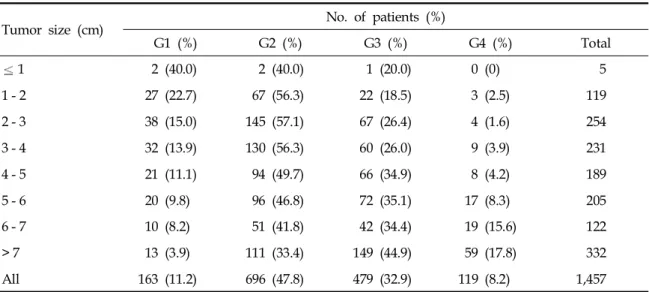
Among 1,457 RCC patients for whom Fuhrman's nuclear grade was available, 11.2% were classed as grade 1, 47.8% as grade 2, 32.9% as grade 3, and 8.2% as grade 4 (Table 4). When these tumors were stratified according to size, the percentages of grades 1 and 2 increased as size decreased whereas the percentages of grades 3 and 4 decreased as size decreased.
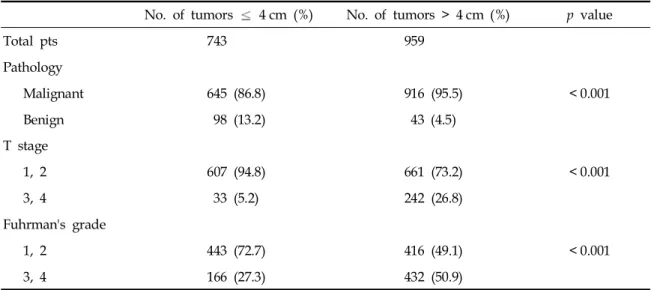
Of the tumors, 743 (43.7%) were 4 cm or less in diameter and 959 (56.3%) were larger than 4 cm in diameter (Table 5). The percentage of benign tumors was significantly greater for tumors equal to or less than 4 cm in diameter (13.2%) than for tumors larger than 4 cm in diameter (4.5%) (p <
0.001). Furthermore, in a subset of 99 tumors 4 cm or less in diameter that were treated by partial nephrectomy, 36 (36%) were benign.
Among RCC patients, the percentage of tumors classed as equal to or higher than stage T3 was significantly lower for tumors equal to or less than 4 cm in diameter (5.2%) than for tumors larger than 4 cm in diameter (26.8%, p < 0.001).
The percentage of Fuhrman's nuclear grades 3 and 4 was significantly less for tumors equal to or less than 4 cm in diameter (27.3%) than for tumors larger than 4 cm in diameter (50.9%) (p <
0.001).
Pathological diagnoses for benign tumors equal to or less than 4 cm in diameter were (in de- creasing order of frequency) AML (41), benign cyst (20), oncocytoma (19), cystic nephroma (2), hemangioma (2), metanephric adenoma (2), and others (9) (Table 6).
Oncological outcome data were available for 1,290 patients. Five-year overall and cancer- Table 1. Clinicopathologic Characteristics of Patients
with Clinical Impression of Renal Malignancy Treated Surgically
Characteristic Patients (n) %
Total 1,702
Mean age (yrs) (range) 55.0 (16 - 86) Gender (male : female ratio) 2.04 : 1
Male 1,142 67.1
Female 560 32.9
Mode of presentation 1,618
Incidental 1,181 73.0
Symptomatic 437 27.0
Treatment 1,702
Radical nephrectomy 1,591 93.5
Partial nephrectomy 111 6.5
Tumor size (cm) (range) 5.4 (0.7 - 24.0)
Benign pathology 141 8.3
T stage (RCC) 1,543
1 1,049 68.0
2 217 14.1
3 254 16.5
4 21 1.4
N stage (RCC) 1,316
0 1,245 94.6
1 71 5.4
M stage (RCC) 1,316
0 1,218 92.6
1 98 7.4
Fuhrman's nuclear grade 1,457
G1 163 11.2
G2 696 47.8
G3 479 32.9
G4 119 8.2
Histological subtype 1,316
Clear cell 1,104 83.9
Papillary 79 6.0
Chromophobe 66 5.0
Collecting duct 5 0.4
Granular 20 1.5
Mixed 12 0.9
Sarcomatoid 9 0.7
Unclassified 2 0.2
Unknown 18 1.4
RCC, renal cell carcinoma.
specific survival was 81.0% and 82.7%, respec- tively (Fig. 1). The presence of symptom at initial presentation, pathological T stage, N stage, M stage, Fuhrman's nuclear grades and tumor size were significant predictors of cancer- specific survival (p < 0.001) whereas tumor subtype was not (p = 0.779). On multivariate analysis, T stage (p < 0.001), N stage (p < 0.001) M stage (p = 0.025) and Fuhrman's nuclear (p <
0.001) grades were significant predictors of cancer-specific survival.
DISCUSSION
In our patients, who constituted 13 - 15% of all surgically managed renal tumor patients in Korea during the past 11 years, the recent global trend toward early diagnosis of small tumors in the absence of symptoms was evident (73.0% of tumors were incidental and 43.7% were equal to or less than 4 cm in diameter). Moreover, the clinical benefit of this trend was clearly evidenced by an increase in the percentage of benign tumors Table 3. Pathological T Stage T3 in Renal Cell Carcinoma Stratified according to Tumor Size*
Tumor size (cm) No. of patients
Cumulative % T3
Total T3 (%)
1 5 0 (0) 0
1 - 2 124 8 (6.5) 6.2
2 - 3 266 10 (3.8) 4.6
3 - 4 245 15 (6.1) 5.2
4 - 5 197 27 (13.7) 7.2
5 - 6 216 37 (17.1) 9.2
6 - 7 128 31 (24.2) 10.8
> 7 362 147 (40.6) 17.8
All 1,543 275 (17.8)
*Among 1,546 renal cell carcinomas, pathological T stage data were available for 1,543.
Table 2. Histopathology of Renal Tumors Stratified according to Tumor Size
Tumor size (cm) No. of patients (%)
Cumulative % benign
Malignancy Benign Total
1 5 (35.7) 9 (64.3) 14 64.3
1 - 2 124 (78.0) 35 (22.0) 159 25.4
2 - 3 268 (89.3) 32 (10.7) 300 16.1
3 - 4 248 (91.9) 22 (8.1) 270 13.2
4 - 5 199 (97.5) 5 (2.5) 204 10.9
5 - 6 217 (96.0) 9 (4.0) 226 9.5
6 - 7 130 (95.6) 6 (4.4) 136 9.0
> 7 370 (94.1) 23 (5.9) 393
All 1,561 (91.7) 141 (8.3) 1,702
and a decrease in the percentage of tumors with high T stages and grades as tumor size decreased.
In a review of 131 consecutive patients who underwent radical nephrectomy between 1989 and 1993, Jayson and Sanders1 found that 61% of tumors were discovered incidentally, which contrasts sharply with the results of Skinner et al.,10 who reported that 7% of tumors were in- cidental between 1935 and 1965 (when CT scanning was not available). A progressive increase of
incidental tumors was also observed in Europe:
from 13.0% in 1982 - 1983 to 59.2% in 1996 - 1997.3 Patard et al.2 reviewed 729 cases of surgically treated renal tumors conducted over 20 years that were divided into 3 distinct periods and observed significant change in the percentage of incidental tumors (24.4% to 51.1%), tumors of 4 cm or smaller in diameter (12.2% to 26.9%), and benign tumors (4.6% to 9.8%) from period 1 to 3. In a retrospective analysis of patients in whom renal tumors were managed surgically, Lee et al.5noted a significant 32% decrease in tumor size from the first to the last year of a recent decade. In that study, 252 of 670 (38%) patients had tumors equal to or less than 4 cm in diameter of which 79%
were discovered incidentally. Similarly, in a recent prospective analysis of 106 renal tumors, 47%
were 4 cm in diameter or smaller.8
Our finding that the incidence of benign tumors among those 4 cm or less in diameter (13.2%) was significantly greater than the incidence of benign tumors among those larger than 4 cm in diameter corroborates with similar reports by others,2,4,8 while the incidence is less than that reported recently by some institutions. Unlike previous studies, our study is a multi-institutional study of 14 training hospitals and, to our knowledge, is the largest of its kind. Multi-institutional studies may reflect the true epidemiology of diseases more accurately than single-institution studies. The
Table 4. Fuhrman's Nuclear Grade in Renal Cell Carcinoma Stratified according to Tumor Size*
Tumor size (cm) No. of patients (%)
G1 (%) G2 (%) G3 (%) G4 (%) Total
1 2 (40.0) 2 (40.0) 1 (20.0) 0 (0) 5
1 - 2 27 (22.7) 67 (56.3) 22 (18.5) 3 (2.5) 119
2 - 3 38 (15.0) 145 (57.1) 67 (26.4) 4 (1.6) 254
3 - 4 32 (13.9) 130 (56.3) 60 (26.0) 9 (3.9) 231
4 - 5 21 (11.1) 94 (49.7) 66 (34.9) 8 (4.2) 189
5 - 6 20 (9.8) 96 (46.8) 72 (35.1) 17 (8.3) 205
6 - 7 10 (8.2) 51 (41.8) 42 (34.4) 19 (15.6) 122
> 7 13 (3.9) 111 (33.4) 149 (44.9) 59 (17.8) 332
All 163 (11.2) 696 (47.8) 479 (32.9) 119 (8.2) 1,457
*Among 1,546 renal cell carcinomas, data on Fuhrman's nuclear grade were available for 1,457.
Fig. 1.Cancer-specific survival curve of 1,290 patients who underwent radical or nephron-sparing surgery for renal cell carcinoma.
percentage of benign tumors reported by indivi- dual institutions may be subject to inter-institu- tional bias due to differences in policies regarding the management of renal tumors. For instance, some surgeons may prefer surgery for small incidentally detected complex renal cysts or solid renal tumors with equivocal fat density while others will opt for regular follow-up assessment.
Consequently, a more aggressive attitude toward equivocal tumors may result in a higher per- centage of benign tumors. On the other hand,
multi-institutional studies reflect average results from several institutions. Valeri et al.11 presented data on 2139 renal solid tumors from surgeries at 5 French tertiary centers during 1 year. The percentage of benign tumors in the subset of 722 tumors equal to or less than 4 cm in diameter varied widely between centers and ranged from 4.7 to 29% with a mean of 8.7%, which was lower than our result.
An association between nephron-sparing surgery and a particularly high percentage of benign tumors has been observed in our as well as other studies. According to Marszalek et al.7 who reviewed a recent series of 129 patients who underwent nephron-sparing surgery for renal tumors less than 5 cm in diameter, 32.6% were benign. Similarly, the incidence of benign pathology in our subset of tumors equal to or less than 4 cm in diameter and treated by partial nephrectomy was 36%. The reason for this association is not clear and could be criticized for the lack of supporting evidence. However, it is likely that small renal tumors exhibiting radiolo- gical features favoring benignity that would have been closely observed in the past are increasingly being treated by nephron-sparing approach.
Accumulation of results from contemporary nephron-sparing surgery series could elucidate this issue in the future.
Table 5. Histopathological Characteristics of Renal Tumors according to Tumors 4 cm and > 4 cm*
No. of tumors 4 cm (%) No. of tumors > 4 cm (%) p value
Total pts 743 959
Pathology
Malignant 645 (86.8) 916 (95.5) < 0.001
Benign 98 (13.2) 43 (4.5)
T stage
1, 2 607 (94.8) 661 (73.2) < 0.001
3, 4 33 (5.2) 242 (26.8)
Fuhrman's grade
1, 2 443 (72.7) 416 (49.1) < 0.001
3, 4 166 (27.3) 432 (50.9)
*Data on pathological T stage and Fuhrman's nuclear grade were available for all 1,543 and 1,457 renal cell carcinomas, respectively.
Table 6. Histopathology of Renal Tumors 4 cm in Diameter
Patients (n) %
RCC 640 86.1
Other malignancy 5 0.7
AML 41 5.5
Benign cyst 20 2.7
Oncocytoma 19 2.6
Cystic nephroma 2 0.3
Hemangioma 2 0.3
Metanephric adenoma 2 0.3
Others 12 1.6
RCC, renal cell carcinoma; AML, angiomyolipoma.
The ethnic difference between Asians and Caucasians may be a potential cause for the relatively low incidence of benign tumors in our study. To our knowledge, however, no study has properly addressed this issue. The percentage of oncocytoma was 2.6% in our subset of renal tumors equal to or less than 4 cm in diameter and 2.0% for all tumors (data not shown).
Oncocytoma is the most common benign renal cortical tumor and represents 3-7% of renal tumors although the incidence seems to have increased recently.7,8,12 Although the reason for the distinctively low percentage of oncocytoma in our series is unclear, this may indicate that there are ethnic differences in the incidence of oncocytoma. To our knowledge, the greatest number of renal oncocytomas treated at a single center in Asia (16) was reported in the English literature by Yen et al.,13 who emphasized the relative rarity of oncocytoma in Eastern compared to Western countries and suggested genetic or environmental differences or simple underdiagnosis as possible causes. However, the proportional contribution of oncocytoma to all treated renal tumors was not given in their study. Our study presents objective evidence to support the view that Eastern people are, for unknown reasons, less prone to oncocytoma than Western people. A low incidence of oncocytoma may have contributed to the comparatively low percentage of benign tumors in our study.
In contrast to the low incidence of oncocytoma, a relatively high incidence of AML in our patients could be pointed out. As with oncocytoma, ethnic difference could be suggested as a possible reason but no pertaining evidence is found in the literature. On the other hand, our 5.5% incidence of AML may not be unusually high as the reported incidence of AML among surgically treated renal tumors ranges from 2 to 5.9%.4,7,14,15 In 14% of AMLs, fat cannot be identified with CT scan, presumably related to a reduced proportion of mature adipose tissue.16 In these fat poor AMLs, a definite diagnosis cannot be made by the imaging technique and should be managed like all enhancing renal masses.17 It is unlikely that fat content was not well examined by preoperative CT scan in our small AML cases. It is probable that most of these tumors fall under
the category of fat poor AML although patholo- gical review was not performed to confirm that these tumors satisfy the pathological definition of fat poor AML as suggested by Milner et al.17
The increased incidence of benign small renal tumors revealed by our series and those of others may increase awareness of the usefulness of preoperative biopsy. It can be assumed that the preoperative biopsy rate was negligible in our series because none of the participating centers were performing it routinely at the time of the study. In a prospective study on 100 consecutive preoperative renal tumor biopsies, accuracy was 72%, sensitivity was 83%, and specificity was 33%.18 In another retrospective study of percutaneous renal mass biopsy, the sensitivity and negative predictive value for masses 3 cm and less in diameter were 84% and 60%, respectively.19Because the accuracy of renal mass biopsy is low, we believe that the value of preoperative biopsy for obviating unnecessary surgical excision of small renal masses is questionable.
Our study is limited by the failure to have the pathological slides reviewed and agreed upon by the pathologists of the participating centers.
Although the reviewing process would have strengthened the validity of the pathological data, it was not considered vital for our study. In a previous multicenter study of RCC conducted by pathologists at 20 institutes in Korea, the pro- portions of each histological subtype 86.3% clear cell, 7.3% papillary, and 6.16% chromophobe were similar to ours, supporting the validity of our subtype data.20
In this multi-institutional study on the histopa- thology of surgically removed renal tumors, small tumor size was an important predictor of favorable histopathologies such as benign tumors, low T stages, and low Fuhrman's nuclear grades.
These data and the high incidence of benign pathology in our small subset of partial nephrec- tomy performed for tumors equal to or less than 4 cm should facilitate the adoption of partial nephrectomy by Korean urologists. However, accumulation of experience and oncological outcome data will be necessary to firmly support partial nephrectomy as a viable surgical option for small renal tumors.
REFERENCES
1. Jayson M, Sanders H. Increased incidence of serendipi- tously discovered renal cell carcinoma. Urology 1998;
51:203-5.
2. Patard JJ, Tazi H, Bensalah K, Rodriguez A, Vincendeau S, Rioux-Leclercq N, et al. The changing evolution of renal tumours: a single center experience over a two-decade period. Eur Urol 2004;45:490-4.
3. Luciani LG, Cestari R, Tallarigo C. Incidental renal cell carcinoma-age and stage characterization and clinical implications: study of 1092 patients (1982-1997). Urology 2000;56:58-62.
4. Duchene DA, Lotan Y, Cadeddu JA, Sagalowsky AI, Koeneman KS. Histopathology of surgically managed renal tumors: analysis of a contemporary series. Urology 2003;62:827-30.
5. Lee CT, Katz J, Shi W, Thaler HT, Reuter VE, Russo P. Surgical management of renal tumors 4 cm. or less in a contemporary cohort. J Urol 2000;163:730-6.
6. Lam JS, Shvarts O, Pantuck AJ. Changing concepts in the surgical management of renal cell carcinoma. Eur Urol 2004;45:692-705.
7. Marszalek M, Ponholzer A, Brössner C, Wachter J, Maier U, Madersbacher S. Elective open nephron-sparing surgery for renal masses: single-center experience with 129 consecutive patients. Urology 2004;64:38-42.
8. Dechet CB, Sebo T, Farrow G, Blute ML, Engen DE, Zincke H. Prospective analysis of intraoperative frozen needle biopsy of solid renal masses in adults. J Urol 1999;162:1282-4; discussion 1284-5.
9. Guinan P, Sobin LH, Algaba F, Badellino F, Kameyama S, MacLennan G, et al. TNM staging of renal cell carcinoma: Workgroup No. 3. Union International Contre le Cancer (UICC) and the American Joint Committee on Cancer (AJCC). Cancer 1997;80:992-3.
10. Skinner DG, Colvin RB, Vermillion CD, Pfister RC, Leadbetter WF. Diagnosis and management of renal cell carcinoma. A clinical and pathologic study of 309 cases. Cancer 1971;28:1165-77.
11. Valeri A, Lang H, Taccoen X, Skowron O, Descotes JL, Coulange C, et al. Pathological features of kidney masses 4 cm or less: the less frequently malignant nature compared to larger tumours, supports the interest of nephron sparing surgery (abstract). Eur Urol Suppl 2005;4:49.
12. Lieber MM. Renal oncocytoma. Urol Clin North Am 1993;20:355-9.
13. Yen TH, Chen Y, Lin JL, Ng KF. Renal oncocytoma in Taiwan. Ren Fail 2006;28:141-7.
14. Jeschke K, Peschel R, Wakonig J, Schellander L, Bartsch G, Henning K. Laparoscopic nephron-sparing surgery for renal tumors. Urology 2001;58:688-92.
15. Snyder ME, Bach A, Kattan MW, Raj GV, Reuter VE, Russo P. Incidence of benign lesions for clinically localized renal masses smaller than 7 cm in radiological diameter: influence of sex. J Urol 2006;176:2391-5;
discussion 2395-6.
16. Lemaitre L, Claudon M, Dubrulle F, Mazeman E.
Imaging of angiomyolipomas. Semin Ultrasound CT MR 1997;18:100-14.
17. Milner J, McNeil B, Alioto J, Proud K, Rubinas T, Picken M, et al. Fat poor renal angiomyolipoma:
patient, computerized tomography and histological findings. J Urol 2006;176:905-9.
18. Dechet CB, Zincke H, Sebo TJ, King BF, LeRoy AJ, Farrow GM, et al. Prospective analysis of computerized tomography and needle biopsy with permanent sectioning to determine the nature of solid renal masses in adults. J Urol 2003;169:71-4.
19. Rybicki FJ, Shu KM, Cibas ES, Fielding JR, vanSonnenberg E, Silverman SG. Percutaneous biopsy of renal masses: sensitivity and negative predictive value stratified by clinical setting and size of masses.
AJR Am J Roentgenol 2003;180:1281-7.
20. Kim H, Cho NH, Kim DS, Kwon YM, Kim EK, Rha SH, et al; Genitourinary Pathology Study Group of the Korean Society of Pathologists. Renal cell carcinoma in South Korea: a multicenter study. Hum Pathol 2004;35:
1556-63.