Polydeoxyribonucleotide Improves Peripheral Tissue Oxygenation and Accelerates Angiogenesis in Diabetic Foot Ulcers
8
0
0
전체 글
(2)
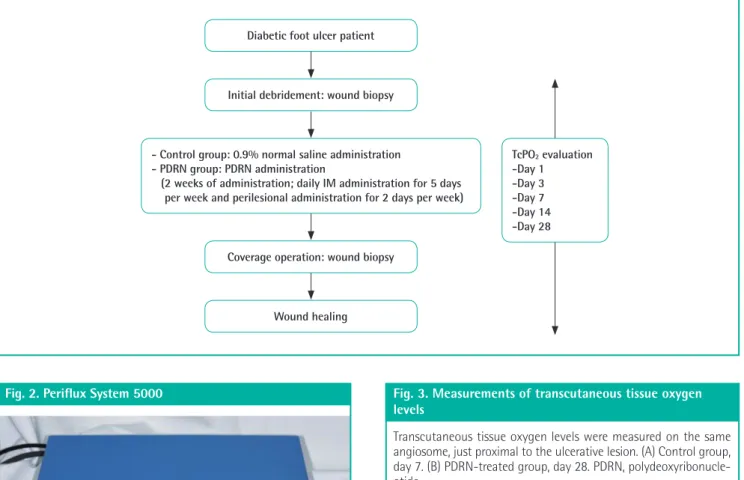
(3)
(4)
(5)
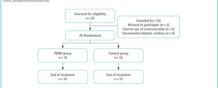
(6)
(7)
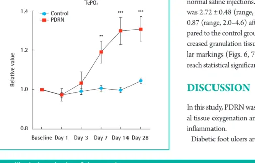
(8)
수치
관련 문서
The purpose of this study was to investigate how the 8-week aerobic exercise program affects the physical strength and stress of obese women in the
The aim of this study was not only to compare the differences of ego-resilience between students with high experience and students with low experience in
The aim of this study was to investigate the removal of phosphorus in combined sewer overflow(CSO) using rapid coagulation system(plug flow reactor +
The aim of this research was to investigate the differences of intentions and satisfactions to classes according to middle school students’ gender
This study was conducted to investigate the effects of a 12-week swimming exercise program on body composition, blood lipids and inflammatory indicators in
Objective: The purpose of this study was to investigate the expression of syndecan-1, E-cadherin, and beta-catenin in tissue sections of normal sun-damaged skin,
In this study, front kicks and take action: The time required for, toe kicks back the speed, trajectory of the foot, floor 30 feet to measure the capacity
The aim of this study was to develop a training program for swallowing and to test its effect on swallowing capacity and nutritional status in stroke
