The Effect of Delayed Administration of Green Tea
Polyphenol, (-)-epigallocatechin-3-gallate, on the Change of Putrescine Level and Hippocampal Neuronal Cell
Damage after Transient Global Ischemia in Gerbil
Seong-Ryong Lee, M.D., Ph.D.,1 Sang-Pyo Kim, M.D., Ph.D.,2 Man-Bin Yim, M.D., Ph.D.3
Departments of Pharmacology,1 Pathology,2 Neurosurgery,3 School of Medicine Keimyung University, Daegu, Korea
Objective:(-)-Epigallocatechin gallate(EGCG) ‘a green tea polyphenol’ is a potent antioxidant and known to reduce the free radical-induced lipid peroxidation. In our previous study, systemic administration of EGCG immediately after ischemia has been shown to inhibit the hippocampal neuronal damage in the gerbil model of global ischemia. Polyamines, especially putrescine(PU) is thought to be important in the generation of brain edema and neuronal cell damage associated with various types of excitotoxic neuronal injury. We investigate the effects of delayed administration of EGCG on the changes in polyamine levels and neuronal damage after transient global ischemia in gerbils.
Methods:To produce transient global ischemia, both common carotid arteries were occluded for 3 min with micro-clips. The gerbils were treated with EGCG(50mg/kg, i.p.) immediately or 2hr after ischemia. Putrescine levels were examined in the cerebral cortex and hippocampus 24 hours after ischemia using high per-formance liquid chromatography.
Results:PU levels in the cerebral cortex and hippocampus were increased significantly after the ischemia.
The administrations of EGCG immediately after the ischemia attenuated the ischemia-induced increase of PU level, however, 2 hr delayed EGCG administration did not reduce the increase of PU level. EGCG administered immediately or 2 hr after ischemia significantly reduced neuronal damage in the hippocampal CA1 region, res- pectively.
Conclusion:These findings suggest that EGCG may has a promise in the management of stroke.
KEY WORDS:Global ischemia·Gerbil·Neuroprotection·Green tea·Polyphenol·(-)-Epigallocatechin gallate.
Introduction
The naturally occurring polyamines in mammalian cells are putrescine(PU), spermidine(SD), and spermine(SM) that play an essential role in the process of cellular growth, de- velopment, and differentiation40,45). Endogenous polyamines have multiple effects in the central nervous system and have been suggested to be neurotransmitters or neuromodulators47). Various kinds of stressful stimuli including stresses, seizures, excitotoxic conditions, and traumatic brain injuries increase the polyamines responses2-4,17,18,30,33). The changes in brain polyamine levels after brain ischemia have been studied4,15,38,39)
and polyamine, especially putrescine is thought to be im- portant in the generation of brain edema, blood-brain barrier breakdown, and neuronal cell damage associated with vari- ous type of brain injury including brain ischemia and trauma.
Strategies including the inhibition of polyamine metabolism have been reported to have neuroprotective effect against ischemic neuronal injury4,15,22,23).
The chemical composition of green tea contains many po- lyphenolic compounds, generally known as catechins. The main catechins in green tea are (-)-epicatechin(EC), (-) -epicatechin gallate(ECG), (-)-epigallocatechin(EGC), and (-)-epigallocatechin gallate(EGCG). Among them, EGCG is the most active major polyphenol of green tea and prima- rily responsible for the green tea effect. In addition, EGCG has been demonstrated to display a potent antioxidant pro- perties18). EGCG possesses two triphenolic groups in its structure, which are thought to be important for its stronger
• Received:April15, 2002 • Accepted:May29, 2002
• Address for reprints:Man-Bin Yim, M.D., Ph.D., Department of Neu- rosurgery School of Medicine Keimyung University 194 Dong-san dong, Taegu, 700-712 Korea
Tel:053)250-7332, Fax:053)250-7356 E-mail:[email protected]
antioxidant action34). It is important to note that EGCG acts as an antioxidant in biological systems. Although the specific mechanisms of its antioxidant actions remain unclear, several pharmacological antioxidant properties of EGCG have been identified such as:(a) free radical scavenging activity or attenuation of lipid peroxidation due to various forms of radicals19,26,42);(b) inhibition of xanthine oxidase activity1); and (c) blockade of inducible nitric oxide synthase(iNOS) and neuronal nitric oxide synthase(nNOS) induction9,32). Th- ese action mechanisms may contribute to the potent antioxi- dant and putative neuroprotective actions of EGCG.
Oxygen free radical-induced lipid peroxidation has been strongly suggested to play an important role in the pathoge- nesis of delayed neuronal damage after global ischemia21). Using in vitro and in vivo models, recent studies suggest the protective effects of green tea extract and EGCG on neuronal damage induced by free radical attack19,31). In previous study, we reported systemic administration of EGCG reduced neu- ronal damage following transient global ischemia29). In the present study, we examined whether EGCG reduces PU level changes in brain regions and neuronal damage in the gerbil hippocampus after transient global ischemia.
Materials and Methods
Animals
Male Mongolian gerbils(Meriones ungiculatus) weighing 60-80g were used in this study. These animals were housed in laboratory cages and maintained on a 12-hour light-dark cycle, with ad libitum access to food and water throughout the study period. The gerbils were treated with EGCG(50mg/
kg, i.p., purchased from Sigma Chemical Co., St. Louis, MO, USA) immediately or 2 hours after ischemia. EGCG was di- ssolved in normal saline. In the ischemic control groups, the vehicle(normal saline, i.p.) was administered immediately or 2 hours after ischemia. In this study, we used 62 gerbils totally and the animals were divided according to the experi- mental groups as follows. (1) sham-operated group:all of procedures were same with other except arterial occlusion (n=10, 5 for polyamine assay and 5 for histology). (2) veh- icle 0 group:ischemic damaged group treated with vehicle immediately after ischemia(n=12, 6 for polyamine assay and 6 for histology). (3) EGCG 0 group:ischemic damaged gr- oup treated with EGCG immediately after ischemia(n=13, 5 for polyamine assay and 8 for histology). (4) vehicle 2 gr- oup:ischemic damaged group treated with vehicle 2 hours after ischemia(n=12, 7 for polyamine assay and 5 for histology). (5) EGCG 2 group:ischemic damaged group treated with EGCG 2 hours after ischemia(n=15, 6 for polyamine assay
and 9 for histology).
Surgery
The gerbils were anesthetized with chloral hydrate(400mg/
kg, i.p.). In the supine position, a midline ventral incision of 2cm was made in the neck. Both common carotid arteries were exposed, separated carefully from the vagus nerve, and occluded for 3 minutes with micro-clips. Blood flow during the occlusion and reperfusion after removal of the clips was confirmed visually and the incision was closed. The rectal temperature was monitored and maintained at 37±0.5℃
with a feedback-controlled heating pad(CMA, Stockholm, Sweden) and an incandescent light was placed over the head from the induction of anesthesia until 3 to 4 hours after ischemia and placed in warm box(at about 30℃) for 3 hours to avoid the biased results by hypothermia28). In the sham group, the neck incision was made only to expose both co- mmon carotid arteries without occlusion. Other procedures were identical to those of other groups.
Polyamine extraction and high performance liquid chromatography(HPLC) analysis
The animals were sacrificed 24 hours after ischemia for polyamine extraction4,39). The brains were removed rapidly from the skull and dissected into cerebral cortex and hippo- campus. The extraction procedure was carried out in ice- chilled conditions. Derivation and HPLC analysis of polya- mines were based upon the method of Spragg and Hutchings43) with some modification. Each brain sample was homogenized with a glass tissue homogenizer in 10 volumes of ice-chilled 0.4M perchloric acid containing 2mM disodium EDTA and 1,8-diaminooctane 4×10⁻⁵M as an internal standard. The homogenate was centrifuged at 12,000g for 10 minutes at 4℃ and 100μl of the supernatant was evaporated by a vacuum drier. The dried tissue was dissolved in 100μl of 1M sodium bicarbonate then deprived with 300μl of 4-fluoro-3- nitrobenzotrifluoride(FNBT) reagent(a mixture of 10μl of FNBT and 1ml of dimethyl sulfoxide) at 60℃ for 20 minutes.
At the end of derivation, 40μl of 1M histidine in 1M so- dium bicarbonate was added to the reaction mixture then the derivation continued for another 5 minutes to scavenge excess FNBT. After cooling the mixture in an ice basket, the N-2- nitro-4-trifluoromethylphenyl derivatives of polyamines were extracted twice with 2ml of 2-methylbutane. After centri- fugation at 3,000g for 10 minutes, the organic phase was evaporated under nitrogen gas flow and the residue was re- constituted with 1.0ml of HPLC grade methanol. The 20μl of the methanol solution was applied to the isocratic reversed phase HPLC system(Gilson Medical Electronics, Villiers-le-
Bel, France), then the separation of NTP-polyamines was accomplished by elution of acetonitrile-water(85:15, v:v) mobile phase at the flow rate of 1.0ml/min within 30 min.
The eluent was monitored by UV/VIS detector set at 242nm and a MicrosorbTM C18 column(5μM, 4.6mm×25cm, Ra- inin instrument Co. Woburn, Mass, USA) was used30).
Histology
The gerbils were sacrificed 5 days after ischemia. They were deeply anesthetized with diethyl ether and perfused transcardially with cold heparinized phosphate-buffered sa- line(PBS, pH 7.2) and 10% formalin in PBS. The brains were removed from the skull and fixed in the same fixative for 24 to 48 hours. Thereafter the brains were embedded in paraffin and representative coronal sections(6-μm thick), which included the dorsal hippocampus, were obtained with a rotary microtome. Tissue sections were stained with hema- toxylin and eosin. A blinded investigator performed the his- tological examination. The hippocampal CA1 damage was determined by counting the surviving pyramidal neurons27). The mean number of CA1 pyramidal neurons per millimeter for both hemispheres in a section of dorsal hippocampus was calculated for each group of the gerbils.
Statistics
Statistical analysis was performed using ANOVA followed by Scheffe’s post-hoc test and significance refers to results where p<0.05 was obtained.
Results
Effect of delayed administration of EGCG on the changes in PU levels
The changes in polyamine levels were examined 24 hours after ischemia. The PU levels of the cerebral cortex increased after ischemia compared with sham-operated group(Fig. 1). In the hippocampus, the PU levels also increased after ische- mia when compared to the sham-operated group(Fig. 2).
EGCG administered immediately after ischemia attenuated the increases of the cortical and hippocampal PU levels(res- pectively p<0.01, Fig. 1 and 2). However, EGCG admini- stered 2 hours after ischemia failed to attenuate the increases of the cortical and hippocampal PU levels(Fig. 1 and 2).
Histology
Histological examination of the nervous system demon- strated marked cell damage in the hippocampal CA1 region in the gerbils treated with a vehicle when compared with the sham-operated group. CA1 pyramidal neurons showed pyk- nosis, eosinophilia, karyorrhexia, and chromosome conden- sation in the vehicle-treated group(Fig. 3). This neuronal cell damage was suppressed by EGCG administration. EGCG administered immediately or 2 hours after ischemic insult significantly reduced neuronal damage(p<0.001 and p<0.001, respectively, Fig. 4).
Relative Concentration of Putrescine(%)
250
200
150
100
050
000
Sham
(n=5) (VEH0 n=6) (VEH2 n=5) EGCG0 (n=7) EGCG2 (n=6)
**
Cortex
Fig. 1. Changes of putrescine(PU) levels in gerbil cerebral cor- tex after global ischemia. Sham, sham-operated(n=5); VEH0, vehicle-treated immediately after ischemia(n=6); VEH2, vehi- cle-treated 2 hours after ischemia(n=5); EGCG0, group treated with EGCG immediately after ischemia(n=7), and EGCG2, gr- oup treated with EGCG 2 hours after ischemia(n=6). Data ex- pressed as mean±SEM. **p<0.01 for the comparison between group treated with EGCG and VEHO.
Relative Concentration of Putrescine(%)
250
200
150
100
050
000 Sham
(n=5) (VEH0 n=6) (VEH2 n=5) EGCG0 (n=7) EGCG2 (n=6)
**
Hippocampus
Fig. 2. Changes of putrescine(PU) levels in gerbil hippocam- pus after global ischemia. Sham, sham-operated(n=5); VEH0, vehicle-treated immediately after ischemia(n=6); VEH2, vehi- cle-treated 2 hours after ischemia(n=5); EGCG0, group treated with EGCG immediately after ischemia(n=7), and EGCG2, group treated with EGCG 2 hours after ischemia(n=6). Data expressed as mean±SEM. **p<0.01 for the comparison bet- ween group treated with EGCG and VEHO.
Discussion
It is suggested that polyamines released from necrotic neurons into the extracellular compartment bind to the NM- DA receptor of cells located in close vicinity and thus render neurons vulnerable to subtoxic levels of excitotoxins. Several researchers examined the changes in brain polyamine levels after focal or global ischemia4,15,39). Various kinds of stimuli or stresses such as seizures, excitotoxicity, and traumatic brain injury modify the ornithine decarboxylase(ODC), the regulatory enzyme in the polyamine biosynthesis2,3,8,16,33)
. These changes may be related to modifications of intrace- llular calcium ion fluxes because polyamines increase the cytosolic amino acids. Some authors have shown discrepan- cies between ODC activity and the concentration of polya- mine46), a finding suggesting that the latter might be more useful than the former.
In this study, PU levels in cortex and hippocampus incre-
ased after transient global ischemia. These changes in PU levels bear a strong similarity to those described by Paschen et al39). The diamine precursor of polyamines, PU is norm- ally in low level and long lasting accumulation of PU may be harmful39). An association between brain damage and high PU levels in the ischemic brain has also been found previ- ously suggesting a role for PU in mediating the ischemic damage. ODC and polyamines are thought to be important in the generation of edema and neuronal cell loss associated with cerebral ischemia39). Baskaya et al3) suggested that poly- amines may play a role in posttraumatic brain edema forma- tion particularly in brain regions.
Polyamines are known to increase cytosolic calcium ion concentration20,24,25) and induce the release of excitatory amino acid8). A remarkable increase of the extracellular concentra- tion of excitatory amino acids including glutamate, induced by cerebral ischemia leading to a large amount of calcium ion influx through glutamate receptor in neurons and neuronal injury6,12,36). PU levels particularly correlate with the density of cell necrosis39). PU might be a reliable marker for acute pathology in brain tissue injury35). Tissue PU increased in the penumbra region that developed brain edema in permanent focal cerebral ischemia4). In addition, the blockade of ODC resulted in a protective effect against focal or global ischemic brain damage22) and partially antagonized the convulsant ac- tivity13) suggesting that polyamine metabolism plays a role in the development of neuronal injuries following brain ische- mia or epileptic seizure. In regarding the effect of EGCG on the PU level, although there is no definite evidences, we can suggest two possibilities. First, EGCG attenuates the harmful accumulation of PU by influence on the polyamine metabo- lism. Second, EGCG-induced neuroprotection due to potent antioxidant effect may decrease the PU response to excito- toxicity.
In this study, SD and SM levels in the cortex and hip- pocampus showed no significant changes after ischemia and EGCG did not show any influences on the SD and SM levels (data not shown). Activation of interconversion pathway enzymes, SD/SM N1-acetyltranseferase48) and PA oxidase5)
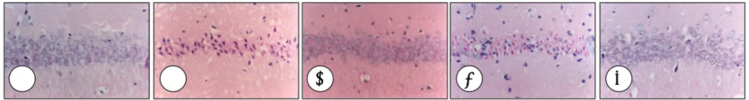
Fig. 3. Microphotographs of the hippocampal CA1 region in the gerbil 5 days after global ischemia(hematoxylin and eosin staining).
CA1 region in sham-operated(A), vehicle-treated immediately after ischemia(B), vehicle- treated 2 hours after ischemia(C), EGCG- treated immediately after ischemia(D), and EGCG-treated 2 hours after ischemia(E). After ischemic insult, only a few normal cells are seen with round cell bodies and clear nuclei and nucleoli. Damaged cells are shrunken and distorted, with small dense nuclear remnants. Bar=50μm.
A A A
A BBBB CC CC DD DD EEEE
Surviving Neuronal Cells in CA1 region per mm
300
250 200 150 100 050
000
Sham
(n=5) (VEH0 n=6) EGCG0 (n=8) (VEH2 n=5) EGCG2 (n=9)
***
***
Fig. 4. Effect of EGCG on the number of surviving cells in the hippocampal CA1 region in the gerbil 5 days after global ische- mia. Sham, sham-operated(n=5); VEH0, vehicle-treated imme- diately after ischemia(n=6); VEH2, vehicle- treated 2 hours after ischemia(n=8); EGCG0, group treated with EGCG immediately after ischemia(n=5), and EGCG2, group treated with EGCG 2 hours after ischemia(n=9). Data are expressed as mean±SEM.
***p<0.001 for the comparison between group treated with EG- CG and their corresponding vehicle-treated groups, respectively.
which convert SM to SD and SD to PU, is a probable major factor in PU accumulation41). Tissue PU levels change to a remarkable degree than those of SD and SM after various pathological conditions2-4,14,24,25,30,38).
The results of this study suggest that administration of EGCG immediately after ischemia can reduce the accumula- tion of PU levels. However, 2 hours delayed administration of EGCG did not reduce PU accumulation. It seemed that polyamine biosynthesis increased rapidly after ischemia.
Therefore, EGCG of delayed administration failed to reduce the ischemia-induced PU accumulation. However, the role of EGCG in inhibition of PU level or polyamine metabolism is not clear in this study and needs to be further studied.
In the present study, the delayed administration of EGCG reduced hippocampal pyramidal neuronal cell damage follo- wing global ischemia. Many researchers have described the effectiveness of green tea in inhibition of carcinogenesis, inflammation, and free radical-induced injury. Recently, the antioxidant effects of EGCG were extensively studied. EGCG shows protective effects against oxidative stress-induced lipid peroxidation in synaptosome19) and the scavenging effect of peroxyl radicals26). Chiou et al10) reported EGCG-induced facilitation of retinal function recovery after retinal ischemia.
We reported the protective effect of EGCG against beta- amyloid protein-induced neuronal damage11). In addition, green tea extract has been shown to protect the liver, kidney and brain from lipid peroxidation injury31,42). EGCG has been demonstrated to pass the blood brain barrier and reach brain parenchyma in animal study44). In previous study, we re- ported protective effect of EGCG against neuronal damage induced by transient global ischemia in the gerbils when ad- ministered immediately after ischemic insults29). In this study, we tried EGCG administration at 2 hours after ischemia. In addition to EGCG treated immediately after ischemia, ani- mals treated EGCG 2 hours after ischemia displayed a signi- ficant increase in the number of surviving neurons in the hippocampal CA1 region.
It has been well known that oxygen radical-induced lipid peroxidation has been strongly suggested to play a role in postischemic neuronal damage9,21). Recently, a variety of studies have examined the neuroprotective properties of antioxidants in brain ischemia7,9,37). Green tea contains many polyphenolic antioxidants and EGCG is the key polyphe- nolic antioxidant responsible for cancer chemoprevention, anti-inflammation, and neuroprotection. Although the mech- anisms underlying the neuroprotective effect of EGCG are not fully understood, in summary, this study demonstrated that EGCG has a neuroprotective effect against hippocampal neuronal damage in a gerbil model of global ischemia.
Conclusion
The present results show that the administrations of EGCG early after ischemia can inhibit the transient global ischemia- induced increase of PU levels in brain regions. EGCG is neuroprotective against neuronal damage even when admini- stered up to 2 hours after global ischemia. Because 2 hours delayed administration of EGCG failed to reduce PA level, PU may partially attribute to neuronal damage process in ischemia. However, the role of polyamines, especially PU, in the pathogenesis of brain ischemia is not clear and needs to be further studied. It seems that the potent antioxidant effects of EGCG contributed to its neuroprotective effect in this study.
These findings suggest that EGCG may have a promise in the acute treatment of stroke.
• Acknowledgement
This work was supported by the research promoting grant from the Keimyung University Dongsan Medical Center in 2000
References
1. Aucamp J, Gaspar A, Hara Y, Apostolides Z:Inhibition of xanthine oxidase by catechins from tea(Camellia sinensis). Anticancer Res 17:4381-4386, 1997
2. Baskaya MK, Rao AM, Prasad MR, Dempsey RJ:Regional activity of ornithine decarboxylase and edema formation after traumatic brain injury. Neurosurgery 38:140-145, 1996
3. Baskaya MK, Rao AM, Puckett L, Prasad MR, Dempsey RJ:Effect of difluoromethylornithine treatment on regional ornithine decarbo- xylase activity and edema formation after experimental brain injury.
J Neurotrauma 13:85-92, 1996
4. Baskaya MK, Rao AM, Dogan A, Donaldson D, Gellin G, Dempsey RJ:Regional brain polyamine levels in permanent focal cerebral is- chemia. Brain Res 744:302-308, 1997
5. Baudry M, Najm I:Kainate-induced seizure activity stimulates the polyamine interconversion pathway in rat brain. Neurosci Lett 171: 151-154, 1997
6. Benveniste H, Drejer J, Schousbe A, Deimer NH:Elevation of extra- cellular concentrations of glutamate and aspartate in rat hippocampus during transient cerebral ischemia monitored by intracerebral micro- dialysis. J Neurochem 43:1369-1374, 1984
7. Block F, Schmitt W, Schwarz M:The antioxidant LY 231617 amelio- rates functional and morphological sequelae induced by global ische- mia in rats. Brain Res 694:308-311, 1995
8. Bondy SC, Walker CH:Polyamines contribute to calcium-stimulated release of aspartate from brain particulate fractions. Brain Res 371: 96-100, 1986
9. Chan PH, Kawase M, Murakami K, Chen SF, Li Y, Calagui B, et al:
Overexpression of SOD1 in transgenic rats protects vulnerable ne- urons against ischemic damage after global cerebral ischemia and reperfusion. J Neurosci 18:8292-8299, 1998
10. Chiou GC, Li BH, Wang MS:Facilitation of retinal function recovery by natural products after temporary ischemic occlusion of central retinal artery. J Ocul Pharmacol 10:493-498, 1994
11. Choi YT, Jung CH, Lee SR, Bae JH, Baek WK, Suh MH, et al:(-)- Epigallocatechin gallate attenuates beta-amyloid-induced neurotoxi- city in cultured hippocampal neurons. Life Sci 70:603-614, 2001 12. Choi DW, Rothman SM:Role of glutamate neurotoxicity in hypoxic-
ischemic neuronal death. Annu Rev Neurosci 13:171-182, 1990 13. de Vera N, Artigas F, Serratosa J, Martinez E:Changes in polyamine
levels in rat brain after systemic kainic acid administration:relation-
ship to convulsant activity and brain damage. J Neurochem 57:1-8, 1991
14. Dempsey RJ, Combs DJ, Olson JW, Maley M:Brain ornithine decar- boxylase activity following transient cerebral ischemia:relationship to cerebral edema development. Neurol Res 10:175-178, 1988 15. Dogan A, Rao AM, Hatcher J, Rao VLR, Baskaya MK, Dempsey
RJ:Effects of MDL 72527, a specific inhibitor of polyamine oxidase, on brain edema, ischemic injury volume, and tissue polyamine levels in rats after temporary middle cerebral artery occlusion. J Neurochem 72:765-770, 1999
16. Gardiner IM, de Belleroche J:Reversal of neurotoxin-induced orni- thine decarboxylase activity in rat cerebral cortex by nimodipine, A potential neuroprotective mechanism. Stroke 21:93-94, 1990 17. Gilad GM, Gilad VH:The brain polyamine-stress-response:recurr-
ence after repetitive stressor and inhibition by lithium. J Neurochem 67:1992-1996, 1992
18. Gilad GM, Gilad VH:Polyamines in neurotrauma;Ubiquitous mole- cules in search of a function. Biochem Pharmacol 44:401-407, 1992 19. Guo Q, Zhao B, Li M, Shen S, Xin W:Studies on protective mechan- isms of four components of green tea polyphenols against lipid pe- roxidation in synaptosomes. Biochim Biophys Acta 1304:210-222, 1996
20. Iqbal Z, Koenig H:Polyamines appear to be second messengers in mediating Ca++ fluxes and neurotransmitter release in potassium- depolarized synaptosomes. Biochem Biophys Res Commun 133: 563-573, 1985
21. Kawase M, Murakami K, Fujimura M, Morita-Fujimura Y, Gasche Y, Kondo T, et al:Exacerbation of delayed cell injury after transient global ischemia in mutant mice with CuZn superoxide dismutase deficiency. Stroke 30:1962-1968, 1999
22. Kindy MS, Hu Y, Dempsey RJ:Blockade of ornithine decarboxylase enzyme protects against ischemic brain damage. J Cereb Blood Flow Metab 14:1040-1045, 1994
23. Kish SJ, Wilson JM, Fletcher PJ:The polyamine synthesis inhibitor alpha- difluoromethylornithine is neuroprotective against N-methyl- D-aspartate- induced brain damage in vivo. Eur J Pharmacol 209: 101-103, 1991
24. Koenig H, Goldsteine AD, Lu CY:Polyamines regulate calcium flu- xes in a rapid plasma membrane response. Nature 305:530-534, 1983
25. Koenig H, Goldstone AD, Lu CY, Trout JJ:Polyamines and Ca++
mediate hyperosmolal opening of the blood-brain barrier:in vitro stu- dies in isolated rat cerebral capillaries. J Neurochem 52:1135-1142, 1989
26. Kondo K, Kurihara M, Miyata N, Suzuki T, Toyoda M:Scavenging mechanisms of (-)-epigallocatechin gallate and (-)-epicatechin gallate on peroxyl radicals and formation of superoxide during the inhibitory action. Free Radical Biol Med 27:855-863, 1999 27. Lee SR, Cheun JK:Propofol administration reduces hippocampal ne-
uronal damage induced by kainic acid in rats. Neurol Res 21:225-228, 1999
28. Lee SR, Kim SP, Kim JE:Protective effect of topiramate against hippocampal neuronal damage after global ischemia in the gerbils.
Neurosci Lett 281:183-186, 2000
29. Lee SR, Suh SI, Kim SP:Protective effects of the green tea polyphenol (-)-epigallocatechin gallate against hippocampal neuronal damage after transient global ischemia in gerbils. Neurosci Lett 287:191-194, 2000
30. Lee YK, Lee SR, Kim CY:Melatonin attenuates the changes in po-
lyamine levels induced by systemic kainate administration in rat brains. J Neurol Sci 178:124-131, 2000
31. Lin AM, Chyi BY, Wu LY, Hwang LS, Ho LT:The antioxidative property of green tea against iron-induced oxidative stress in rat brain.
Chin J Physiol 41:189-194, 1998
32. Lin YL, Lin JK:(-)-Epigallocatechin-3-gallate blocks the induction of nitric oxide synthase by down-regulating lipopolysaccharide-indu- ced activity of transcription factor nuclear factor-B. Mol Pharmacol 52:465-472, 1997
33. Martinez E, de Vera N, Artigas F:Differential response of rat brain polyamines to convulsant agents. Life Sci 48:77-84, 1991
34. Matsuo N, Yamada K, Shoji K, Mori M, Sugano M:Effect of tea pol- yphenols on histamine release from rat basophilic leukemia(RBL- 2H3) cells: the structure-inhibitory activity relationship. Allergy 52: 58-64, 1997
35. Najm I, el-Skaf G, Massicotte G, Vanderklish P, Lynch G, Baudry M:Changes in polyamine levels and spectrin degradation following kainate-induced seizure activity:effect of difluoromethylornithine.
Exp Neurol 116:345-354, 1992
36. Olney JW:Excitatory amino acids and neuropsychiatric disorders.
Ann Rev Pharmacol Toxicol 30:47-71, 1990
37. O’Neill MJ, Hicks C, Ward M, Panetta JA:Neuroprotective effects of the antioxidant LY231617 and NO synthase inhibitors in global ce- rebral ischaemia. Brain Res 760:170-178, 1997
38. Paschen W, Schmidt-Kastner R, Djuricic B, Meese C, Linn F, Ho- ssmann, KA:Polyamine changes in reversible cerebral ischemia. J Neurochem 49:35-37, 1987
39. Paschen W, Rohn G, Meese CO, Djuricic B, Schmidt-Kastner R:
Polyamine metabolism in reversible cerebral ischemia: effect of al- pha - difluoromethylornithine. Brain Res 453:9-16, 1988 40. Pegg AE:Recent advances in the biochemistry of polyamines in euk-
aryotes. Biochem J 234:249-262, 1986
41. Rao AM, Hatcher JF, Baskaya MK, Dempsey RJ:Simultaneous assay of ornithine decarboxylase and polyamines after central nervous sys- tem injury in gerbil and rat. Neurosci Lett 256:65-68, 1998 42. Sano M, Takahashi Y, Yoshino K, Shimoi K, Nakamura Y, Tomita I,
et al:Effect of tea(Camellia sinensis L.) on lipid peroxidation in rat liver and kidney:a comparison of green and black tea feeding. Biol Pharm Bull 18:1006-1008, 1995
43. Spragg BP, Hutchings AD:High-performance liquid chromatographic determination of putrescine, spermidine, and spermine after deprivation with 4-fluoro-3-nitrobenzotrifluoride. J Chromatogr 258:289-292, 1983
44. Suganuma M, Okabe S, Oniyama M, Tada Y, Ito H, Fujiki H:Wide distribution of [3H]-(-)-epigallocatechin gallate, a cancer preven- tive tea polyphenol, in mouse tissue. Carcinogenesis 19:1771-1776, 1998
45. Tabor CW, Tabor H:Polyamines. Annu Rev Biochem 53:749-790, 1984
46. Trout JJ, Koenig H, Goldstone AD, Lu CY:Blood brain barrier br- eakdown by cold injury. Polyamine signals mediate acute stimulation of endocytosis, vesicular transport, and microvillus formation in rat cerebral capillaries. Lab Invest 55:622-631, 1986
47. Williams K, Romano C, Dichter MA, Molinoff PB:Modulation of the NMDA receptor by polyamines. Life Sci 48:469-498, 1991 48. Zoli M, Pedrazzi P, Zini I, Agnati LF:Spermidine/spermine N1-acetyl
transferase mRNA levels show marked and region-specific changes in the early phase after transient forebrain ischemia. Mol Brain Res 38:122-134, 1996