1467
난치성 간질을 유발하는 뇌혈관 병변의 수술
*
계명대학교 의과대학 신경외과학교실, 간질센타
이 성 열·손 은 익
= Abstract =
Surgery of Cerebrovascular Lesions Causing Intractable Epilepsy
Sung Yeal Lee, M.D., Eun Ik Son, M.D.
Department of Neurosurgery and Epilepsy Center, Keimyung University School of Medicine, Taegu, Korea
bjective:Traditionally, the main indications for surgery in vascular-related lesion were based upon reduction or control of seizures, reversal of symptoms of deficits related to mass effect, and prevention of hemorrhage or recurrent hemorrhage. However, the results of surgical treatment for seizure control are disappointing in some reports. Here we describe surgical strategies and our experience in treating patients with intractable seizures associated with vascular-related lesions according to sophisticated presurgical and intraoperative evaluation.
Methods:Twelve(4.5%) patients were selected for this study out of total 264 patients with resective epilepsy surgery at our epilepsy center during four years since 1992. All were treated with anticonvulsant agents but became refractory.
These patients operated on under local or general anesthesia for resection surgery, underwent presurgical and intraoperative evaluation for identification of adjacent, beyond or remote epileptogenic area and the eloquent area.
Results:Of these 12 patients, vascular malformations(AVM, cavernous angioma) were 7, overt hemorrhage due to vascular lesion were 2 and intractable ongoing seizure after vascular surgery were 3. Other vascular lesion including occlusive disease, moyamoya disease or previous hemorrhage were excluded in this study. The location of the lesion was mainly temporal and peri-Rolandic areas, and dual pathology was verified in 2 cases of 6 temporal lesion. The surgical outcome(class I;7, II;3, III;1, IV;1) was excellent by Engel’s classification.
Conclusion:Control of seizures related to vascular lesions remains strong indication for surgical resection. For this reason, careful presurgical evaluations are essential to evaluate the remote epileptogenic area, especially in temporal lesion. Intraoperative acute recording(ECoG) and functional mapping by electrical stimulation or SSEP are important for maximal resection of epileptogenic area with minimal sequellae.
KEY WORDS:Arteriovenous malformation(AVM)・Cavernous angioma・Dual pathology・Intractable epilepsy・Elec- trocorticography(ECoG)・Functional brain mapping.
서 론
전통적으로 뇌혈관 질환에 대한 수술은 종괴효과(mass effect)에 의한 신경학적 장애의 호전, 출혈 혹은 재출혈의 방지 외에도 간질의 조절에도 주요 목적이 있다. 그러나 혈 관성 질환의 단순 제거수술후의 간질조절의 결과는 일부에 서 만족스럽지 못한 것으로 알려져 있다. 특히, Piepgras
18)등은 뇌동정맥기형 수술전에 발작이 전혀 없던 환자도 술후 6%에서 새로운 간질이 생기며, 수술전에 발작이 있었던 환 자는 수술후 17%에서 간질이 지속된다고 보고하며, 한편으 로 뇌동정맥기형의 위치에 따른 술후 간질위험에 대한 Cr- awford
4)등의 보고에서 전두-두정엽 부위(peri-Rolandic) 는 57%, 측두엽은 37%로 매우 높은 것으로 되어 있다.
최근 간질의 수술적 치료의 활성화와 함께 그 원인으로 잠 재성 뇌혈관질환과 연관된 경우와 기존 뇌혈관질환의 수술 후에 병발되는 경우가 있는데, 이때 술후 간질의 위험이 높
OOOO
*이 연구는 계명대학교 대학원 학생학술연구 장학금에 의한 것임.
을 때는 술전 또는 수술중에 간질 유발부위와 고도 뇌기능 부위에 대한 검사와 규명을 통한 부가적인 기능수술로서 술 후 환자 상태와 삶의 질의 향상에 도움을 줄 수 있다.
따라서 난치성 간질이 동반된 뇌혈관질환의 치료에서 미 세수술을 통한 단순 병변제거 외에 간질조절을 위한 검사와 수술 전략 및 그 적응증에 대하여, 본 교실의 수술경험을 중 심으로 문헌고찰과 함께 보고하고자 한다.
대상 및 방법
본 연구는 1993년부터 4년간 본 교실에서 시행한 264례 의 난치성간질 수술환자중, 뇌혈관 조영술 및 병리조직 검 사상 혈관성 병변으로 확인된 12례(4.5%)를 대상으로 하 였다. 이중 간질의 원인이 뇌동정맥 기형과 해면상 혈관종등 의 혈관기형이 7례, 뇌혈관 질환에 의한 뇌출혈후 유발된 2 례, 뇌혈관 질환의 수술후 병발된 경우가 3례였다. 그러나, 폐 쇄성 뇌혈관 질환, 모야모야병 혹은 고혈압성 뇌내출혈, 지주 막하 출혈, 뇌경막 동정맥기형, Sturge-Weber 증후군에 의 한 출혈이 원인이 된 경우는 제외하였다.
수술전 검사로는 간질의 발작양상(semiology), 신경영상 검사(MRI, SPECT 등), 뇌파 집중검사(video-EEG moni- toring) 외에 신경심리검사 등을 통해 간질유발부위와 고도 뇌피질 기능부위와의 연관성을 규명하며, 경우에 따라 뇌경 막하 전극을 이용하여 좀더 명확한 검사가 필요한 경우도 있다.
수술중에는 우선 뇌피질의 해부학적 구조와 혈관분포를 자세히 관찰하여 병변의 위치를 확인하며, 뇌피질파검사
(ECoG)와 전기자극 및 SSEP를 이용한 뇌기능 지도화를 통해 후유증을 최소화하면서 일차적인 뇌혈관병변과 간질 유발부위는 최대한 제거하는 재단절제(tailored resection) 를 시행하였다.
이상의 방법으로 간질을 동반한 뇌혈관질환 수술환자 12 례에 대한 일반적 특성과 술전검사, 병변부위에 따른 수술중 검사 및 수술방법, 병리소견 그리고 수술후 결과 등을 임상 분석하였다.
결 과
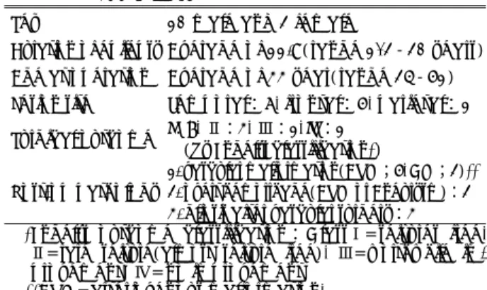
난치성 간질을 동반한 뇌혈관병변 환자 12명의 수술례에 서 일반적 특성을 보면 성별 분포는 남자가 10명으로 많았 고, 간질의 기간은 6개월에서 20년까지로 평균 11.8년이었 으며, 수술당시의 연령은 24세에서 51세까지로 평균 33세
Table 1. Description of 12 patients with vascular lesionrela- ted epilepsy
Sex 10 male and 2 female
Duration of epilepsy Average of 11.8(range 1/2-20 years) Age at operation Average of 33 year(range 24-51) Lesion site Temporal:6, frontal:5, parietal:1 Surgical outcome I:7, Ⅱ:3, Ⅲ:1, IV:1
(by Engel’s classification*) Post-op pathology
1. vascular malformation(AVM;5, CM;2)**
2. overt hemorrhage(AVM or Aneurysm):2 3. gliosis after vascular surgery:3
*Engel’s outcome classification: ClassⅠ=seizures free,
Ⅱ=rare seizure(almost seizure free), Ⅲ=worthwhile im- provement, Ⅳ=no improvement
**AVM=arteriovenous malformation, CM=cavernous malformation
Table 2. Summary of 12 patients with vascular lesion and epilepsy
Case Sex/Age Site* SZ(yr) Pathology** Anesthesia+ Operation++ Outcome
1 M 36 Rt.F 1/2 Small AVM L/P L+C I
2 M 26 Rt.T 11 Small AVM L/P L+ATL+AH I
3 M 51 Rt.T 10 (ICH)MCAaneurysm L/P Clip+ATL+AH II
4 M 27 Rt.T 13 Small AVM L/P L+ATL+AH I
5 M 28 Lt.F 15 Cavernous angioma Gen L II
6 F 24 Lt.P 11 s/p(ICH, AVM) L/P C+MST(LM) IV
7 M 37 Lt.F 20 Large AVM Gen L+C I
8 M 26 Rt.F 20 s/p(AVM, ICH) L/P C+MST(MM) II
9 F 26 Rt.T 6 s/p(ICH, AVM) L/P L+ATL+AH I
10 M 43 Rt.T 14 Small AVM L/P L+ATL+AH III
11 M 40 Rt.T 20 Cavernous angioma Gen L+ATL+AH I
12 M 27 Rt.F 1 s/p(AVM) Gen C+MST I
*F=frontal, T=temporal, P=parietal
**AVM=arteriovenous malformation, ICH=intracerebral hemorrhage
+Gen=general, L/P=local/propofol anesthesia
++L=lesionectomy, C=corticectomy, ATL=anterior temporal lobectomy, AH=amygdalohippocampectomy,
MST=multiple subpial transection, s/p=previous surgery, LM=Language mapping, MM=motor mapping
J Korean Neurosurg Soc/Volume 28/October, 1999 1469 였다. 수술례중 4례는 전신 마취로, 8례는 국소 및 propofol
정맥마취하에서 시행하였다(Table 1, 2).
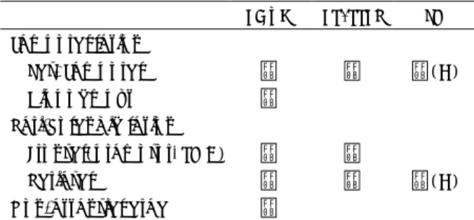
병변 부위는 측두엽이 6례로 가장 많았고, Rolandic area 주변부인 전두엽 5례와 두정엽 1례 순이었다. 이들 부위에 따른 수술중 검사의 종류는 외측-측두엽의 경우 뇌피질파 검사와 전기자극(ES)이나 체감각유발전위검사(SSEP)를 이 용한 뇌기능 지도화(우위측두엽인 경우 언어검사 포함)를, 내 측인 해마부위인 경우는 뇌피질파 검사만 필요하였다. Rol- andic 주변 병소중 전두엽인 전운동영역(premotor area)과 부운동영역(supplementary motor area)은 뇌피질파 검사 와 전기자극이나 체감각유발전위 검사가 필요하며, 두정엽 부위는 뇌피질파 검사, 전기자극이나 체감각유발전위 검사와, 우위반구인 경우 언어 지도화(language mapping)를 시행하 였고, 비특발성 부위(non-essential area)는 뇌피질파 검 사만 필요하였다(Table 4).
이들 수술중 검사를 기준으로 재단절제를 하게되는데, 수 술방법은 측두엽 6례중 외측-측두엽의 병변인 3례는 병변 을 포함한 전측두엽절제술(anterior temporal lobectomy) 과 동시에 편도해마절제술(amygdalo-hippocampectomy) 을 시행하였는데 이중 2례는 병리조직검사상 이중병리(dual
pathology)로 확인되었다. 측두엽 내측인 해마(hippocam- pus)부위에 병변이 있던 3례는 최소침습 전측두엽절제술을 통한 편도해마절제술을 시행하였고, Rolandic area 주변 병 변인 6례중에서 병변제거(lesionectomy)만 시행한 경우는 1 례, 병변제거와 피질절제술(corticectomy)을 같이 시행한 경 우가 5례였고, 이중 3례는 연막하 피질다절술(multiple sub- pial transection)도 같이 시행하였다(Table 3).
병리결과는 뇌동정맥기형이 5례, 해면상 혈관종이 2례, 국 소출혈에 의한 원인이 뇌동정맥기형과 뇌동맥류로 확인된 각 각 1례, 뇌동정맥기형 수술후의 신경교증(gliosis)이 3례였다 (Table 1).
수술후 간질조절은 Engel의 분류상 class I이 7례, II가 3 례, III가 1례로 좋은 경과를 보였으며, 언어영역에 간질유발 병소가 중복되어 있었던 1례가 IV였다(Table 1, 2).
증 례
1. 증 례 1:
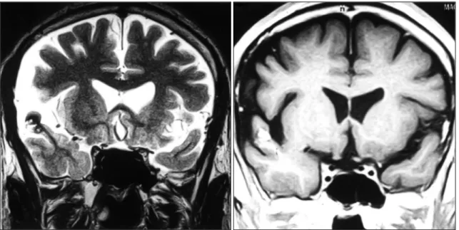
20세경부터 발작이 시작된 40세 남자 환자로서, 14년간 항 경련제를 복용하였으나 1주에 1-2회의 복합 부분발작이 지 속되어, 술전에 발작간, 발작시 뇌파 검사와 CT, MRI, SPE- CT 및 두개강내 뇌파검사를 시행하였다. MRI상(Fig. 1) 우 측 상측두회에 작은 종괴 소견을 보였으며 두개강내 strip을 이용한 뇌파검사상 우측 근심측두엽 간질을 시사하는 소견 을 보였다. Wada 검사상 언어는 좌측 우성이었고 기억력은 우측 5/8, 좌측 7/8 이었다.
술중 뇌피질파 검사와 뇌기능 지도화를 시행하였고 상측 두회에 종괴제거후에 시행한 뇌피질파 검사에서 측두엽 근 심부(mesial)의 해마부위에 비정상 간질뇌파가 지속되어 보였으며, 이를 근거로 전측두엽의 소절제와 편도-해마부 위의 일부제거후 시행한 뇌피질파 검사상 간질파의 소실을 보였다. 술후 병리소견에서 상측두회의 종괴는 해면상 혈 관종이었고 해마부위는 근심 측두엽경화(mesial scle- rosis)로 이중병리(dual pathology)를 나타냈고(Fig. 2), 술후 간질조절결과는 Engel의 분류상 Class I으로 전혀 발 작이 없었다.
2. 증 례 2:
20세 남자환자로 단순 부분발작과 이차성 전신발작을 주 소로 응급실 내원후 시행한 MRI와 혈관조형술상 우측전두 엽에 뇌동정맥기형소견을 보여(Fig. 3) 수술적 가료를 시행하 였다. 술중 뇌동정맥기형 제거시 출혈로 aneurysm clip도 사용하였으나 술후 심한 출혈로 반신마비와 언어장애가 나타
Table 3. Surgical interventions with altenative procedure in 12 cases
Temporal lesion 6 Lesionectomy Lateral temporal 3
Hippocampus 3 With ATL + AH(dual pathology:2)
Peri-rolandic lesion 6 Lesionectomy alone 1 Lesionectomy + Cortisectomy 5 with MST 3
*ATL=Anterior Temporal Lobectomy AH=AmygdaloHippocampectomy MST=Multiple Subpial Transection
Table 4. Categorization of the seizure-related vascular le- sion by location for intraoperative assessment
ECoG ES/SSEP LM
Temporal lesion
Lat. Temporal + + + (D) Hippocmpus +
Peri-Rolandic lesion
Frontal(premotor, SMA) + +
Parietal + + + (D) Non-essential area +
ECoG=Electrocortiography, ES=Electrical stimulation SSEP=Somatosensory evoked potentials
LM=Language mapping, D=dominant,
SMA=Supplementary motor area
났으며 고식적 치료로 호전은 되었으나, 부분발작과 간헐적 전신발작은 다양한 약물치료에도 지속되어 난치성이었다. 술 전 뇌파검사, grid를 이용한 두개강내 뇌파기록을 시행하여 수술 주변부위의 간질유발부위를 확인하고 수술범위를 정한 후 간질수술을 시행하였다.
술 중 뇌피질파 검사와 SSEP를 시행한 후, 간질파가 보이 는 교증(gliosis)부위등의 간질유발 병소는 피질제거술과 함 께 1차 수술시의 clip들을 제거하였고, 간질파가 나타나는 일 부 운동영역에는 연막하 피질 다절술(MST)을 시행하였으며 (Fig. 4), 시술 후 비정상 간질파의 소실을 확인하였다. 술 후 병리 결과는 교증과 함께 hemosiderin의 침착소견을 보였 으며, 환자는 수술 후 Engel의 분류 class I으로 항경련제 없 이 전혀 발작이 없는 상태이다.
Fig. 1. T2 & T1-weighted coronal MRI of 40-year old male presenting complex partial seizures show a cavernous malformation in the right superior temporal gyrus.
Fig. 2. Dual pathology of cavernous malformation(A) and hippocampal neuronal loss(B)(H & E stain, ×100).
Fig. 3. Intraoperative drawing(case 8) based on the ECoG and brain mapping showed the extent of cortical resection and multiple subpial transection(MST) for seizure control.
A A A
A B B B B
J Korean Neurosurg Soc/Volume 28/October, 1999 1471
고 찰
난치성 간질을 동반한 뇌혈관 병변 환자의 치료에서, 간질수 술의 기법을 적용했을 경우에 술 후 간질조절에 실제적인 도 움이 될 수 있을지 여부가 중요한 관건이 되고 있다
8)19)24). 1987년 Wyllie 등
28)은 간질수술을 부가적으로 시행한 13명 의 환자를 단순병변만 제거수술한 결과와 비교를 통해, 술 후 간질조절은 병변의 제거정도 보다는 오히려 간질 유발부위 의 완전제거 여부에 달려있다고 보고하였다. 특히 해면혈관 종인 경우에는 간질조절 자체가 수술의 주된 목적이기도 하 다. 저자의 경험으로도 단순 병변제거외에 부가적인 간질유 발부위 제거를 적절히 시행함으로서 간질조절에 상당한 호 전을 보였다.
간질을 유발하는 뇌혈관 질환중에서 뇌동정맥기형은 전체 인구의 0.14%를 차지하며 20~40대에 호발하는데
18)26), 뇌출혈과 간질등의 주증상을 보이며 이는 청장년기나, 월경 기간, 임신때 심해진다
1)3)16). 이중 간질은 두번째로 많은 임상 증상으로서 뇌내 출혈 없이 간질만 있는 경우는 17~40%로 알려져 있다
4)14)17)18). 간질 유발 기전으로는 혈관의 steal 작용에 의한 병소의 초점성 대뇌허혈(focal cerebral ische- mia), 신경 교증(gliosis), 2차성 간질유발(secondary epile- ptogenesis) 등이 있다
30).
뇌동정맥기형 환자의 간질 유발율에 관한 문헌 보고들을 보면, Crawford 등
4)은 수술을 시행하지 않은 환자 153명 의 추적 관찰에서 20년후 평균 18%이며 특히 측두엽 침범시 37%로 높았으며, 진단 당시의 나이가 10~19세이면 20년후 44%, 30세 이후이면 6%로 어릴수록 간질발작 가능성이 높다고 하였다. 더욱이 간질을 동반한 뇌동정맥기형환자 중 많은 환자들이 처음에는 약물치료에 효과가 있지만 장기간 추적관찰시 결국은 항경련제로 조절이 안되는 것으로 보고
2)되므로 수술치료를 고려하여야 한다.
뇌동정맥기형 수술 자체가 술후 간질 유발의 가장 위험한 요소가 될뿐 아니라, 난치성 간질이 뇌동정맥기형의 수술적 응이 되는 상반되는 점이 있는데, Foy 등
10)은 일반적인 천막 상 개두술을 시행한 환자에서는 17%의 간질 발생 빈도를 보이는데 비해 뇌동정맥 기형 수술후에는 50%의 간질을 동 반한다고 하였고, Crawford 등
5)의 보고에는 뇌동정맥기형 수술후 20년간 추적 관찰상, Peri-Rolandic 부위인 전두엽 과 두정엽 수술시는 57%, 측두엽 수술시는 37%의 높은 간질 발생빈도를 보였다.
Piepgras 등
18)은 술전 간질이 없는 뇌동정맥 기형의 경우 술후 새로운 간질 발생이 6%이나, 술전 간질이 있던 동정맥
기형의 경우 술후 계속되는 간질은 17%로 보고하였으나, Foster 등
9)은 간질을 동반한 뇌동정맥 기형 환자의 수술후 14%에서만 간질 감소의 낮은 빈도를 보인데 비해, Rass- musen
20)은 2/3에서 간질 소실의 빈도를 보인다고 하였고, Yeh 등
29)30)은 뇌동정맥 기형 제거와 함께 간질 발작 병소제 거를 같이 시행하여 3/4에서 좋은 결과를 보여 간질 자체가 수술 적응이라 보고하였으며 간질병소가 국한되어 접근 하 기 쉬운 경우에는 피질절제술이 간질조절을 위한 표준치료 라 하였다. 이때 혈관 기형 주변의 gliosis와 헤모시데린(he- mosiderin) 침착 부위는 간질발작 활성도와 관련이 있으므로 제거하여야 하며, 병변 주위에 대한 뇌피질파등의 수술중 검 사로 정확한 간질부위를 평가한 후 제거 범위를 정하여야 된 다. 또한 병변주위에 관한 검사외에도, 이차성 간질유발 병소 로 근심 측두엽 부위(mesial temporal structure)가 복합 부 분발작의 가장 흔한 부위로 알려져 있는데
15)28), 본 연구에서 도 측두엽 외피질병변에 의한 빈번한 발작으로 인해 이차성 병소로 해마나 편도부위에 경화증(sclerosis)이 보이는 이 중 병리(dual pathology) 소견이 3례중 2례 있었다. 따라서 이러한 병소의 규명을 위한 세밀한 검사도 필요하다.
수술적 치료 이외에 간질조절을 위한 방법으로 Forster 등
9)은 뇌동정맥기형 색전술을, Lunsford 등
12)은 감마나이프(ga- mmaknife) 방사선 수술(radiosurgery)을, Heikkinen 등
11)은 정위적 양성자 치료(Stereotactic proton-beam irradia- tion)등을 이용하여 간질 빈도 감소를 보고하였으나 이는 뇌동 정맥기형 주위의 간질병소에 대해서는 불확실한 목표(target) 로 인해 치료효과에 신빙성이 없는 것으로 알려져 있다.
해면상 혈관종의 경우도 전체 인구의 0.02~0.13%를 차 지하며, 주요증상은 간질, 국소 신경학적 결손, 두통 등으로 알려져 있고, 전두엽 측두엽에 병변시 간질의 위험이 높은 것 으로 보고되고 있다
6)7)23). 해면상 혈관종의 수술후 간질 동 반은 술전 간질의 기간이 길거나 간질 빈도가 많은 경우와 여 자에서 많다고 보고되고 있으며
6)일반적으로 해면상 혈관종 에 대한 Robinson의
21)22)수술적응을 보면 난치성 간질이 있 는 경우(50%)와 이전에 출혈(0.7%)이 있었던 경우는 반드 시(absolute indication) 해야하며, 진행되는 신경증상이 있는 중요부위(eloquent area)의 병변이 접근 가능한 부위에 있 다면 수술이 고려 될 수 있다
22).
해면상 혈관종의 수술방법에서도, 수술후 보다 나은 간질 조
절을 위해서는 단순 병변 제거외에도 부가적인 절제술(addi-
tional corticectomy)이 필요하며, 특히 측두엽 병소인 경
우에는 근심부에 대한 원격절제도 고려되어야 하며, 뇌혈관
병변의 출혈이나 술후 교질화로 인한 간질의 경우도 정확한
간질 유발병소의 이해와 수술적 가료를 하여야 좋은 결과를
얻을수 있다.
이상을 종합하면 난치성 간질을 동반한 혈관성 병변인 경 우의 수술전략은 본 연구에서 시행한 임상증후의 분석, 간질 뇌파등의 전기생리학적검사, MRI나 SPECT 등의 신경영상 검사 그리고 Wada 검사 등을 기본으로 수술을 결정하며, 병 변부위에 따른 특성에 따라, 술중 뇌피질파(ECoG)검사, 기 능적 지도화(functional mapping)를 시행하여(Table 4) 수 술범위를 정할 수 있다. 본 연구에서도 뇌동정맥기형 수술후 간질이 지속되거나 악화된 환자에서 뇌경막하 전극 등을 이용 한 술전, 수술중 검사를 통하여 간질 유발부위를 제거함으 로서 간질 조절이 가능하였다.
결론적으로 간질수술 기법의 적응으로는, 1) 수술 시간이 비교적 적게 걸리는 3cm 이하의 작은 뇌동정맥 기형이나, 2) 해면상 혈관종의 경우이며, 이때 간질유발부위의 확인을 위해 혈관성 병변의 주위(adjacent) 피질에 대한 수술중 검 사가 필요하며, 특히 측두엽 외측부위 병변인 경우에는 원 격(remote) 부위의 이중병변의 가능성도 고려되어야 하며, 3) 뇌 중요부위(eloquent)의 병변인 경우에는 후유증을 최 소화 하기위해 수술중 뇌기능 지도화검사를 반드시 시행하 여야 할 것이다. 이와는 달리 Spetzler grade
25)가 높은 거 대 뇌동정맥기형인 경우나, 술자의 미숙으로 수술 시간이 오래 걸리거나, clip을 사용해야 할 정도의 대량 출혈의 가 능성이 높은 뇌동정맥기형인 경우는 구명(life saving)을 위 한 단순 병변제거가 우선되어야 한다. 그러나 4) 단순병변 제거술후에도 간질이 난치성으로 지속되면, 간질조절을 위한 2차수술을 시행할 수 있다.
결 론
간질을 동반한 뇌혈관 병변의 환자중에서, 병변이 측두엽인 경우나 Spetzler grade가 낮으며 수술 시간이 짧게 걸리는 작 은 뇌동정맥 기형이나 해면상 혈관종의 경우에는 세심한 술 전 수술중 검사를 통해 뇌혈관 병변의 크기와 위치 및 간질양 상 등의 환자 개개인의 특성에 따른 간질수술 전략과 함께, 후 유증의 최소화를 위해 중요 뇌기능 피질부위의 기능적 뇌지도 화를 이용하여 병변주위 혹은 원격부위의 부가적인 피질절제 술(additional corticectomy)을 시행함으로서 새로운 간질의 방지와 술후 간질 조절에 좋은 결과를 보일 것으로 생각된다.
•
논문접수일:1999년 1월 27일•
심사완료일:1999년 7월 19일•
교신저자:손 은 익700-712 대구광역시 중구 동산동 194 계명대학교 의과대학 신경외과학교실
전화:053) 250-7306, 전송:053) 250-7356
References
1) Amacher AL, Allcock JM, Drake CG:Cerebral angiomas:
the sequelae of surgical treatment. J Neurosurg 37
:571-575, 1972
2) Aminoff MJ:Treatment of unruptured cerebral arteriovenous
malformations. Neurology 37
:815-819, 1987
3) Brown RD, Wiebers D, Forbes G, et al:The natural history of
unruptured intracranial arteriovenous malformations. J Ne- urosurg 68
:352-357, 1988
4) Crawford PM, West CR, chadwick PW, et al:Arteriovenous
malformations of the brain
:natural history in unoperated pa- tients. J Neurol Neurosurg Psychiatry 49
:1-10, 1986
5) Crawford PM, West CR, Shaw MD, et al:Cerebral arteriov-enous malformations and epilepsy
:Factors in the develo- pement of epilepsy. Epilepsia 27
:270-275, 1986
6) Cohen DS, Zubay GP, Goodman RR:Seizure outcome after
lesionectomy for cavernous malformations. J Neurosurg 83
:237-242, 1995
7) Curling OD, Kelly DL, Elster AD:An analysis of the natural
history of cavernous angiomas. J Neurosurg 75
:702-708, 1991
8) Fish D, Andermann F, Olivier A:Complex partial seizuresand small posterior temporal or extratemporal structural lesi- ons
:Surgical management. Neurology 41
:1781-1784, 1991
9) Forster DMC, Steiner L, Hakanson S:Arteriovenous malfo-rmations of the brain. A long-term clinical study. J Neurosurg 37
:562-570, 1972
10) Foy DM, Copeland GP, Shaw MDM:The incidence of posto-
perative seizures. Acta Neurochir 55
:253-264, 1981
11) Heikkinen ER, Konnov B, Melnikov L, et al:Relief of epil-
epsy by radiosurgery of cerebral arteriovenous malformations.
Stereotact Funct Neurosurg 53
:157-166, 1989
12) Lunsford LD, Kondziolka D, Flickinger JC, et al:Stereota-
ctic radiosurgery for arteriovenous malformations of the brain.
J Neurosurg 75
:512-524, 1991
13) Michelsen WJ:Natural history and pathophysiology of arte-
riovenous malformations, In
:clinical neurosurgery
:procee- dings of the Congress of Neurological Surgeons. Baltimore
:Williams and Wilkins, 1979, pp307-313
14) Moody RA, Poppen JL:Arteriovenous malformations. J Ne-
urosurg 32
:503-511, 1970
15) Morrell F:Varieties of human secondary epileptogenesis. J
clin Neurophysiol 6
:227-275, 1989
16) Parkinson D, Bachers G:Arteriovenous malformations:
Sum- mary of 100 consecutive supratentorial cases. J Neurosurg 53
:285-299, 1980
17) Perret G, Noshioka H:Report of the Cooperative study of In-
tracranial aneurysms and subarachnoid Hemorrhage. Se-
ction VI. Artetiovenous malformations. An analysis of 545
cases of cranio-cerebral artetiovenous malformations and
J Korean Neurosurg Soc/Volume 28/October, 1999 1473 fistulae re-ported to the Cooperative study. J Neurorosurg
25
:467-490, 1966
18) Piepgras DG, Sundt TM, Ragoowansi AT, et al:Seizure out-
come in patients with surgically treated cerebral arteriovenous malformations. J Neurosurg 78
:5-11, 1993
19) Pilcher WH, Silbergeld DL, Berger MS, et al:Intraoperative
electrocorticography during tumor resection
:impact on sei- zure outcome in patients with gangliogliomas. J Neurosurg 78
:891-902, 1993
20) Rasmussen T:Surgery of epilepsy associated with brain tum-
ors. Adv Neurol 8
:227-239, 1975
21) Robinson JR, Awad IA, Little JR:Natural history of the
carvenous angioma. J Neurosurg 75
:709-714, 1991
22) Robinson JR, Awad IA, Magdinec M:Factors predisposingto clinical disability in patients with cavernous malformations of the brain. Neurosurg 32
(5
):730-736, 1993
23) Simard JM, Bengochea FG, Ballinger WE, et al:Cavernous
Angioma
:A Review of 126 Collected and 12 New Clinical Ca- ses. Neurology 18
(2
):162-172, 1986
24) Spencer DD, Spencer SS, Mattson RH:Intracerebral masses
in patients with intractable partial epilepsy. Neurology 34
:432-436, 1984
25) Spetzler RF, Martin NA:A proposed grading system for
arteriovenous malformation. J Neurosurg 65
:476, 1986
26) Wilkins RH:Natural history of intracranial vascular malfor-mation
;A review. Neurosurg 16
:421-430, 1985
27) Williamson PD, Wiese-HG, Delgado-Eseneta AV:Clinical
characteristics of partial seizures. In
:Engel J Jr. ed. Surgical Treatment of the Epilepsies. New York NY
:Raven Press, 1987, pp101-120
28) Wyllie E, Lders H, Morris HH, et al:Clinical outcome after
complete or partial cortical resection for intractable epilepsy.
Neurology 37
:1634-1641, 1987
29) Yeh HS, Kashiw S, Tew JM, et al:Surgical management of
epilepsy associated with cerebral arteriovenous malformations.
J Neurosurg 72
:216-223, 1990
30) Yeh HS, Tew JM, Gartner M:Seizure control after surgery