Ⅰ. 서 론
임플란트 식립 시 여러 가지 불리한 조건에 흔히 직면하게 된다. 특히 발치 후 장기간 무치악으로 지낸 환자들에 있어 수직적인 골소실 뿐만 아니라 수평적인 골소실로 인해 임플 란트 식립 시 여러 가지 한계점을 나타낸다. 이러한 경우 골 이식은 필수적인 과정이나 골 이식 방법을 선택함에 있어서 도 많은 한계점을 드러낸다. 특히 협, 설로 악골이 심하게 위축되었을 경우 골 이식과 동시에 임플란트 식립은 더욱더 어렵다. 이를 극복하기 위해서 Ridge splitting technique
1)을 고려할 수 있다.
일반적인 GBR이나 골이식은 협,설로 골이 심하게 흡수되
었을 경우 많은 양의 골을 사용해야 하며, 추가적인 titani- um mesh 나 흡수 혹은 비흡수성 차단막이 필요하며, 골 이식과 동시에 임플란트 식립이 거의 불가능하다
2). 또한 Block bone graft를 시행할 경우 이차적인 공여부 술식이 필요하므로 더 많은 시간과 술식이 필요하며 환자가 느끼는 불안과 통증 또한 더 할 것이다. 그러나 Ridge splitting 은 2-3mm정도의 골 폭에서 골을 협-설로 증대시켜 보다 쉽게 임플란트 식립 공간을 확보 해 줄 뿐만 아니라 compaction 효과도 가져다줘 임플란트의 초기 고정을 증가시킬 수 있다 또한 협면과 설면의 피질골이 이식된 골의 housing effect 를 가져다 주며 혈행공급이 충분하므로 osseointegration 또한 증진시킨다
3).
여덕성∙임소연∙이현진∙안미라∙손동석 대구가톨릭대학병원 구강악안면외과학교실
심한 협-설골 위축에서 치조골 수평 확장술을 이용한 골 재건
RECONSTRUCTION OF SEVERE BUCCO-LINGUAL BONE RESORPTION AREA USING
“RIDGE SPLITTING TECHNIQUE
”Duck-Sung Yeo, So-Yeon Lim, Hyun-Jin Lee, Mi-Ra Ahn, Dong-Seok Sohn Dept. of Dentistry, Oral and Maxillofacial Surgery, Daegu Catholic university Hospital
Dental implant has become one of the important option for completely or partially edentulous patients, But it is challenging to reconstruct the severely atrophic ridge. Insufficient bone volume could restrict to place the wide and long implant and because of excessive interocclusal clearance, improper prosthetics could be produced. In this case bone augmentation for implant dentistry is necessary procedure to improve the insufficient bone volume. Therefore, bone augmentation or GBR is the most important procedure for successful implant placement and for ideal crown- root ratio. There are various bone augmentation tech- niques have been introduced recently; like block bone graft, distraction osteogenesis, inlay graft, onlay graft, etc….
In severe bucco-lingual resorption area, ridge splitting is the first choice of the treatment, because it pro- vides a place for implantation and also has compaction effect. This technique may be indicated for sharp mandible and maxillary ridges in patients whose bone quantity is inadequate for primary stabilization. We report that the clinical experience of bone augmentation using ridge splitting technique in bucco-lingual bone resorption area.
Key words: Ridge splitting, Bone augmentation, Implant placement
Abstract
본 증례에서는 상악 전치부
4), 상악 소구치부
5), 하악 대구 치부
6)에서 협, 설로 심하게 위축된 치조골에서 ridge split- ting을 이용한 골 증대술 시행과 동시에 임플란트 식립을 한 증례들을 보고하는 바이다.
Ⅱ. 증례보고
1. 증례1: 상악 전치부
45세의 여성 환자로 특별한 의과적 병력은 없었다. 상악 에 총의치를 장착하고 있으나 유지력 부족을 호소하였고, 방사선 사진에서 좌측 정중부에서 매복된 과잉치가 관찰된 다. 하악의 경우 심한 치주질한으로 인해 모든 치아가 불량 한 예후를 보이고 있다. 또한 Cephalo lateral 사진 상 상 악 치아의 조기 상실로 인한 심한 치조골 흡수로 pseudo class III 양상을 나타내고 있다(Fig. 1~3). 상악 전치부에 심한 수직 및 수평골 흡수를 나타내고 있어 ridge splitting 후 임플란트를 식립하여 Implant supported overdenture 를 계획하였다. 상악 치조골에 midcrestal incision을 가한 후 전층판막을 형성하여 치조골을 노출시켰다. 치조골은 3~4mm의 골폭을 나타냈다(Fig. 4). piezoelectric
device를 이용하여 치조골의 순설측 두께의 정중간에 깊이 약 5mm의 수평적 골절개를 형성한 후 수평적 골절개의 전 후방에 수직적 골절개를 형성하여 수평적 골절개와 연결한 후 (Fig. 5)와 osteotome로 협측 cortical bone을 분리하 였다(Fig. 6). Ridge splitting 후 endopore implant가 식 립되었으며(Fig. 7), 치조골 사이에 존재하는 골결손 부위 는 irradiated allograft(Tutoplast, Germany)를 이식한 후(Fig. 8) 흡수성 차단막으로 Pericardium(Tutoplast, Germany)을 이용하였다(Fig. 9). 약 6개월 후 2차 수술을 시행하였으며 상악 전치부에 충분한 골이 형성 되어 있는 것을 확인할 수 있다(Fig. 10). 동시에 근단 변위 판막술을 시행하여 부족한 부착치은을 보강하였으며(Fig. 11), 2차 수술 2개월 후 최종 보철물을 장착하였다(Fig. 12, 13).
2. 증례2: 상악 소구치부
46세의 남성 환자로 전반적인 구강상태는 양호 하였으나 상악 좌측 제1소구치의 조기 상실로 협,설측으로 심한 흡 수가 보이며 및 제 2소구치의 근심이동이 나타나고 있다 (Fig. 14). 전층 판막을 형성한 후 치조골을 확인한 결과 굉 장히 얇은 치조골 폭이 나타났다(Fig. 15). piezoelectric
Fig. 1. Panoramic view shows severe periodontitis and multiple missing teeth.
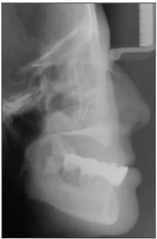
Fig. 2. Cephlaolateral graph shows pseudo class III relationship due to severe atrophied maxilla.
Fig. 3. Intraoral photograph shows residual maxillary second molar.
Fig. 4. Maxillary alveolar bone shows only 3~4mm.
Fig. 5. Osteotomy was done with piezoelectric device.
Fig. 6. Expanded the bone with
osteotome.
device를 이용하여 골 절개를 가한 후(Fig. 16), chisel로 협측 cortical bone을 분리하는 과정에서 buccal cortical bone에 incomplete fracture가 있었으나(Fig. 17), implant 식립 후 fracture site 에 추가적인 irradiated allograft(orthoblast II isotis, USA)으로 bone graft한
후(Fig. 18) 흡수성 차단막인 Pericardium을 이식부에 덮 고 봉합하였다(Fig. 19). 술 후 파노라마 사진에서 임플란 트가 잘 식립된 것을 확인할 수 있다(Fig. 20). 이 후 보철 물은 의뢰한 의원에서 시행하였다.
Fig. 7. Endopore implants were placed. Fig. 8. Bone graft was done with irradi- ated allograft.
Fig. 9. Coverd with resorbable men- brane.
Fig. 10. Photograph shows sufficient bone volume at second surgery.
Fig. 11. Apically repositioning flap was done.
Fig. 12. Intraoral photograph of final prosthetics.
Fig. 13. Panoramic view of final pros- thetics.
Fig. 14. Left maxillary first premolar area shows severe bone resorption.
Fig. 15. Alveolar bone shows only 3mm.
Fig. 16. Osteotomy was done with piezoelectric device.
Fig. 17. Expanded the bone with osteotome.
Fig. 18. Implant was placed.
3. 증례3: 하악 대구치부
68세의 여성 환자로 상악 구치부에 임플란트를 시립 한 상태이며, 하악 좌측 제1,2 대구치 상실 및 knife edged ridge crest를 나타내고 있다(Fig. 21). 역시 Piezoelectric devicel를 이용하여 cortical bone을 분리하였다(Fig. 22).
확장된 치조골 사이에 Implant를 식립하였으며(Fig. 23), blood supply을 증진 시키기 위하여 decortication을 시행 하였으며(Fig. 24), 자가골 과 irradiated allograft (Tutoplast, Germany)으로 bone graft 시행하였고(Fig.
25), 흡수성 차단막인 pericardium으로 이식부를 덮은 후 봉합하였다(Fig. 26). 약 4개월 후 2차 수술을 시행하였으
Fig. 19. Bone graft was done with irra- diated allograft.
Fig. 20. Post operative panoramic view.
Fig. 21. Intraoral photograph shows knife edged ridge crest on left man- dible 1,2 molar area.
Fig. 22. Osteotomy was done with pie- zoelectric device.
Fig. 23. Implants were placed.
Fig. 24. Decortication was done. Fig. 25. Bone graft was done with autogenous bone and irradiated allo- graft.
Fig. 26. Coverd with resorbable men- brane.
Fig. 27. Photograph shows sufficient bone volume at second surgery.
Fig. 28. Intraoral photograph of final prosthetics.
Fig. 29. Panoramic view of final pros-
thetics.
며 충분한 골이 형성 된 것을 볼 수 있다(Fig. 27), 2차 수 술 1개월 후 최종보철물이 장착되었고 (Fig. 28), 파노라마 사진 상에도 잘 식립된 임플란트와 보철물을 확인할 수 있 다(Fig. 29).
Ⅲ. 총괄 및 고찰
임플란트의 발전으로 인해 임플란트 식립을 위한 골 이식 또한 많은 발전을 가져다왔다. 그러나 수직적 골 소실에 대 한 골 증대술은 많으나 상대적으로 수평골 흡수에 대한 골 증대술은 그 선택의 폭이 좁다고 할 수 있다. Ridge split- ting은 osteotome과 chisel 등을 이용하여 green stick fracture을 일으켜 implant bed 및 골 이식부를 형성하는 술식으로 가장 큰 장점은 가장 적절한 위치에 wide implant 의 식립이 가능하다는 것이다, 특히 심하게 위축된 하악 구 치부에서의 초기고정을 얻을 수 있다. 또한 vascularization 이 좋으므로 조직치유 능력이 좋으므로 상대적으로 적은 양 의 골로 충분한 osseointegration을 증진 시킬 수 있다.
Simion
7)등은 ridge spltting technique이 self?space- making structure를 형성하여 osteogenic cell의 이동을 허락하여 골 재생을 촉진한다고 보고하였다. Shimovana 등은 ridge splitting 후 즉시 임플란트 식립을 추천하였으 며, 또한 이식부의 천공을 방지하여 osteogenesis를 방해 하지 않도록 membrane의 사용을 추천하였다. Oika- rinen
8)등은 ridge splitting technique로 임플란트를 식립 한 경우 86-97%에 이른다고 보고 하였다.
협, 설의 cortical bone 분리 시 사용되는 도구 중 saw
9), bur, piezoelectric device(Piezosurgery
�, Mectron, Italy) 등 많은 기구들이 있으나 상기 증례에서 사용한 piezoelectric device
10,11,12)의 경우 다른 기구에 비해 시간이 다소 걸리는 단점이 있으나 기구의 직경이 짧아 좁은 치조 골에 쉽게 적용이 가능하고, micro vibration으로 규칙적이 며 정교한 골 삭제가 가능하다. 또한 우발적으로 연조직과 접촉하여도 연조직에 손상을 주지 않으면서 쉽게 접근이 가능하고 소음과 진동이 적으므로 환자가 느끼는 불안이 훨 씬 줄어든다.
이처럼 Ridge splitting technique은 협, 설로 심하게 위 축을 보이는 치조골에서 짧은 기간 내에 양호한 골 형성을 가져다 주는 술자와 환자 모두에게 만족스러운 결과를 나타 내는 술식이라 사료된다.
Ⅳ. 결 론
본 증례에서 나타나듯이 ridge splitting technique은 상, 하악 전치부 및 구치부 등의 위치에 관계없이 협, 설로 심하 게 위축된 치조골에서 상대적으로 적은 양의 이식골로 골 이식과 동시에 임플란트 식립이 가능한 술식으로 시술시 피 질골을 최대한으로 보존하고 주의 깊은 시술을 시행한다면 양호한 임플란트의 초기고정을 얻을 수 있고 또한 양호한 골유착을 확인 할 수 있다.
참고문헌