I. 서론
수직 수평적으로 3차원적인 골결손이 있는 부위에 임프 란트를 식립하는 것은 임프란트 주변의 충분한 골을 확 보하기 어렵다는 점에서 임프란트의 장기적 안정성을 보장하기 어렵다. 따라서 임상에서는 최대한 잔존골을 이용하여 식립하는 경우가 많은데 이럴 경우 보철물 제 작에 불리한 위치와 각도로 식립되는 경우가 발생하여 심미적으로 중요한 부위에서는 문제를 야기할 수 있다. 따라서 부족한 골량을 보완하기 위한 외과적 술식으로 다양한 종류의 골이식재를 흡수성, 비흡수성 차단막과 함께 사용하는 방법들이 제시되어 왔으나1)현재로서는 자가골의 이식이 황금률로 알려져 있다2, 3). 특히 수평 수 직적 골결손이 심한 경우에는 구강 내에서 block bone을 채득하는 것이 추천되고 있다4-9). 자가골 채취를 위한 구강 내 부위는 흔히 retromolar area와 symphysis가 추천되는데 이는 간단한 국소 마취 로도 수술이 가능하며 비교적 술식도 간단하기 때문이 다. 수술 부위의 선택은 필요한 골의 양과 골결손부의 형 태와 골질에 따라 달라진다10). 일반적으로 retromolar area는 symphysis에서 얻을 수 있는 양의 1/2 정도로서 단일 치아 수복 시에 적합하다고 알려져 있다4). 따라서 광범위한 골결손부의 수복 시에는 symphysis에서 골절 편을 채득하는 것이 추천된다. 하지만 symphysis에서 골 채취 후 일부에서 장기간의 감각 이상이 보고된 바 있 어 주의를 요한다10, 11). 반면 retromolar area는 하악 신경 관의 위치와 같은 해부학적 구조물을 피해야하는 어려 움과 접근성이 떨어지는 단점은 있으나 감각 이상의 발 생은 적은 것으로 보고되어 있다12).Block bone의 채득 시에는 외과적 bur나 saw와 같은 다 양한 기구를 이용할 수 있다. 하지만 이들 기구는 신속하 고 정교한 절단이 가능하지만 연조직 접촉 시 절상이나 burning을 발생할 가능성이 있고, 조직학적 분석 시 절단 면에서 necrosis가 관찰되고 있어13)최근 이를 피할 수 있는 piezoelectric surgery 장비(PiezosurgeryⓇ, Metron, Italy)가 주목받고 있다. 이 장비는 미세 진동을 통해 골 을 선택적으로 삭제할 수 있어 골삭제나 골분할 시에 사 용할 수 있고14), 연조직에 대한 영향을 최소로 하면서 삭 제를 할 수 있어 상악동 내의 Schneiderain membrane, 혈관 및 신경과 같은 연조직의 손상을 피할 수 있다14, 15). 또한 조직학적 분석 결과 일반적인 외과 삭제 기구보다 piezoelectric device를 사용한 술식을 시행하였을 때 골 치유와 골개조가 양호하다고 보고 되었다16). 본 증례는 하악 전치부 결손 및 수평적 골결손이 심한 환 자에서 PiezosurgeryⓇ장비를 이용한 하악 정중부의 block bone 채득 후 임프란트 식립 및 보철을 시행한 결 과를 보고하고자 함이다.
Piezosurgery
Ⓡ를 이용한 하악 정중결합부의
block bone 시술 및 임프란트 식립 치험례
1연세대학교 치과대학 치주과학교실, 치주조직재생연구소 2연세대학교 치과대학 보철과학교실 박정철1, 정재욱2, 김지환2, 엄유정1, 정의원1, 김성태2, 김창성1, 이재훈2, 심준성2, 문홍석2, 최성호1*Corresponding author: Seong-Ho Choi
Department of Periodontology, College of Dentistry, Yonsei University, 134 Shinchon-dong, Seodaemun-gu, Seoul, 120-752,Korea
E-mail: [email protected]
II. 증례 보고
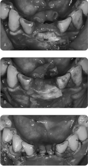
특별한 전신 병력이 없는 19세 남성 환자가 하악 전치의 결손부 수복을 주소로 연세대학교 치과대학병원 치주과 에 내원하였다. #32, 42번 치아의 선천적 결손으로 #31, 41 치아의 distal angulation이 관찰되는 상태였다 (Fig. 1). 우선적으로 환자에게 교정을 통한 치축의 회복 및 임프란트 수복 치료를 권유하였으나 교정 치료 기간 과 비용의 문제로 교정 치료는 제외하였다. 이 후 보철적 으로 고정성 보철 제작을 권유하였으나 지대치의 치축 경사가 심하여 근관 치료의 가능성이 있었고 환자는 자 연치의 의도적 삭제를 원하지 않아 고정성 보철물을 이 용한 치료 역시 계획에서 제외하였다. 결국 단일 임프란 트 식립 후 cantilever crown의 형태로 수복하기로 환자 와 최종 상의 후 결정하였다. 불편감을 최소화하고 수술을 단순화 하기 위하여 수술Fig. 1. Clinical photos and periapical radiograph taken at initial visit.
A. Labial view.
B. Occlusal view. Moderate bone resorption was noted on the buccal side.
C. Note the distoangulated teeth leaving narrow space for only one implant installation.
Fig. 2. A. Full thickness flap was retracted to show moderate bone loss in bucco-lingual dimension. B, C. Osteotomy was performed using
PiezosurgeryⓇ
and hole was drilled before removal of the block. The block bone was removed using surgical curette.
A B A B C C
부위와 인접한 symphysis에서 자가골을 block 형태로 채취하기로 하였다. 하악 전달 마취 및 국소 마취 후 sulcular incision을 하악 양측 견치까지 연장하여 절개한 뒤 전층판막을 거상하여 광범위한 결손부를 확인하였다 (Fig. 2-A). Symphysis 부위까지 골막을 모두 박리하여 시야를 확보 한 뒤 하악 전치로부터 최소 5mm의 수직 거리를 확인 한 뒤 defect의 형태에 맞게 절편을 design 하였다17). 이 후 PiezosurgeryⓇ장비를 이용해 절편을 절개하였고, 핀 고정을 위한 drilling을 절편 상에 시행하였다(Fig. 2-B, C). 일반적으로 절편을 공여부로부터 분리한 뒤 구강 외 에서 drilling을 할 경우 절편의 안정된 고정이 어려워 drill의 회전 시 절편이 미끄러져 떨어질 가능성이 높다. 따라서 공여부에서 완전히 분리되기 전 미리 screw 고 정을 위한 drilling을 시행하는 것이 안전하다. 이 후 핀의 돌출을 막기 위해 절편 상에 countersink drilling을 시행 하였다. 그 후 cortico-cancellous 절편을 분리하고 7mm 핀(Cheil corporation, Seoul, Korea)을 이용하여 골절편 을 최대한 결손부에 밀착하여 고정하고(Fig. 3-A), 공여 부로부터 망상골을 채득하여 절편과 결손부 사이에 보 강하였다. 공여부에는 흡수성 콜라겐 스폰지(CollaPlugⓇ, Zimmer Dental, CA, USA)을 충전했고 흡수성 막(Bio-Gide Ⓡ, Geistlich, Wolhusen, Switzerland)을 block bone 이 모두 덮히도록 피개한 뒤(Fig. 3-B) 협측 판막에 이 완절개 시행 후 일차 봉합하였다(Fig. 3-C). 10일 후에 발사하였고 감각 이상과 같은 특이한 소견은 발견되지 않았다. 술 후 2개월 내원 시 초진보다 양호한 골 증강 소견을 보였다(Fig. 4-A). 술 후 4개월 시점에 서 임프란트 수술을 시행하였다. 판막 형성 후 수평적으 로는 증강된 골량을 확인할 수 있었으나 수직적으로는 핀이 위치하는 부위까지 골이 흡수된 것을 확인할 수 있 었다(Fig. 4-B). 핀을 제거한 뒤 이동식 방사선 장치를 이용해 임프란트 드릴의 방향을 확인하며 임프란트를 식립하였다(Replace NP ø3.5x13mm)(Fig. 4-C). 5mm 높이의 치유지대주를 체결하고 봉합 한 뒤(Fig 4-D) 방사선 촬영으로 식립 상태를 확인하였다(Fig. 5). 발사를 위한 내원 시 환자는 개인적인 사정이 생겨 2달 이내에 보철을 완성하기를 원하였다. 식립 후 최소 2개 월 이상의 치유기간이 필요함을 설명하였으나 불가피한 사정으로 환자에게 조기 보철 제작으로 인한 문제점을 설명 한 뒤 식립 후 1달 시점에서 final impression을 채 득하였다. 환자에게는 연조직의 치유가 계속 일어나고 있으므로 보철물의 경계가 노출될 수 있음을 미리 주지 시켰고 안정적인 골유착을 위해 교합 접촉이 안 되도록 보철을 제작할 것임을 또한 설명하였다. 임프란트 식립 후 6주 시점에서 최종 보철물을 체결 할 당시 이미 연조
Fig. 3. A. Block bone was fixed with 7mm screw onto the recipient bed.
B. Resorbable collagen membrane was covered over the block bone.
C. Primary suture was performed after releasing incision.
A
B
직의 치유로 인해 보철물의 경계가 보이는 상태였으나 환자는 만족하였고 추후에 보철물을 재제작 받도록 권 유하였다(Fig. 6).
III. 고찰 및 결론
PiezosurgeryⓇ는 초음파 진동을 이용하여 경조직을 절 단하는 장치로서 연조직에는 손상을 최소화하며 환자에 게 강한 진동이나 소음을 주지 않아 외과적 bur나 trephine bur를 대체할 수 있는 유용한 장치이다. 특히 조 직학적으로 관찰 했을 때 drill이나 bur가 야기하는 미세 골괴사를 감소시킬 수 있어 골의 치유를 돕고 정상적인 골개조를 방해하지 않는다는 장점이 있다. 또한 상악동 거상술 시 선택적인 절개를 통해 Schneiderian 막을 천공 하지 않고 측방벽을 개통할 수 있어 유용하게 사용될 수 있다18). 또한 팁의 각도가 여러 가지로 구비되어 있어 좁 은 공간에서도 용이하게 사용할 수 있어 임상적으로 장 점을 갖는다.Fig. 5. Periapical radiograph after implant fixture installation.
Fig. 4. A. Clinical photo at 2 month check up. B. Clinical photo at implant first stage surgery.
Note horizontally augmented bone volume and vertically reduced block bone. C. Implant fixture was successfully installed. D. 5mm high healing abutment was connected
and flap was sutured.
Fig. 6. Implant prosthesis was successfully installed.
A
B
C
본 증례에서처럼 하악 정중부 결합으로부터 골을 채득 할 경우 하악 전치부의 감각 이상이 발생할 수 있음이 이 미 보고된 바 있다. 2001년 Nkenke 등은 20명의 환자에 게 술식을 시행한 후 12개월 간 추적을 하였는데 술 후 22%의 치아에서 치수 감각이 사라졌음을 보고하였다. 하지만 그 수치는 6개월 경과 후 13%, 12개월 경과 후 11%로 감소하였다. 1999년 Chiapasco 등19)의 연구에서 는 수술 후 80%의 환자에서 치수 감각이 소실된 것으로 나타났으나, 12개월 시점에서는 13%로 줄었다고 보고 하였다. Misch 등4)은 안전 거리 5mm를 확보하면 이러한 감각 이상을 예방할 수 있다고 권장하고 있다. 하지만 von Arx 등17)은 치수 감각의 소실은 골절편의 삭제 부위 가 치근첨으로부터 5mm의 안전 거리를 확보하지 못 해 서라기 보다는 mental foramen으로부터의 수평 거리가 충분히 확보되지 않았을 경우에 발생하는 것이라고 보 고하였다. 따라서 하악 정중부 결합으로부터의 골절편 채득 시에 는 반드시 방사선 및 임상적 검사를 충분히 해야 하며 술 전에 mental nerve 기능과 치수 검사를 시행하고 기록해 두는 것이 필요하다. 환자에게는 술 후 일시적 또는 장기 적 합병증이 생길 수 있음을 고지해야 하며, 절개선은 견 치의 원심면 후방으로는 연장을 피해야 한다. 술자와 보 조자는 협측 점막의 견인 시 후방으로 과도하게 견인하 여 이신경을 자극하지 않도록 주의해야 한다. 또한 하악 전치 치근단으로부터의 안전 거리인 5mm는 치수 감각 소실 예방을 보장할 수는 없으나 최소한 치근첨으로 들 어가는 혈류를 차단하여 치수 괴사가 발생하는 것을 예 방할 수 있는 거리이므로 반드시 이를 지키는 것이 필요 하다17). 위와 같은 주의점을 숙지하고 술식을 진행하였 으며, 본 증례에서는 감각 이상 등의 합병증은 없었다. 본 증례에서는 하악 중절치가 선천적 결손 상태였으므 로 치근첨으로부터의 안전 거리 확보에 유리한 상황이 었다. 따라서 충분한 골절편 채득이 가능하였고 대다수 의 골결손부를 절편만으로 충분히 충전이 가능하였다. 하지만 골량의 증대를 최대화 하기 위해 절편과 이식부 위 사이에 발생한 공간에는 공여부에서 bone roungeur 를 이용하여 망상골을 채득하여 충전하였다. 술 후 2개 월까지는 임상적으로 상당한 골량이 확보된 것으로 확 인되었으나 이식 후 4개월 시점에서 노출 시킨 결과 수 평적으로는 상당량이 증가되었으나 수직적으로는 pin의 수준까지 골이 흡수된 것을 관찰했다. Cordaro 등20)에 의 하면 symphysis에서 block bone 채득 시 최고 42%까지 골이 흡수되지만 membrane을 사용할 경우 이를 예방할 수 있다고 한다. 본 증례에서도 수직적으로는 골이 다소 흡수되었지만 수평적으로는 임프란트 식립에 양호한 골 량과 골질이 확보되었다. 이를 통해 볼 때 선택된 증례에서 하악 정중결합으로부 터 골절편을 채득하는 술식은 감각 이상 같은 큰 합병증 없이 성공적인 임프란트를 식립할 수 있는 술식이 될 수 있을 것으로 사료된다. 시술 전 술자는 정확한 골의 상태 와 필요량 및 해부학적 구조에 대한 주의를 기울일 필요 가 있고 이를 준수한다면 예측성 있는 술식으로서 사용 될 수 있을 것이다.
REFERENCES
1. Raghoebar GM, Batenburg RH, Vissink A, Reintsema H. Augmentation of localized defects of the anterior maxillary ridge with autogenous bone before insertion of implants. J Oral Maxillofac Surg 1996;54:1180-5; discussion 1185-6.
2. Boyne PJ, James RA. Grafting of the maxillary sinus floor with autogenous marrow and bone. J Oral Surg 1980;38:613-6.
3. Wood RM, Moore DL. Grafting of the maxillary sinus with intraorally harvested autogenous bone prior to implant placement. Int J Oral Maxillofac Implants 1988;3:209-14.
4. Misch CM. Comparison of intraoral donor sites for onlay grafting prior to implant placement. Int J Oral Maxillofac Implants 1997;12:767-76.
5. Misch CM. The harvest of ramus bone in conjunction with third molar removal for onlay grafting before placement of dental implants. J Oral Maxillofac Surg
1999;57:1376-9.
6. Misch CM. Use of the mandibular ramus as a donor site for onlay bone grafting. J Oral Implantol 2000;26:42-9.
7. Pikos MA. Facilitating implant placement with chin grafts as donor sites for maxillary bone augmentation--Part I. Dent Implantol Update 1995;6:89-92.
8. Pikos MA. Block autografts for localized ridge augmentation: Part II. The posterior mandible. Implant Dent 2000;9:67-75.
9. Pikos MA. Atrophic posterior mandibular reconstruction utilizing mandibular block autografts: risk management. Int J Oral Maxillofac Implants 2003;18: 765-6.
10. Raghoebar GM, Louwerse C, Kalk WW, Vissink A. Morbidity of chin bone harvesting. Clin Oral Implants Res 2001;12:503-7.
11. Nkenke E, Schultze-Mosgau S, Radespiel-Troger M, et al. Morbidity of harvesting of chin grafts: a prospective study. Clin Oral Implants Res 2001;12:495-502.
12. Nkenke E, Radespiel-Troger M, Wiltfang J, et al. Morbidity of harvesting of retromolar bone grafts: a prospective study. Clin Oral Implants Res 2002;13:514-21.
13. Aro H, Kallioniemi H, Aho AJ, Kellokumpu-Lehtinen P. Ultrasonic device in bone cutting. A histological and scanning electron microscopical study. Acta Orthop Scand 1981;52:5-10.
14. Vercellotti T. Piezoelectric surgery in implantology: a case report--a new piezoelectric ridge expansion technique. Int J Periodontics Restorative Dent 2000;20:358-65.
15. Robiony M, Polini F, Costa F, et al. Piezoelectric bone cutting in multipiece maxillary osteotomies. J Oral Maxillofac Surg 2004;62:759-61.
16. Vercellotti T, Nevins ML, Kim DM, et al. Osseous response following resective therapy with piezosurgery. Int J Periodontics Restorative Dent 2005;25:543-9. 17. von Arx T, Hafliger J, Chappuis V. Neurosensory disturbances following bone harvesting in the symphysis: a prospective clinical study. Clin Oral Implants Res 2005;16:432-9.
18. Vercellotti T, De Paoli S, Nevins M. The piezoelectric bony window osteotomy and sinus membrane elevation: introduction of a new technique for simplification of the sinus augmentation procedure. Int J Periodontics Restorative Dent 2001;21:561-7. 19. Chiapasco M, Abati S, Romeo E, Vogel G. Clinical outcome of autogenous bone blocks or guided bone regeneration with e-PTFE membranes for the reconstruction of narrow edentulous ridges. Clin Oral Implants Res 1999;10:278-88.
20. Cordaro L, Amade D, Cordaro M. Clinical results of alveolar ridge augmentation with mandibular block bone grafts in partially edentulous patients prior to implant placement. Clin Oral Implants Res 2002;13:103-111.
Block bone graft from mandibular symphysis using Piezoelectric osteotomy and
implant restoration; Case report
Jung-Chul Park1, Jae-Wook Jung2, Ji-Hwan Kim2, Yoo-Jung Um1, Jee-Hwan Kim2,
Ui-Won Jung1, Sung-Tae Kim2, Young-Bum Park2, Chang-Sung Kim1, Jae-Hoon Lee2,
June-Sung Shim2, Hong-Seok Moon2, Seong-Ho Choi1*
1Department of Periodontology, Oral Science Research Center, College of Dentistry, Yonsei University
2Department of Prosthodontics, College of Dentistry, Yonsei University
Purpose: The aim of this case report is to describe block bone graft from mandibular symphysis using Piezosurgery¢Á and implant installation procedure.
Material and Methods: A 19 year old male patient walked in with complaint of missing tooth on #31, 41 due to congenital missing. Block bone was grafted from mandibular symphysis and fixed with screws. After 4 months, implant was installed(Replace NP ©™3.5x13mm). The patient requested early restoration and the final restoration was delivered 6 weeks after implant 1stsurgery.
Results: Relatively enough bone was acquired using block bone even though there was considerable bone loss in vertical dimension. Bone quality was good enough to place implant fixture. Slight gingival recession took place during soft tissue healing period after 1stsurgery, and
the patient agreed re-visit for final restoration check up.
Conclusion: Block bone graft from symphysis using Piezosurgery¢Á can be used as a predictable procedure for ridge augmentation when certain precautions are observed.[THE JOURNAL OF THE KOREAN ACADEMY OF IMPLANT DENTISTRY 2009;28(2):36-42]