Agmatine pretreatment attenuates retinal
ganglion cell death by an oxidative stress
IIZUKA YOKO
Department of Medical Science
The Graduate School, Yonsei University
Agmatine pretreatment attenuates retinal
ganglion cell death by an oxidative stress
Directed by Professor Yang Woo Ick
The Doctoral Dissertation
submitted to the Department of Medical Science,
the Graduate School of Yonsei University
in partial fulfillment of the requirements for the degree
of Doctor of Philosophy of Medical Science
IIZUKA YOKO
December 2008
This certifies that the Doctoral
Dissertation of IIZUKA YOKO is
approved.
---
Thesis Supervisor: Yang Woo Ick
---
Thesis Committee Member#1: Seong Gong Je
---
Thesis Committee Member#2: Lee Jong Eun
---
Thesis Committee Member#3: Kim Chan Yun
---
Thesis Committee Member#4: Kim Chul Hoon
The Graduate School
Yonsei University
ACKNOWLEDGEMENTS
The author wishes to express her profound gratitude
to Dr. Yang Woo Ick, professor, Department of Pathology,
and Dr. Seong Gong Je, professor, Department of
Ophthalmology for their guidance and constructive
criticism during the course of the study. The author also
would like to express appreciations to Dr. Lee Jong Eun,
professor, Department of Anatomy, Dr. Kim Chan Yun,
associate professor, Department of Ophthalmology, and Dr.
Kim Chul Hoon, assistant professor, Department of
Pharmacology, for excellent and helpful discussions and
advices.
<TABLE OF CONTENTS>
ABSTRACT……….10
Ⅰ
. INTRODUCTION………..12
Ⅱ
. MATERIALS AND METHODS………14
1.
Chemicals………14
2.
Cell culture and agmatine pretreatment……….14
3.
Oxidative stress ………14
4.
Cell viability assays………14
5.
Morphological analysis……….15
6.
Apoptosis assays………15
7.
Western immunoblot……….16
8.
Real time RT-PCR……….17
9.
Statistical analysis……….18
Ⅲ
. RESULTS………...19
1.
Differentiation of RGC-5 cells………..19
2.
Oxidative stress induced cytotoxicity of differentiated RGC-5
cells………22
3.
Protective effects of agmatine pretreatment………..22
4.
TUNEL staining………24
5.
Caspase activities on RGC-5 cells pretreated by agmatine……...25
6.
Agmatine as an alpha 2-adrenergic receptor agonist……….26
7.
Agmatine as a NMDA receptor antagonist...26
8.
E x pr es s i o n of t o ta l a n d p h os p h or yl at e d B cl- 2 f a m il y
proteins………..28
9.
Expression of total and phosphorylated MAPKs proteins………29
Ⅳ
. DISCUSSION………30
Ⅴ
. CONCLUSION………33
REFERENCES……….34
LIST OF FIGURES
Figure 1. Staurosporine treatment and recovery of RGC-5
cells………...20
Figure 2. Differentiation of RGC-5 cells by staurosporine….21
Figure 3. Cytotoxicity of hydrogen peroxide (H
2O
2) on RGC-5
cells………...22
Figure 4. Protective effect of agmatine pretreatment to
differentiated RGC-5 cells against oxidative stress by
H
2O
2………...23
Figure 5. Effect of agmatine pretreatment on apoptosis of
differentiated RGC-5 cells……….24
Figure 6. Caspase-3 (A) and -9 (B) activities of oxidative
stressed RGC-5 cells after 2 hours pretreatment with
100
µM agmatine………...25
Figure 7. Inhibitory effect of yohimbine to agmatine
pretreatment………...26
Figure 8. Influence of glutamate (A) and NMDA (B) on
agmatine pretreatment………...27
Figure 9. Results of quantitative real time RT-PCR for Bcl-2
family………....28
Figure 10. Expression of Bcl-2 family proteins at various
incubation periods after the application of 1.0 mM
H
2O
2oxidative
stress
analyzed
by
Western
Figure 11. Expression of MAPKs proteins at various
incubation periods after the application of 1.0 mM
H
2O
2oxidative
stress
analyzed
by
Western
LIST OF TABLE
<ABSTRACT>
Agmatine pretreatment attenuates retinal ganglion cell death by an
oxidative stress
IIZUKA YOKO
Department of Medical Science
The Graduate School, Yonsei University
(Directed by Professor Yang Woo Ick)
Purpose; To investigate the protective effects of agmatine pretreatment
on oxidative stress in immortalized and differentiated rat retinal
ganglion cell line (RGC-5 cells).
Method; RGC-5 cells were differentiated by staurosporine, cultured in
the presence of hydrogen peroxide (H
2O
2) with or without pretreatment
of agmatine. Cell viability was determined by lactate dehydrogenase
(LDH) assay. Apoptosis was examined by TUNEL assay and
caspase-3 and -9 assays. Total and phosphorylated Bcl-2 family
proteins (Bcl-2, Bcl-xl, Bax, and Bad) and mitogen-activated protein
kinases (MAPKs; ERK p44/42, JNK, and p38)
were investigated by
Western blot analysis. Also mRNA expressions of Bcl-2 family were
Results; RGC-5 cells, differentiated by 1.0
µM staurosporine for 6
hours and recovered for 3 days, contained fully developed neurites and
were well connected with each other. Under H
2O
2oxidative stress,
agmatine pretreatment for 2 hours enhanced cell survival dose
dependently. The cytotoxicity assay showed 40.9 % cell loss, which
was reduced to 27.3 % when the cells were pretreated by 100
µM
agmatine for 2 hours followed by 1.0 mM H
2O
2treatment for 16 hours.
This cell loss was due to apoptotic cell death, as established by TUNEL
assay, but was neither caspase-3 nor caspase-9 dependent. The effect
of agmatine pretreatment was completely inhibited by yohimbine,
but
partially decreased by glutamate or NMDA
.Total expression of Bcl-2
family proteins and MAPKs were not influenced by H
2O
2oxidative
stress. Also mRNAs of Bcl-2 family were not changed. In contrast,
the phosphorylation of JNK was suppressed by agmatine pretreatment
followed by H
2O
2oxidative stress.
Conclusion;
The
present
study
shows
that
agmatine
had
neuroprotective effects on oxidative stressed differentiated RGC-5 cells.
It might suggest a therapeutic strategy for many ocular diseases
associated with oxidative stress.
---
Agmatine pretreatment attenuates retinal ganglion cell death by an
oxidative stress
IIZUKA YOKO
Department of Medical Science
The Graduate School, Yonsei University
(Directed by Professor Yang Woo Ick)
I. INTRODUCTION
Glaucoma, one of the leading causes of blindness in the world, is characterized by progressive loss of retinal ganglion cells (RGCs) including their axons, and by tissue remodeling of the optic nerve head. This is followed by visual field defects. Even though the elevated intraocular pressure is the major risk factor associated with glaucomatous visual field loss, oxidative stress has recently been proposed as another important risk factor in the pathogenesis of glaucoma.1-3
Glutamate is the principal excitatory neurotransmitter in the retina. The extracellular concentration of glutamate increases after an ischemic insult and a significant component of ischemic injury to CNS neurons results from glutamatergic excitotoxicity. Glutamate and nitric oxide (NO) promote oxidative damage by reacting with superoxide anion. RGCs express both N-methyl-D-aspartate (NMDA) and non-NMDA type glutamatergic receptors.
The oxidative stress has been shown to play an important role in ischemic retinopathy.4,5 RGCs are susceptible and vulnerable to the oxidative stress. Hydrogen peroxide (H2O2) induces apoptosis through the mitochondrial death pathway. Bcl-2 family has been shown to be involved in cytotoxicity by glutamate and NO.6,7 Also the activity of several mitogen activated protein kinases (MAPKs) have been studied in H2O2 and NMDA induced apoptosis.8,9
Primary cultures of RGCs have some limitations to study RGC pathophysiology. Despite the fact that transformed rat retinal ganglion cell line (RGC-5 cell) has a number of characteristics of normal RGCs, it is mitotically active and is not exactly same as primary RGCs. It has been shown that RGC-5 cells treated by the broad-spectrum protein kinase inhibitor staurosporine are not dividing and maintain neuronal characteristics.10-12
Several molecules having some neuroprotective effects, to the hypoxic injury at CNS, have been reported.13-20 The neuroprotective effects by pretreatment of these molecules against hypoxia-induced neuronal death have not been well understood. Although some drugs have been reported to have a protective role in RGC, most of them have no sufficient data to support their definite role.21-24
Agmatine, a primary amine formed by the decarboxylation of L-arginine synthesized in mammalian brain, is an agonist for the alpha 2-adrenergic and imidazoline receptors and an antagonist at NMDA receptors.25-27 Recent studies have shown that agmatine may be neuroprotective in neuronal ischemic models.28-31
In the present study, I examined the protective effects of agmatine pretreatment on oxidative injuries of RGC-5 cells in vitro, and characterized the mechanisms involved in these activities.
II. MATERIALS AND METHODS 1. Chemicals
Agmatine sulfate, staurosporine (from Streptomyces staurosporeus), glutamate, and yohimbine were purchased from Sigma-Aldrich (St. Louis, MO, USA). NMDA was purchased from Calbiochem (San Diego, CA, USA).
2. Cell culture and agmatine pretreatment
RGC-5 cells (donated by Alcon Research, Forth Worth, TX, USA) were cultured in Dulbecco’s modified Eagle’s medium (DMEM, Gibco, Carlsbad, CA, USA) with 1 g/L glucose, 100 U/mL penicillin, 100 µg/mL streptomycin, and 10 % heat inactivated fetal bovine serum (FBS, Gibco). All experiments were performed at a confluence of 70 to 80 %. RGC-5 cells were differentiated by exposure to 1.0 µM staurosporine. After recovery using 10 % FBS-DMEM,25 the differentiated RGC-5 cells were incubated with various concentration of agmatine.
3. Oxidative stress
Cellular oxidative stress was induced by addition of H2O2 to the culture medium.
4. Cell viability assays
Total cell population and viability was quantified by lactate dehydrogenase (LDH) assay (Promega, Madison, WI, USA) according to the manufacturer's instructions. Briefly, the cells in a culture were lysed by Lysis Solution (0.9 % (v/v) Triton X-100 in water). The LDH activities in
50 µL of the sample supernatant in 96 well-plates were colorized by a
an ELISA plate reader (Vmax; Molecular Device, Sunnyvale, CA, USA). The cell population was proportional to the absorbance values.
The proportion of injured cells among total cell population was quantified by measurement of released LDH to culture media from the cells. Cytotoxicity was expressed as a percentage of the LDH activity in medium to the total (medium and cellular LDH). Data of total cell population and viability are expressed as mean ± SEM (standard error of mean).
5. Morphological analysis
Photomicrographs of cells were taken at 200×, digitalized and stored as JPEG. Axonal outgrowth of differentiated RGC-5 cells was measured and assessed by NeuronJ image software (ImageJ 1.40; http//rsb.info.nih.gov/ij). The axonal length was expressed as the mean ± SEM of 40 to 50 cells.
6. Apoptosis assays
Differentiated RGC-5 cells were plated on
poly-D-lysine/Laminin-coated 12 mm φ cover slide glass (BD Biosciences, Bedford, MA, USA). The TUNEL assay was performed using Apo-BrdU in situ DNA Fragmentation Assay Kit (BioVision, Mountain View, CA, USA) with minor modification.32 Briefly, the cells were fixed in 4 % paraformaldehyde in phosphate-buffered saline (PBS), pH 7.4 for 30 minutes and permealized in 0.1 % Triton X-100 in 0.1 % sodium citrate. After washing in PBS, the cells were stored in 70 % (v/v) ethanol at -20℃ over night. After washing, the cells were carefully covered by the DNA labeling solution containing TdT enzyme and bromolated deoxyuridine triphosphate digoxigenine (Br-dUTP), and incubated in a dark and
humidified 37℃ incubator for 60 minutes. After washing, the cells were covered by the anti-BrdU-FITC antibody solution and incubated in a humidified incubator at room temperature. After removing the antibody solution, the cells were covered by propidium iodide (PI) /Rnase A solution and incubated in a dark in a humidified incubator for 30 minutes. By fluorescence microscopy, apoptotic cells show green staining over an orange-red PI counter-staining. Double stained cells were visualized with a total magnification of 200×. The cells incubated in the labeling solution without TdT enzyme were used as negative control (data was not shown).
Caspase-3 and -9 activities were measured using Caspase-3 and Caspase-9 Fluorometric Assay Kits (BioVision) respectively, according to the manufacturer's instructions. Briefly, RGC-5 cells were suspended in the lysis buffer. In 96 well-plate, cell lysate 50 or 100 µg as total protein was mixed with the reaction buffer containing 10 mM DTT and added either DEVD-AFC substrate for caspase-3 or LEHD-AFC substrate for caspase-9. The reaction mixture was incubated at 37℃ for 2 hours, and then read in a fluorometer equipped with a 400 nm excitation filter and 505 nm emission filter. Increase in caspase activity was determined by comparing the results with the level of untreated control. Amount of total protein was determined by Bradford protein assay (BIO-RAD, Philadelphia, CA, USA). Data are expressed as mean ± SEM of 8 to 10 independent cultures.
7. Western immunoblot
Differentiated RGC-5 cells cultured were washed twice in cold PBS, and then lysed in lysis buffer (Mammmalian Cell Lysis kit, Sigma-Aldrich) containing 1.0 mM Na3VO3 and 1.0 mM phenylmethyonylsulfonyl fluoride. The protein (20 or 30 µg) were separated on 12 % SDS-polyacrylamide gels,
transferred to a polyvinylidene difluoride membrane (Millipore, Bedford, MA, USA), and blocked by 5 % nonfat dry milk in a Tween20-Tris buffered saline for one hour at room temperature. Membranes were then incubated with the following primary antibodies over night at 4℃: mouse anti-Bcl-2 (1:200; Santa Cruz Biotechnology, Santa Cruz, CA, USA), rabbit anti-phospho-Bcl-2 (1:1000; Cell Signaling Technology, Danvers, MA, USA), rabbit anti-Bcl-xl (1:1000; Cell Signaling Technology), rabbit anti-phospho-Bcl-xl (1:500; SAB, Pearland, TX, USA), rabbit anti-Bax (1:1000; Cell Signaling Technology), mouse anti-Bad (1:100; Santa Cruz Biotechnology), rabbit anti-phospho-Bad (1:1000; Cell Signaling Technology) for Bcl-2 family protein, and rabbit anti-SAPK/JNK (1:1000; Cell Signaling Technology), rabbit anti-phospho-SAPK/JNK (1:1000; Cell Signaling Technology), rabbit anti-MAPK p38 (1:1000; Cell Signaling Technology), rabbit anti-phospho MAPK p38 (1:1000; Cell Signaling Technology), rabbit anti-p44/p42 (1:1000; Cell Signaling Technology), rabbit anti-phospho-p44/p42 (1:1000; Cell Signaling Technology) for MAPK family, and mouse anti-β-actin (1:10000; Sigma-Aldrich). After treatment with the secondary goat anti-rabbit (1:2000; Cell Signaling Technology) or horse anti-mouse (1:2000; Cell Signaling Technology) IgG conjugated by horseradish peroxidase for one hour, immunoreactive bands were visualized with the ECL detection system (Thermo Scientific, Waltham, IL, USA).
8. Real time RT-PCR
Total RNA was extracted using RNeasy Mini Kit (QIAGEN, Valencia, CA, USA) and treated with DNase (QIAGEN) to remove contaminating DNA according to manufacturer directions. The cDNAs were synthesized
using Superscript Ⅲ First-Strand Synthesis System for RT-PCR (Invitrogen, Carlsbad, CA, USA). A Real time PCR was performed with 50 ng cDNA per a reaction using Power SYBR Green PCR Master Mix (BIO-RAD) in a total volume of 25 µL of reaction mixture (iCycler iQ, BIO-RAD). The SYBER green data and level of target mRNA were analyzed by iCycler iQ software with a relative standard curve of β-actin. All experiments were performed in triplicate and repeated 4 times. Data were presented as mean ± SEM. Sequences of oligonucleotides used as primers are summarized in Table1.33-35
Table1. Primer sequences for real-time RT-PCR
mRNA Sequence
Bcl-2 sense 5’GTG GTG GAG GAA CTC TTC AGG GAT G3’
antisense 5’GGT CTT CAG AGA CAG CCA GGA GAA ATC3’
Bcl-xl sense 5’GTA GTG AAT GAA CTC TTT CGG GAT GG 3’
antisense 5’ACC AGC CAC AGT CAT GCC CGT CAG G 3’
Bax sense 5’AAT ATG GAG CTG CAG AGG ATG ATT G3’
antisense 5’GCA CTT TAG TGC ACA GGG CCT TGA G3’
Bad sense 5’GAG CGA TGA ATT TGA GGG TTC3’
antisense 5’GAT CCC ACC AGG ACT GGA TAA3’
β-actin sense 5'AGA TGA CCC AGA TCA TGT TTG AGA3'
antisense 5'ACC AGA GGC ATA CAG GGA CAA3'
9. Statistical analysis
The data will be analyzed by 2-tailed Student t-test or one-way ANOVA, followed by post hoc comparisons (Student-Newman-Keuls) using the Statistical Package for Social Sciences 12.0 (SPSS).
III. RESULTS
1. Differentiation of RGC-5 cells
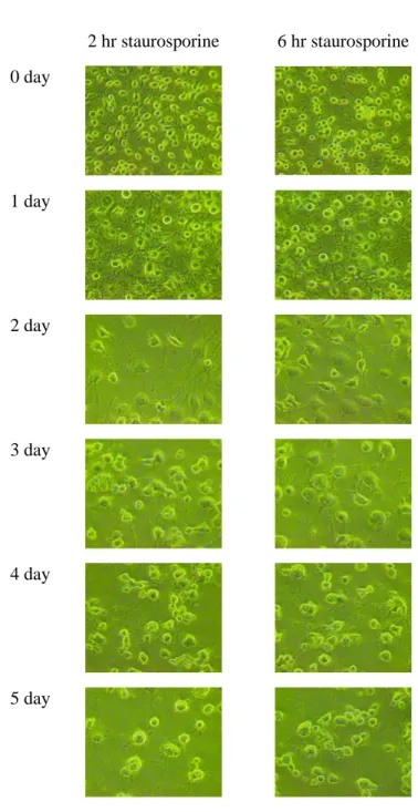
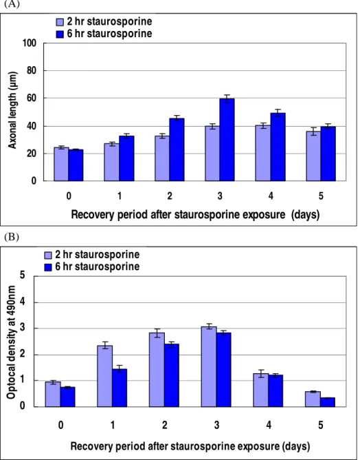
Staurosporine changed the morphology of RGC-5 cells from flat to round shape and induced the outgrowth of neurites (Figure 1). The change in morphology was already observed after 1.0 µM staurosporine treatment for 2 and 6 hours. The neurites were growing through recovery period in 10 % FBS-DMEM for 3 days. These neurites had multiple and long branches, and sufficiently contacted with those from neighbor cells. In 3-days recovered cells, the average of axonal length was 39.5 ± 1.74 µm and 59.6 ± 2.72 µm for the 2 and 6 hours treated cells, respectively (Figure 2A). The absorbance values of the total cellular LDH at 490 nm were increasing, but there were no significant differences between the 2 and 6 hours treated cells on 3-day recovery (Figure 2B). Both of the neurites outgrowth and total cell population decreased after 3 days. Based on these results, RGC-5 cells differentiated by 1.0 µM staurosporine for 6 hours and recovered for 3 days were used for the following oxidative stress experiments.
2 hr staurosporine 6 hr staurosporine 0 day 1 day 2 day 3 day 4 day 5 day
Figure 1. Staurosporine treatment and recovery of RGC-5 cells. After
treatment by 1.0 µM staurosporine for 2 or 6 hours, RGC-5 cells were recovered in 10 % FBS-DMEM up to 5 days.
(A) 0 20 40 60 80 100 0 1 2 3 4 5
Recovery period after staurosporine exposure (days)
A xo n al le n g th (µ m ) 2 hr staurosporine 6 hr staurosporine (B) 0 1 2 3 4 5 0 1 2 3 4 5
Recovery period after staurosporine exposure (days)
O p to ca l d en si ty a t 4 90 n m 2 hr staurosporine 6 hr staurosporine
Figure 2. Differentiation of RGC-5 cells by staurosporine. The RGC-5 cells
were exposed to 1.0 µM staurosporine for 2 and 6 hours, and then recovered up to 5 days. Outgrowth length of the axons (A) and optical density of total cellular LDH (B) over time.
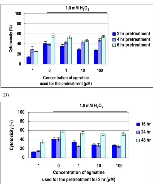
2. Oxidative stress induced cytotoxicity of differentiated RGC-5 cells The concentration of H2O2 below 0.8 mM did not significantly induce cell death. Over 1.0 mM H2O2, both time and concentration dependent cell death were observed (Figure 3). The 16 hours incubation of the differentiated RGC-5 cells with 1.0 mM H2O2 was selected as the oxidative stress condition for further experiments.
0 20 40 60 80 100 120 0 0.5 0.8 1 2.5 H2O2 concentration (mM) C yt o to xi ci ty (% ) 16 hr 24 hr 48 hr
Figure 3. Cytotoxicity of hydrogen peroxide (H2O2) on RGC-5 cells.
3. Protective effects of agmatine pretreatment
After the 2 hours pretreatment of agmatine, the cytotoxic effect of H2O2 on differentiated RGC-5 cells most effectively decreased (Figure 4A). Sixteen hours exposure to 1.0 mM H2O2 induced 40.9 % cytotoxicity, but it decreased to 35.8 %, 28.9 % (p<0.007), and 27.3 % (p<0.003) by 2 hours pretreatment of agmatine of 1, 10, and 100 µM, respectively. Although 1 µM agmatine did not make statistically significant difference, 10 and 100 µM agmatine significantly suppressed the cytotoxic effect of H2O2. And 2 hours incubation demonstrated the most effective protection (Figure 4A). The protective effects lasted for 24 hours (Figure 4B).
(A) 0 20 40 60 80 100 * 0 1 10 100 Concentration of agmatine used for the pretreatment (µM)
C yt o to xi ci ty ( % ) 2 hr pretreatment 4 hr pretreatment 6 hr pretreatment 1.0 mM H2O2 (B) 0 20 40 60 80 100 * 0 1 10 100 Concentration of agmatine
used for the pretreatment for 2 hr (µM)
C yt o to xi ci ty (% ) 16 hr 24 hr 48 hr 1.0 mM H2O2
Figure 4. Protective effect of agmatine pretreatment to differentiated RGC-5
cells against oxidative stress by H2O2. (A) Agmatine treatment for 2, 4, or 6 hours followed by 1.0 mM H2O2 oxidative stress for 16 hours. (B) Agmatine treatment for 2 hours followed by 1.0 mM H2O2 oxidative stress for 16, 24, or 48 hours. * Negative control; the cells were incubated in DMEM without any treatments.
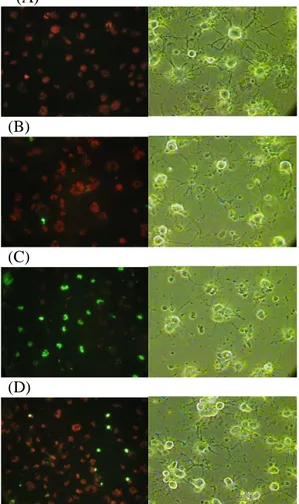
4. TUNEL staining
To characterize the effect of agmatine pretreatment on the cell death induced by oxidative stress, TUNEL assays were performed. TUNEL positive apoptotic cells, showing bright green refringence under the fluorescent microscope, marked increased in RGC-5 cells treated with 1.0 mM H2O2 only. Pretreatment with 100 µM agmatine before 1.0 mM H2O2 application significantly decreased apoptotic cells with green refringence (Figure 5).
(A)
(B)
(C)
(D)
Figure 5. Effect of agmatine pretreatment on apoptosis of differentiated
RGC-5 cells. (A) Control; (B) 100 µM agmatine pretreatment only; (C) 1.0 mM H2O2 oxidative stress only; and (D) 100 µM agmatine pretreatment followed by 1.0 mM H2O2 oxidative stress.
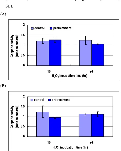
5. Caspase activities on RGC-5 pretreated by agmatine
The RGC-5 cells were pretreated with 100 µM agmatine for 2 hours, and then incubated in 1.0 mM H2O2 for 16 or 24 hours. The caspase-3 activity slightly decreased at 24 hours (p<0.406) (Figure 6A). The caspace-9 activity decreased at the period of 16 hours after agmatine pretreatment, but it was not statistically significant (p<0.373) (Figure 6B). (A) 0 0.5 1 1.5 2 16 24 H2O2 incubation time (hr) C as p a se a ct iv ity (r at io t o c o n tr o l) control pretreatment (B) 0 0.5 1 1.5 2 16 24 H2O2 incubation time (hr) C as p as e ac tiv ity (r at io to c o n tr o l) control pretreatment
Figure 6. Caspase-3 (A) and -9 (B) activities of oxidative stressed RGC-5
6. Agmatine as an alpha 2-adrenergic receptor agonist
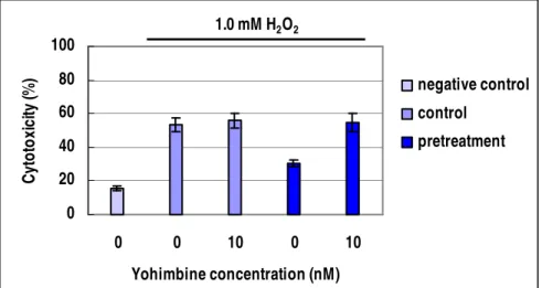
Effect of agmatine was examined with co-treatment of yohimbine (alpha 2 receptor antagonist) and 1.0 mM H2O2 (Figure 7). In RGC-5 cells without agmatine pretreatment, co-treatment of yohimbine demonstrated no difference in cytotoxicity. When the cells were pretreated by 100 µM agmatine, co-treatment of 10 nM yohimbine increased cytotoxicity by 20.4 %, thus showed that protective effect of agmatine pretreatment disappeared in the presence of yohimbine.
0 20 40 60 80 100 0 0 10 0 10 Yohimbine concentration (nM) C yt o to xi ci ty (% ) negative control control pretreatment 1.0 mM H2O2
Figure 7. Inhibitory effect of yohimbine to agmatine pretreatment. Negative
control; the cells in DMEM without any treatments. Control; 1.0 mM H2O2 oxidative stress with or without 10 nM yohimbine. Pretreatment; 1.0 mM H2O2 oxidative stress with or without yohimbine after 100 µM agmatine pretreatments.
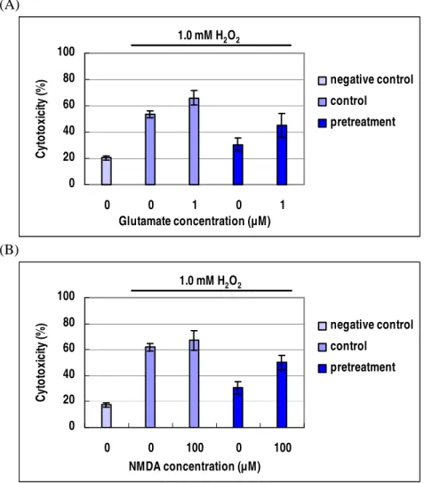
7. Agmatine as a NMDA receptor antagonist
The protective effect of agmatine pretreatment was examined in the presence of glutamate and NMDA under 1.0 mM H2O2 oxidative stress for 16 hours. The cytoprotective effect of agmatine was abolished with the presence of glutamate and NMDA (Figure 8). The mean value of
cytotoxicity assays increased from 53.6 % to 66.1 % in the RGC-5 cells after the application of 1.0 µM glutamate. The agmatine pretreated RGC-5 cells also demonstrated increased cytotoxicity but less mean value after the application of 1.0 µM glutamate (Figure 8A). The application of 100 µM NMDA showed similar results (Figure 8B). (A) 0 20 40 60 80 100 0 0 1 0 1 Glutamate concentration (µM) C yt o to xi ci ty (% ) negative control control pretreatment 1.0 mM H2O2 (B) 0 20 40 60 80 100 0 0 100 0 100 NMDA concentration (µM) C yt o to xi ci ty (% ) negative control control pretreatment 1.0 mM H2O2
Figure 8. Influence of glautamate (A) and NMDA (B) on agmatine
pretreatment. Negative control; the cells in DMEM without any treatments. Control; 1.0 mM H2O2 oxidative stress with or without 1.0 µM glutamate or
100 µM NMDA. Pretreatment; 1.0 mM H2O2 oxidative stress with or
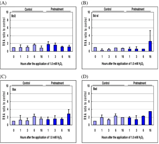
8. Expression of total and phosphorylated Bcl-2 family proteins
Expression of Bcl-2 family proteins was examined by quantitative real time RT-PCR and Western blot assay. Even though the mRNA expression of Bcl-xl from 100 µM agmatine treated RGC-5 cells slightly increased at 16 hours after the application of 1.0 mM H2O2, other Bcl-2 family proteins (Bcl-2, Bax, and Bad) did not show significant changes (Figure 9). The protein contents of Bcl-2 family proteins from the RGC-5 cells did not demonstrate significant changes after the application of the oxidative stress (Figure 10). Bcl-xl seemed to be slightly increased by agmatine pretreatment, but the phosphorylation was not stimulated. (A) 0 2 4 6 8 10 0 1 3 6 16 1 3 6 16
Hours after the application of 1.0 mM H2O2
R N A r at io t o c o n tr o l Control Pretreatment Bcl2 (B) 0 2 4 6 8 10 0 1 3 6 16 1 3 6 16
Hours after the application of 1.0 mM H2O2
R N A r at io t o c o n tr o l Pretreatment Control Bcl-xl (C) 0 2 4 6 8 10 0 1 3 6 16 1 3 6 16
Hours after the application of 1.0 mM H2O2
R N A r at io t o c o n tr o l Pretreatment Control Bax (D) 0 2 4 6 8 10 0 1 3 6 16 1 3 6 16
Hours after the application of 1.0 mM H2O2
R N A r at io t o c o n tr o l Pretreatment Control Bad
Figure 9. Results of quantitative real time RT-PCR for Bcl-2 family. (A)
Control Pretreatment (hr) 0 1 3 6 9 12 16 0 1 3 6 9 12 16 Bcl-2 Phospho-Bcl-2 Bcl-xl Phospho-Bcl-xl Bax Bad Phospho-Bad
Figure 10. Expression of Bcl-2 family proteins at various incubation periods
after the application of 1.0 mM H2O2 oxidative stress analyzed by Western immunoblot.
9. Expression of total and phosphorylated MAPKs proteins
Western immunoblot was performed to examine the effect of agmatine on the expression of three MAPK proteins (JNK, ERK p44/42, and p38) (Figure 11). The expression of phospho-JNK in the agmatine pretreated RGC-5 cells decreased earlier after oxidative stress than that in the RGC-5 cells without agmatine pretreatment. But agmatine pretreatment demonstrated no effect on the expression of phospho-ERK p44/42 MAPKs. Total amount of p38 was not changed thorough oxidative stress.
Control Pretreatment (hr) 0 1 3 6 9 12 16 0 1 3 6 9 12 16 JNK Phospho-JNK ERK p44/42 Phospho-p44/42 p38 Phospho-p38 β-actin
Figure 11. Expression of MAPKs proteins at various incubation periods after
the application of 1.0 mM H2O2 oxidative stress analyzed by Western immunoblot.
IV. DISCUSSION
Researches about RGC pathophysiology have been hampered by difficulties and limitations in using primary RGCs. Cultured RGCs survived in relatively short period and their isolation is difficult. Although immortalized RGC-5 cell line expresses neuronal markers characteristic of RGC, it is mitotically active and morphologically more similar to glial cells. It has been shown that RGC-5 cells differentiated by staurosporine are non-mitotic and express sufficient neurites for a while. 25,28,29 In this study,
RGC-5 cells differentiated by 1.0 µM staurosporine for 6 hours well survived for three days in 10 % FBS-DMEM and contained sufficient growing axons and connected with each other.
Apoptosis is programmed cell death and caspases usually mediate apoptotic neuronal death. However, there have been studies that H2O2 cause apoptosis by either caspase-independent or dependent pathway.36-39 In the present study, oxidative stress was induced by treatment of the differentiated RGC-5 cells with H2O2 and the effects of agmatine pretreatment were examined. The results indicated that the pretreatment with agmatine protected and rescued the RGC-5 cells from apoptosis dose dependently, and this effect was independent of caspase-3 and -9 activities.
Agmatine has been known as an agonist for the alpha 2-adrenergic and imidazoline receptors and an antagonist at NMDA receptors.25-27 Wheeler and Nylander40, and Kalapesi et al. 41 have studied the alpha 2-adrenergic receptor on human ganglion cells and RGC-5 cell line. They demonstrated the presence of alpha 2A-adrenergic receptor on both undifferentiated and succinyl concanavaline-A differentiated RGC-5 cell lines. They also showed an increased expression of alpha 2A-adrenergic receptor on the 7-day differentiated RGC-5 cells compared to the undifferentiated and early differentiated cells. Although staurosporine was used to differentiate RGC-5 cells in the present study, it was shown that the protective effect of agmatine pretreatment was completely abolished by yohimbine (antagonist for alpha 2-adrenergic receptor) in the differentiated and 3-day recovered RGC-5 cells. This result suggests a possibility that pretreatment with agmatine protects RGC-5 cells through alpha 2-adrenergic receptor.
RGCs also express both NMDA and non-NMDA type glutamatergic receptors. NMDA-type glutamatergic excitotoxicity has been implicated as a mechanism for injuries associated with ischemia and other insults to neurons in many regions of the CNS. However, there have been contradictive results about the rule of glutamate and NMDA excitotoxicity to vulnerability of RGCs.42, 43 Although varied evidences have been shown in several species and strains of the experimental animal used, Ullian et al.42 indicated that primary rat RGCs is invulnerable to glutamate and NMDA, in vitro. Their rat primary RGCs survived in the presence of glutamate (500 µM) or NMDA (500 µM) in relatively long period of incubation. In present experiment, the protective effect of agmatine pretreatment under H2O2 oxidative stress was not completely inhibited by glutamate and NMDA. It may suggest that the agmatine pretreatment partially paralyze NMDA.
Bcl-2 family includes both anti-apoptotic (ex. Bcl-2 and Bcl-xl) and pro-apoptotic (ex. Bad and Bax) regulator proteins on mitochondrial membrane. Cytochrome c release is inhibited by Bcl-2 and Bcl-xl. Tamatani et al. 6, 7 estimated that Bcl-2 or Bax were involved in cytotoxicity by glutamate and NO. Li and Osborne38 showed oxidative-induced apoptosis to an immortalized ganglion cell line is caspase independent. The results of the present study showed that Bcl-2 family proteins in the RGC-5 cells did not show dramatic changes under H2O2 oxidative stress. So it is indicated that caspase independent mechanisms irrespective of Bcl-2 family proteins are involved in the H2O2 induced oxidative damage of RGC-5 cells.
Exogenous H2O2 causes intracellular reactive oxygen species accumulation and mitochondrial dysfunction, and activates MAPK pathway.
To elucidate the mechanism of agmatine effects, Hong et al.31 recently demonstrate that agmatine is neuroprotective against hypoxia-induced damage of RGC-5 cells through the JNK and NF-κB signaling pathways. The present study demonstrated that JNK was suppressed in agmatine pretreated RGC-5 cells after the application of oxidative stresses. Although I have not examined NF-κB signaling pathways, results of this study indicates that MAPK seems to be involved in the mechanism of the neuroprotective effects of agmatine.
In present study, I demonstrated the protective effects of agmatine against oxidative stress induced apoptosis of RGC-5 cells. Although more well-designed studies are required to elucidate the precise mechanism of neuroportective effects of agmatine, the present results indicate several underlying mechanisms of the neuroprotective effects of agmatine to RGCs.
V. CONCLUSION
The aim of this study was to evaluate the neuroprotective effects of agmatine pretreatment on oxidative stressed RGCs in vitro and to investigate the underlying mechanism. The RGC-5 cells were sufficiently differentiated by exposure to staurospirine and the oxidative stress was induced by incubation with H2O2. By agmatine pretreatment, the cell viability was increased and apoptosis was decreased under H2O2 oxidative stress. These protective effects seemed to be associated with MAPKs through alpha 2-adrenergic and NMDA receptors. Finally, agmatine might be a therapeutic strategy for many ocular diseases associated with oxidative stress.
REFERENCES
1. Neufeld AH. Nitric oxide: a potential mediator of retinal ganglion cell damage in glaucoma. Surv Ophthalmol 1999; 43:129-35.
2. Ferreira SM, Lerner SF, Brunzini R, Evelson PA, Llesuy SF. Oxidative stress markers in aqueous humor of glaucoma patients. Am J Ophthalmol 2004; 137:62-9.
3. Kumar DM, Agarwal BS, Agarwal N. Oxidative stress in glaucoma; a burden of evidence. J Glaucoma 2007; 16:334-43.
4. Cariello A. New insights on oxidative stress and diabetic complication may lead to a “causal” anti oxidant therapy. Diabetes Care 2003; 26:1589-96. 5. Hardy P, Dumont I, Bhattacharya M, Hou X, Lachapello P, Varma DR, et
al. Oxidants, nitric oxide and prostanoids in the developing ocular vasculature: a basis for ischemic retinopathy. Cardiovasc Res 2000; 47:489-09.
6. Tamatani M, Ogawa S, Niitsu Y, Tohyama M. Involvement of Bcl-2 family and caspase 3-like protease in NO-mediated neuronal apoptosis. J Neurochem 1998; 71:1588-96.
7. Tamatani M, Ogawa S, Nunez G, Tohyama M. Growth factors prevent changes in Bcl-2 and Bax expression and neuronal apoptosis induced by nitric oxide. Cell Death Differ 1998; 5:911-9
8. Manabe S, Lipton SA. Divergent NMDA signals leading to proapoptotic and antiapoptotic pathways in the rat retina. Invest Opthalmol Vis Sci 2003; 44:385-92.
9. Gargi A, Kumar N, Dey CS. Differential regulation of map kinase isoforms by H2O2 in neuronal cells. Neurosci Res Com 2003; 33:17-29.
the death of differentiated retinal ganglion cells and stabilized their neurite network in vitro. Invest Opthalmol Vis Sci 2007; 48:1884-91.
11. Lievin CJ, Millet LE, Hoegger MJ, Levin LA. Induction of axon and dendrite formation during early RGC-5 cell differentiation. Exp Eye Res 2007; 85:678-83
12. Frassetto LJ, Schlieve CR, Lieven CJ, Utter AA, Jones MV, Agarwal N, et al. Kinase-dependent differentiation of a retinal ganglion cell precursor. Invest Opthalmol Vis Sci 2006; 47:427-38.
13. Liu W, Zhang ZQ, Zhao XM, Gao YS. Protective effect of Uncaria rhynchophylla total alkaloids pretreatment on hippocampal neurons after acute hypoxia. Zhongguo Zhong Yao Za Zhi. 2006; 31:763-5
14. Zhao HB, Wang SZ, He QH, Yuan L, Chen AF, Lin ZB. Ganoderma total sterol (GS) and GS1 protect rat cerebral cortical neurons from hypoxia/reoxygenation injury. Life Sci 2005; 76:1027-37.
15. Han MK, Kim M, Bae SY, Kang L, Han SY, Lee YS, et al. VEGF protects human cerebral hybrid neurons from in vitro ischemia. NeuroRep 2004; 15:847-50.
16. Dardzinski BJ, Smith SL, Towfighi J, Williams GD, Vannucci RC, Smith MB. Increased plasma beta-hydroxybutyrate, preserved cerebral energy metabolism, and amelioration of brain damage during neonatal hypoxia ischemia with dexamethasone pretreatment. Pediatr Res 2000; 48:248-55. 17. Ginis I, Schweizer U, Brenner M, Liu J, Azzam N, Spatz M, et al.
TNF-alpha pretreatment prevents subsequent activation of cultured brain cells with TNF-alpha and hypoxia via ceramide. Am J Physiol 1999; 276:1171-83.
hypoxia on hippocampal epileptic after discharges in immature rats. Physiol Res 1999; 48:389-94.
19. Smith DS, Keykhah MM, O'Neill JJ, Harp JR. The effect of etomidate pretreatment on cerebral high energy metabolites, lactate, and glucose during severe hypoxia in the rat. Anesthesiology 1989; 71:438-43.
20. Di Trapani G, Lazari M, Sbriccioli A, Cavaliere F, Sabato AF. Experimental acute hypoxia. Brain preservation by phenobarbital pretreatment. Histological study in guinea pigs. Resuscitation 1984; 11:47-55.
21. Yamada H, Chen YN, Aihara M, Araie M. Neuroprotective effect of calcium channel blocker against retinal ganglion cell damage under hypoxia. Brain Res 2006; 1071:75-80.
22. Goto W, Ota T, Morikawa N, Otori Y, Hara H, Kawazu K, et al. Protective effects of timolol against the neuronal damage induced by glutamate and ischemia in the rat retina. Brain Res 2002; 958:10-9.
23. Melamed S. Neuroprotective properties of a synthetic docosanoid, unoprostone isopropyl: clinical benefits in the treatment of glaucoma. Drugs Exp Clin Res 2002; 28:63-73.
24. Lagreze WA, Muller-Velten R, Feuerstein TJ. The neuroprotective properties of gabapentin-lactam. Graefes Arch Clin Exp Ophthalmol 2001; 239:845-9.
25. Li G, Regunathan S, Barrow CJ, Eshraghi J, Cooper R, Reis DJ. Agmatine: an endogenous clonidine-displacing substance in the brain. Science 1994; 263:966-9.
26. Piletz JE, Chikkala DN, Ernsberger P. Comparison of the properties of agmatine and endogenous clonidine-displacing substance at imidazoline and
alpha-2 adrenergic receptors. J Pharmacol Exp Ther 1995; 272:581-7. 27. Reynolds IJ. Arcaine uncovers dual interactions of polyamines with the
N-methyl-D-aspartate receptor. J Pharmacol Exp Ther 1990; 255:1001-7. 28. Kim JH, Yenari MA, Giffard RG, Cho SW, Park KA, Lee JE. Agmatine
reduces infarct area in a mouse model of transient focal cerebral ischemia and protects cultured neurons from ischemia-like injury. Exp Neurol 2004; 189:122-30.
29. Feng Y, LeBlanc MH. Effect of agmatine on the time course of brain inflammatory cytokines after injury in rat pups. Ann N Y Acad Sci 2003; 1009:152-6.
30. Feng Y, Piletz JE, Leblanc MH. Agmatine suppresses nitric oxide production and attenuates hypoxic-ischemic brain injury in neonatal rats. Pediatr Res 2002; 52:606-11.
31. Hong S, Lee JE, Kim CY, Seong GJ. Agmatine protects retinal ganglion cells from hypoxia-induced apoptosis in transformed rat retinal ganglion cell line. BMC Neurosci 2007; 8:81-91
32. Fan W, Agarwal N, Kumar MD, Cooper NGF. Retinal ganglion cell death and neuroprotection: Involvement of the CaMKIIα gene. Mole Brain Res 2005; 139: 306-16
33. Yamasaki M, Mishima HK, Yamashita H, Kashiwagi K, Murata K, Minamoto A, et al. Neuroprotective effects of erythropoietin on glutamate and nitric oxide toxicity in primary cultured retinal ganglion cells. Brain Res 2005; 1050:15-26
34. Yono M, Yamamoto Y, Yoshida M, Ueda S, Latifpour J. Effects of doxazosin on blood flow and mRNA expression of nitric oxide synthase in the spontaneously hypertensive rat genitourinary tract. Life Sci 2007;
81:218-22
35. Itoh T, Itoh A, Pleasure D. Bcl-2-related protein family gene expression during oligodendroglial differentiation. J Neurochem 2003; 85:1500-12 36. Chen TA. Yang F, Cole GM, Chan SO. Inhibition of caspase-3-like
activity reduces glutamate induced cell death in adult rat retina. Brain Res 2001; 904: 177-88
37. Moore P, El-Sherbeny A, Roon P, Schoenlein PV, Ganapathy V, Smith SV,. Apoptotic cell death in the mouse retinal ganglion cell layer is induced in vivo by the excitatory amino acid homocysteine. Exp Eye Res 2001; 73: 45-57.
38. Li GY, Osborne NN. Oxidative-induced apoptosis to an immortalized ganglion cell line is caspase independent but involves the activation of poly (ADP-ribose) polymerase and apoptosis-inducing factor. Brain Res 2008; 10:35-43.
39. Spalding KL, Dharmarajan AM, Harvey AR. Caspase-independent retinal ganglion cell death after target ablation in the neonatal rat. Eur J Neurosci 2005; 21: 33-45.
40. Wheeler JC, Woldemussie E, Alpha-2 adrenergic receptor agonists are neuroptotective in experimental models of glaucoma. Eur J Ophthalmol 2001; 11 Suppl 2:30-5
41. Kalapesi FB, Coroneo MT, Hill MA. Human ganglion cells express the alpha-2 adrenergic receptor: relevance to neuroprotective. Br J Ophthalmol 2005; 89:758-63
42. Ullian EM, Barkis WB, Chen S, Diamond JS, Barres BA. Invulnerability of retinal ganglion cells to NMDA excitotoxicity. Neuroscience 2004; 26:544-57
43. Lipton, S. Retinal ganglion cells, glaucoma and neuroprotection. Prog Brain Res 2001; 131:712-8
< ABSTRACT (IN KOREAN)>
아그마틴
아그마틴
아그마틴
아그마틴 전처치가
전처치가
전처치가
전처치가 산화성
산화성
산화성 스트레스
산화성
스트레스에
스트레스
스트레스
에
에
에 의해
의해
의해 유발되는
의해
유발되는
유발되는
유발되는
망막
망막
망막
망막신
신
신
신경절
경절
경절
경절 세포사에
세포사에 미
세포사에
세포사에
미
미치는
미
치는
치는
치는 영향
영향
영향
영향
<지도교수 양 우익>
연세대학교 대학원 의과학과
이주카 요코
본 연구에서는 쥐의 망막신경절세포주(RGC-5 cell)를 대상으로 산화스트레스에 대한 L-아르기닌의 탈탄산화에 의해 형성되는 풀리아민인 아그마틴의 전처치 보호효과를 살펴보고자 하였다. 스타우로스포린의 첨가에 의해 분화시킨 RGC-5 세포에 아그마틴 전처치를 시행하고 세포배양액에 과산화수소를 첨가하여 산화스트레스를 유발하였다. 세포사멸은 유산염탈수소효소 분석을 이용하여 평가하였으며, 세포고사는 TUNEL분석과 caspase-3 및 -9 분석으로 평가하였다. 또한, Bcl-2 family (Bcl-2, Bcl-xl, Bax와 Bad)와 MAPKs (ERK p44/42, JNK, p38) 단백질의 총량과 인산화 정도는 웨스턴면역분석법을 이용하여 확인하였고, Bcl-2 family의 mRNA스타우로스포린에 6시간 동안 노출하여 분화시킨 RGC-5 세포를 2시간 동안 100 µM 아그마틴으로 전처치한 후 1.0 mM 과산화수소에 16시간 동안 노출시켜 산화스트레스를 유발하였을 때, 세포사멸은 40.9 %에서 27.3 %로 감소하였다. TUNEL분석을 이용하여 이러한 산화스트레스로 인한 RGC-5 세포의 사멸이 세포고사에 의한 것임을 알 수 있었으며 아그마틴 전처치에 의해 감소됨을 알 수 있었다. 하지만, 아그마틴 전처치에 의한 caspase-3 및 -9의 활성 변화는 관찰되지 않았다. 이러한 아그마틴 전처치의 효과는 yohimbine에 의해 완전하게 억제되었지만, 글루타민산염 또는 NMDA에 의해서는 부분적으로만 억제되었다. 또한, Bcl-2 family 및 MAPKs 단백질의 총량은 산화스트레스의 영향을 받지 않았고, Bcl-2 family의 mRNA도 변화하지 않았다. 하지만 아그마틴 전처치 후 산화스트레스를 유발하였을 때는 MAPKs 중 JNK의 인산화가 억제되었다. 본 연구를 통하여 분화된 RGC-5 세포에서 산화스트레스를 유발하였을 때 아그마틴 전처치가 신경보호 효과를 나타냄을 알 수 있었다. --- 핵심되는 말: 망막신경절세포, 아그마틴, 산화스트레스, 신경보호