JKSCI
The effect of scopoletin on Aβ-induced neuroinflammatory response in microglial BV-2 cells
1)Hui-Jin Mun*, Hyun-Jeong Cho*
*Graduate student, Dept. of Biomedical Laboratory Science, Konyang University, Daejeon, Korea
*Professor, Dept. of Biomedical Laboratory Science, Konyang University, Daejeon, Korea [Abstract]
In this paper, it was confirmed that scopoletin inhibits neuroinflammation induced by amyloid beta oligomer (Aβ1-42) in microglial BV-2. The mechanisms of inflammatory cytokines and inflammatory mediators by scopoletin were identified. Alzheimer's disease is the most common neurodegenerative disease, but it is a disease whose specific etiology is unknown, and many studies are trying to solve it. We first measured the cell viability with the CCK-8 assay method to confirm that scopoletin and Aβ1-42 are toxic to BV-2 cells.
Expression levels of interleukin 1 beta (IL-1β), cyclooxygenase-2 (COX-2), inducible nitric oxide synthase (iNOS), and nuclear factor-κB (NF-κB) in inflammatory reactions induced by Aβ1-42 with western blot were analyzed. The ANOVA assay was used to compare protein expression differences between BV-2 cells treated with Aβ1-42 alone and BV-2 cells pretreated with Aβ1-42 and scopoletin. Therefore, this study suggested that scopoletin is worth developing as a neuroinflammatory protection agent for Alzheimer's disease in the future.
▸Key words: Alzheimer's disease (AD), Amyloid beta oligomer (Aβ1-42), Scopoletin, Neuroinflammation, Microglial BV-2 cell
[요 약]
본 논문에서는 스코폴레틴이 알츠하이머병 신경염증보호제로서의 가능성을 제안하기 위해 미세 아교세포 BV-2에서 아밀로이드베타 올리고머 (Aβ1-42)로 유도된 염증을 억제하는지 확인하였다. 또 한, 염증관련 사이토카인 및 염증매개인자가 어떠한 메커니즘으로 조절되는지 확인하였다. 알츠하 이머병은 가장 흔한 신경 퇴행성 질환이지만, 특정 병인을 알 수 없는 질환이며, 이를 해결하기 위해 많은 연구에서 노력을 기울이고 있다. 우리는 먼저 스코폴레틴과 Aβ1-42가 BV-2 세포에 독성 을 보이는지 확인하기 위해 CCK-8 assay 방법으로 세포 생존율을 측정하였다. Western Blot을 통 해 Aβ1-42로 유도된 염증반응에서 interleukin 1 beta (IL-1β), cyclooxygenase-2 (COX-2), inducible nitric oxide synthase (iNOS), nuclear factor-κB (NF-κB)의 발현정도를 분석하였다. ANOVA 분석법을 통해 Aβ1-42를 단독 처리한 BV-2 세포와 스코폴레틴을 전 처리한 BV-2 세포에서 단백질 발현 차 이를 비교하였다. 그 결과 스코폴레틴을 전 처리한 BV-2 세포에서 IL-1β, COX-2, iNOS, NF-κB 발 현수준이 유의미하게 감소되었다 (p value < 0.05). 따라서 본 연구는 향후 스코폴레틴이 알츠하이 머병의 신경염증보호제로서 개발 가치가 있음을 제시하였다.
▸주제어: 알츠하이머병, 아밀로이드베타 올리고머, 스코폴레틴, 신경염증, 미세아교세포 BV-2
∙First Author: Hui-Jin Mun, Corresponding Author: Hyun-Jeong Cho
*Hui-Jin Mun ([email protected]), Dept. of Biomedical Laboratory Science, Konyang University *Hyun-Jeong Cho ([email protected]), Dept. of Biomedical Laboratory Science, Konyang University
∙Received: 2020. 05. 14, Revised: 2020. 06. 10, Accepted: 2020. 06. 11.
Copyright ⓒ 2020 The Korea Society of Computer and Information http://www.ksci.re.kr pISSN:1598-849X | eISSN:2383-9945
I. Introduction
알츠하이머병(Alzheimer's disease, AD)은 신경퇴행성 질환 중 가장 흔하게 발병하는 질환이다. AD의 정확한 병리 학적 기전은 명확하지 않지만 노인성 플라크, 신경섬유 엉킴, 뉴런의 상실 및 인지 기능 장애를 특징으로 한다[1]. AD의 주요 병리 중 하나는 아밀로이드 베타 (Aβ)의 비정상 축적이 다[2-4]. 39-42개의 아미노산으로 구성된 Aβ 펩티드는 아밀 로이드 전구체 단백질 (APP)의 분해로 형성된다. 최근 연구 에서는 AD관련 병리가 Aβ oligomer 형태의 펩티드로 인한 것으로 확인되었다[5]. 뇌 실질에서 Aβ의 침착은 염증반응과 관련이 있으며, 아밀로이드플라크 주변에 존재하는 면역세 포인 미세아교세포의 활성화에 의해 매개된다[6,7]. 미세아 교세포는 Aβ를 제거하기 위해 면역반응 중 하나인 식균작용 을 통해 활성화되며[8], 세포 외 환경으로부터의 신호에 쉽게 반응하여 뇌 염증에서 중요한 역할을 한다[9]. Aβ로 활성화 된 미세 아교 세포는 염증 매개 인자인 cyclooxygenase-2 (COX-2), inducible nitric oxide synthase (iNOS)를 생성 하고, 염증성 사이토카인 interleukin-1 beta (IL-1β), interleukin-6 (IL-6), tumor necrosis factor-α (TNF-α) 의 합성 및 분비를 증가시키기 때문에 활성화에도 조절이 필요하다. 이전 연구에 따르면 이러한 염증과정을 항염증제 의 장기적인 사용으로 조절하는 것이 AD의 진행을 억제하고 개선할 수 있다고 입증되었다[10-12].
Scopoletin (6-methoxy-7-hydroxycoumarin)은 페놀 성 쿠마린으로 많은 식물에서 분리되는 광범위한 천연 화학 물질이며[13,14], 다른 유형의 세포와 동물모델에서 염증성 매개인자의 생산을 감소시키는 것으로 알려져 있다[15,16].
이 연구에서는 scopoletin의 항염증 효과와 미세아교세 포 BV-2에서의 메커니즘을 조사하였다. 미세아교세포 BV-2 cell에서 Aβ1-42 oligpmer로 인한 염증반응을 유도 하고, scopoletin으로 염증 매개인자와 염증성 사이토카 인이 조절 및 억제되고, NF-kB 신호 전달경로의 활성화가 억제되는 것을 확인하고자 하였다.
따라서, 본 연구의 결과로 scopoletin은 AD를 예방할 수 있고, 신경염증 보호제로서의 개발 가능성을 제안하고 자 한다.
II. Materials and methods
1. Materials
Scopoletin (Sigma-Aldrich, St Louis, MO, USA), Aβ1-42 Hexafluoroisopropanol (HFIP) peptide
(#AS-64129) (AnaSpec, Fremont, Ca, USA), Dulbecco’s modified Eagel’s medium (DMEM) (Welgene, 경산, 대한민국), fetal bovine serum (FBS) 및 항생제 (penicillin and streptomycin) (gibco, Grand Island, NY, USA), Cell Counting Kit-8 (CCK-8) (Dojindo, kumamoto, Japan), RC DC™ Protein Assay Kit (BIO-RAD, Hercules, CA, USA), 일차 항체 Actin (Santa Cruz Biotechnology, CA, USA), IL-1β (Abcam, Cambridge, UK), Nuclear factor-κB (NF-κB) (Cell Signaling, Beverly, MA, USA), 이차 항체 HRP (Anti-rabbit IgG) (Cell Signaling, Beverly, MA, USA), 10X TBS buffer (elpisbio, 대전, 대한민국), Immobilon® PVDF membrane (Merck millipore, USA), 5X Blocking solution (BIOFACT, 대전,대한민국), ECL (GE health care, Chalfont St Giles, Buckinghamshire, UK)에서 구입하여 사용하였다.
2. Culture of BV-2 cells
연구에 사용된 쥐의 미세아교세포 BV-2는 ㈜바이오오 케스트라에서 분양받아 사용하였다. 세포배양은 10% FBS 와 0.1% penicillin and streptomycin을 첨가한 DMEM 에서 37℃, 5% CO2 조건을 유지하며 배양하였다.
3. Cell viability
Scopoletin과 amyloid beta oligomer (Aβ1-42)의 농도, 시간에 따라서 BV-2에 미치는 세포독성 효과를 측정하고, 생존율을 확인하기 위해 CCK-8 assay를 실시하였다.
BV-2 세포를 96 well plate에 1×10⁶cells/well로 seeding 하고 overnight 시간동안 안정화하였다. Aβ1-42를 0.1, 0.5, 1, 5, 10 µM 농도로 처리한 것을 3, 6, 12 ,24 시간 동안 배양하였고, scopoletin은 1, 5, 10 µM 농도로 처리 후 3, 6, 12, 24 시간 동안 배양하였다. 세포배양액의 1/10만큼의 CCK-8 solution을 농도, 시간에 따라 다르게 처리한 각각 의 well에 첨가하고, 다시 1시간 동안 배양하였다. 이 후 SpectraMax iD3 microplate reader (Molecular Devices, Sunnyvale, CA, USA)를 이용하여 450 nm에서 흡광도를 측정하였다. 아무것도 처리하지 않은 대조군의 흡 광도를 100% 기준으로 하여 세포생존율(%)을 계산하였다.
4. Western Blot
단백질 발현을 측정하기 위해 BV-2 세포를 6 well plate 에 3 × 10⁵ cells/well씩 seeding하여 overnight 시간동안 안정화하였다. Scopoletin을 BV-2 세포에 1, 5, 10 µM 농 도로 3시간 전처리 한 뒤, Aβ1-42(1 µM/ml)를 처리하고 6시
간 동안 배양하였다. 처리가 끝난 BV-2 세포를 protease/phosphatase 저해제가 함유된 50 mM Tris-HCl buffer (2% SDS, PH 7.5)로 용해시켜 13,000 rpm으로 15분간 원심 분리하여 상층액을 실험에 사용하였 다. 단백질의 정량은 Bio-Rad protein assay kit를 사용한 BCA assay법으로 정량하였으며, 단백질 (10 ㎍)을 12%
SDS-PAGE에서 전기영동한 뒤 polyvinylidene fluoride (PVDF) membrane에 transfer 하였다. Membrane은 Blocking solution에 1시간 동안 Blocking하고, 일차 항체 를 TBST (1X TBS, Tween 20)에 1:2000의 농도로 희석하 여 4 ℃에서 overnight 반응시킨 후, 1:2000으로 희석한 이 차 항체 (HPR)와 1시간 반응시켰다. 이 후 Image Quant LAS 500 (GE health care, Chalfont St Giles, Buckinghamshire, UK)을 이용하여 염증 관련인자 IL-1β, COX-2, iNOS 및 NF-κB의 단백질 발현을 확인하였다.
5. Statistical analysis
모든 실험결과는 평균 ± 표준편차로 표기하였고, 그룹 간의 통계적 유의성 분석은 분산분석 (One-Way ANOVA) 으로 검정한 후 그룹간의 평균이 유의수준 0.05 이하 일 때 통계적으로 유의성이 있는 것으로 판정하였다.
III. Results
1. Effect of scopoletin and Aβ1-42 on viability of microglial BV-2 cells
Scopoletin과 Aβ1-42가 세포생존율에 영향을 주지 않는 유효 농도를 선정하기 위해 CCK-8 assay를 사용하여 분 석하였다. 그 결과 scopoletin의 농도가 1, 5, 10 µM 일 때 (Fig. 1A), Aβ1-42 농도가 0.1, 0.5, 1, 5, 10 µM (Fig.
1B)일 때 생존율에 변화를 보이지 않고, BV-2 cell에서 독 성효과가 없는 것을 확인하였다. 따라서 scopoletin의 유 효처리 농도를 1, 5, 10 µM로 설정하였다.
Fig. 1. Effect of scopoletin and Amyloid beta oligomer (Aβ1-42) on viability of microglial BV-2 cells. (A) Effect of
Aβ1-42 on viability. (B) Effect of scopoletin on viability.
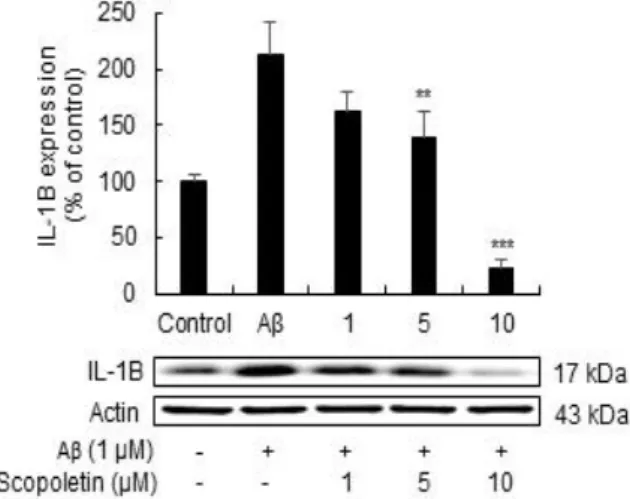
2. Effect of scopoletin on the production of IL-1β in Aβ1-42-induced microglical BV-2 cells
Scopoletin의 항염증 효과를 확인하기 위해 western blot을 이용하여 BV-2 세포에서 Aβ1-42로 유발된 염증성 사이토카인 IL-1β를 확인하였다. Aβ1-42는 세포생존율에 유의적인 변화 없이 염증만을 유발하는 최소농도인 1 µM 을 실험에 사용하였다.
Fig. 2. The effect of scopoletin on the production of interleukin 1 beta (IL-1β) in amyloid beta oligomer (Aβ1-42)-stimulated microglia cells BV-2. BV-2 cells were pretreated with different concentrations of scopoletin as indicated for 3 hours before treatment of 1 μM Aβ1-42 for 6 hours. IL-1β levels were measured using western blot analysis. ***p< 0.001, **p< 0.01: compared with Aβ1-42
alone treatment group.
Scopoletin을 농도에 따라 다르게 처리한 군과 Aβ1-42
단독 처리군을 비교하였고, IL-1β의 발현이 농도 의존적으 로 감소하는 것을 확인하였다. Scopoletin의 농도가 10 µM 일 때 IL-1β의 농도가 22.77%까지 감소하는 것으로 보아 Aβ1-42 처리한 BV-2세포에서 염증성 사이토카인의 생성을 억제하는 것을 확인하였다 (Fig. 2).
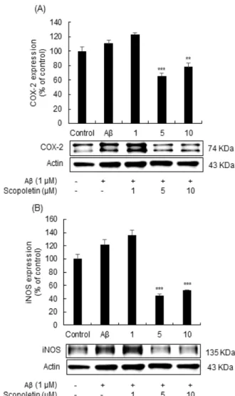
3. Effect of scopoletin on the production of COX-2 and iNOSIL-1β in Aβ1-42-induced microglical BV-2 cells
염증 반응시 염증매개인자인 COX-2, iNOS 발현이 증 가하는데 이에 scopoletin이 미치는 영향을 western blot 으로 확인하였다. Aβ1-42는 세포생존율에 유의적인 변화 없이 염증만을 유발하는 최소농도인 1 µM을 실험에 사용 하였다. Aβ1-42 단독 처리군에 비해 scopoletin을 5, 10 µM 농도로 전 처리한 군에서 염증 매개 인자인 COX-2는 65.8%, 78.19% (Fig. 3A), iNOS는 45.03%, 53.21%
(Fig. 3B)로 감소되어 단백질의 발현수준이 유의하게 억제 된 것을 확인하였다.
Fig. 3. The effect of scopoletin on the production of cyclooxygenase-2 (COX-2) and inducible nitric oxide
synthase (iNOS) in amyloid beta oligomer (Aβ1-42)-stimulated microglia cells BV-2. (A) The effect scopoletin on COX-2 production. (B) The effect scopoletin
on iNOS. BV-2 cells were pretreated with various concentrations of scopoletin as indicated for 3 hours before adding 1 μM Aβ1-42 for 6 hours. Each levels were
measured using western blot analysis. ***p < 0.001,
**p < 0.01: compared with Aβ1-42alone treatment group.
4. Effect of scopoletin on the production of NF-kB in Aβ1-42-induced microglical BV-2 cells
Aβ1-42로 염증반응이 유도된 BV-2 세포에서 scopoletin의 항염증 효과를 확인하고, 염증성 유전자 발 현에서 중요한 신호 전달 경로인 NF-κB의 활성을 확인하 였다. Aβ1-42는 세포생존율에 유의적인 변화 없이 염증만 을 유발하는 최소농도인 1 µM을 실험에 사용하였고, scopoletin을 1, 5, 10 µM 농도로 전 처리 후 Aβ1-42를 6
시간 동안 처리하였다. Aβ1-42 단독 처리 군에서는 세포핵 내 NF-kB의 유의미한 증가를 보였고, scopoletin 전 처 리 군에서는 NF-kB 핵 내 축적이 농도 의존적으로 감소 된 것을 확인하였다 (Fig. 4).
Fig. 4. Effect of scopoletin on Nuclear factor-κB (NF-κB) activity induced by amyloid beta oligomer (Aβ1-42) in
microglial BV-2 cells. Microglial BV-2 cells were pretreated with various concentrations of scopoletin as indicated for 3 hours prior to 1 μM Aβ1-42treatment for 6
hours. NF-κB levels were measured using Western blot analysis. ###p < 0.001 : compared to basal cell, ***p <
0.001 : compared with Aβ1-42 alone treatment group.
IV. Discussion
만성 염증반응은 알츠하이머병 환자의 뇌에서 중요한 역할을 하는데, 과 활성화 된 미세아교세포는 염증성 사이 토카인을 포함한 신경 독성 인자를 생성하여 AD의 발병에 기여하는 것이 확인 되었다[17-19]. Aβ에 의해 형성된 노 인성 플라크 주변에 존재하는 활성화 된 미세아교세포는 염증성 사이토카인 IL-1β와 독성 인자를 생성함으로써 신 경 변성을 촉진한다[20]. 또한 COX-2는 뇌의 신경 독성과 관련이 있고, 억제됨에 따라 AD의 진행이 늦춰지는 것으 로 알려졌다[21]. iNOS의 발현도 Aβ로 인해 상향 조절되 지만, 반대로 억제될 경우 뉴런을 보호하는 것으로 알려져 있다[22]. 본 연구에서는 Aβ1-42 oligomer (Aβ1-42)로 유도 된 미세아교세포 BV-2 세포의 염증반응에서 scopoletin 이 염증성 사이토카인 IL-1β의 발현과 상향 조절되었던 염증 매개인자 COX-2, iNOS를 억제할 수 있는 것을 확인 하여, 염증반응 감소에 유의한 효과가 있는 것을 보여주었 다. 또한, 미세아교세포에서 염증성 사이토카인, COX-2 및 iNOS의 발현은 NF-kB의 전사 활성에 의한 것으로 [23], BV-2 세포에서도 scopoletin이 NF-kB 활성 메커니
즘을 통해 염증반응을 조절한다는 것을 알 수 있었다.
그러나 지금까지 미세아교세포 BV-2에서 Aβ1-42으로 유 도된 신경염증에 대한 scopoletin의 효과는 아직까지 연 구가 되지 않고 있어, 본 연구결과를 통해 scopoletin의 항염증 효과에 기초하여 AD 예방 및 신경염증보호제로서 정보를 제공하고자 한다.
결과적으로 이 연구결과는 scopoletin이 Aβ로 인한 염 증반응을 줄여 미세아교세포의 활성을 억제하고, 염증성 사이토카인 및 염증매개인자생성을 감소시켜 염증반응이 개선되기에 향후 AD의 예방 및 신경염증보호제의 후보로 서 가능성이 있음을 제시한다.
ACKNOWLEDGEMENT
This research was supported by the Basic Science Research Program through the National Research Foundation of Korea (NRF), funded by the Ministry of Education, Science and Technology (2016R1C1B2007025); and by the National Dementia Research and Development Program of the Korea Health Industry Development Institute (KHIDI), funded by the Korean government (Ministry of Health and Welfare) (HI18C1671).
REFERENCES
[1] Alzheimer’s Association, “2020 Alzheimer’s disease facts and figures”, Alzheimer’s and Dementia, Vol. 16, No. 3, pp. 391-460, Mar 2020, DOI: 10.1002/alz.12068
[2] Chandra Sekhar Kuruva, and P Hemachandra Reddy, “Amyloid Beta Modulators and Neuroprotection in Alzheimer's Disease: A Critical Appraisal”, Drug Discovery Today, Vol. 22, pp. 223-233, Feb 2017, DOI: 10.1016/j.drudis.2016.10.010
[3] C. Cheignon, M..Tomas, D.Bonnefont-Rousselot, P. Faller, C.
Hureau, and F. Collin, “Oxidative stress and the amyloid beta peptide in Alzheimer’s disease”, Redox Biology, Vol. 14, pp.
450-464, Apr 2018, DOI: 10.1016/j.redox.2017.10.014 [4] Saeed Sadigh-Eteghad, Babak Sabermarouf, Alireza Majdi, Mahnaz
Talebi, Mehdi Farhoudi, and Javad Mahmoudi “Amyloid-Beta: A Crucial Factor in Alzheimer's Disease”, Medical Principles and Practice, Vol. 24, No. 1, pp. 1-10, Nov 2014, DOI: 10.1159/00 0369101
[5] Barbara Mroczko, Magdalena Groblewska, Ala Litman-Zawadzka, Johannes Kornhuber, and Piotr Lewczuk, “Amyloid β oligomers (AβOs) in Alzheimer’s disease”, Journal of Neural Transmission, Vol. 125, No. 2, pp. 177-191, Feb 2018, DOI: 10.1007/s00702 -017-1820-x
[6] David V. Hansen, Jesse E. Hanson, and Morgan Sheng, “Microglia in Alzheimer’s disease”, Journal of Cell Biololy, Vol. 217, No.
2, pp. 459-472, Feb 2018, DOI: 10.1083/jcb.201709069 [7] Francesca Regen, Julian Hellmann-Regen, Erica Costantini, and
Marcella Reale, “Neuroinflammation and Alzheimer's Disease:
Implications for Microglial Activation”, Current Alzheimer Research, Vol. 14, No. 11, pp. 1140-1148, Nov 2017, DOI 10.2174/1567205014666170203141717
[8] Elizabeth E. Spangenberg, and Kim N. Green, “Inflammation in Alzheimer’s disease: Lessons learned from microglia-depletion models”, Brain, Behavior, and Immunity, Vol. 61, pp. 1-11, Mar 2017, DOI: 10.1016/j.bbi.2016.07.003
[9] Atsuko Katsumoto, Hideyuki Takeuchi, Keita Takahashi, and Fumiaki Tanaka, “Microglia in Alzheimer's Disease: Risk Factors and Inflammation”, Frontiers in Neurology, Vol. 9, No. 978, Nov 2018, DOI: 10.3389/fneur.2018.00978
[10] Atish Kumar Sahoo, Jagnehswar Dandapat, Umesh Chandra Dash, and Satish Kanhar, “Features and outcomes of drugs for combination therapy as multi-targets strategy to combat Alzheimer's disease”, Journal of Ethnopharmacology, Vol. 215, pp. 42-73, Apr 2018, DOI: 10.1016/j.jep.2017.12.015
[11] Devesh Tewari, Adrian M. Stankiewicz, Andrei Mocan, Archana N. Sah1,Nikolay T. Tzvetkov 5, Lukasz Huminiecki, Jarosław O. Horbanczuk, and Atanas G. Atanasov, “Ethnopharmacological Approaches for Dementia Therapy and Significance of Natural Products and Herbal Drugs”, Front Aging Neuroscience, Vol. 10, No. 3, Feb 2018, DOI: 10.3389/fnagi.2018.00003
[12] Abhijit Dey, Raktim Bhattacharya, Anuradha Mukherjee, and Devendra Kumar Pandey, “Natural products against Alzheimer's disease: Pharmaco-therapeutics and biotechnological interventi ons”, Biotechnology Advances, Vol. 35, No. 2, pp. 178-216, Mar-Apr 2017, DOI: 10.1016/j.biotechadv.2016.12.005 [13] Hrishikesh Mohan Revankar, Syed Nasir Abbas Bukhari, Gajjela
Bharath Kumar, and Hua-Li Qin, “Coumarins scaffolds as COX inhibitors”, Bioorganic Chemistry, Vol. 71, pp. 146-159, Apr 2017, DOI: 10.1016/j.bioorg.2017.02.001
[14] Mylena Andréa Oliveira Torres, Isadora de Fátima Braga Magalhães, Renata Mondêgo-Oliveira, Joicy Cortez de Sá, Alessandra Lima Rocha, and Ana Lucia Abreu-Silva “One Plant, Many Uses: A Review of the Pharmacological Applications of Morinda citrifolia”, Phytotherapy Research, Vol. 31, NO. 7, pp.
971-979, Jul 2017, DOI: 10.1002/ptr.5817
[15] Negin Ahmadi, Suhaila Mohamed, Heshu Sulaiman Rahman, and Rozita Rosli, “Epicatechin and scopoletin‐rich Morinda citrifolia
leaf ameliorated leukemia via anti‐inflammatory, anti‐angiogen esis, and apoptosis pathways in vitro and in vivo”, European Journal of Pharmacology, Vol. 43, No. 7, Jul 2019, DOI:
10.1111/jfbc.12868
[16] Gilbert Kirsch, Ahmed Bakr Abdelwahab, and Patrick Chaimbault
“Natural and Synthetic Coumarins with Effects on Inflammation”, Molecules, Vol. 21, No. 10, pp. 1322, Oct 2016, DOI: 10.3390/m olecules21101322
[17] Shane A. Liddelow, Kevin A. Guttenplan, Laura E. Clarke, Frederick C. Bennett, Christopher J. Bohlen, Lucas Schirmer, Mariko L. Bennett, Alexandra E. Münch, Won-Suk Chung, Todd C. Peterson, Daniel K. Wilton, Arnaud Frouin, Brooke A. Napier, Nikhil Panicker, Manoj Kumar, Marion S. Buckwalter, David H. Rowitch, Valina L. Dawson, Ted M. Dawson, Beth Stevens, and Ben A. Barres, “Neurotoxic reactive astrocytes are induced by activated microglia”, Nature, Vol. 541, No. 7638, pp. 481-487, Jan 2017, DOI: 10.1038/nature21029
[18] Kelly S. Kirkley, Katriana A. Popichak, Maryam F. Afzali, Marie E. Legare, and Ronald B. Tjalkens, “Microglia amplify inflam matory activation of astrocytes in manganese neurotoxicity”, Journal of Neuroinflammation, Vol. 14, No. 1, pp. 99, May 2017, DOI: 10.1186/s12974-017-0871-0
[19] Darshpreet Kaur, Vivek Sharma, and Rahul Deshmukh, “Activ ation of microglia and astrocytes: a roadway to neuroinflamm ation and Alzheimer’s disease”, Inflammopharmacology, Vol. 27, No. 4, pp. 663-677, Aug 2019, DOI: 10.1007/s107
87-019-00580-x
[20] Suzanne Hickman, Saef Izzy, Pritha Sen, Liza Morsett, and Joseph El Khoury, “Microglia in neurodegeneration”, Nature Neurosci ence, Vol. 21, No. 10, pp. 1359-1369, Oct 2018, DOI:
10.1038/s41593-018-0242-x
[21] Pei-Pei Guan and Pu Wang, “Integrated Communications Between cyclooxygenase-2 and Alzheimer's Disease”, The FASEB Journal, Vol. 13, No. 1, pp. 13-33, Jan 2019, DOI: 10.1096/fj.2018003 55RRRR
[22] Magali Dumont and M. Flint Beal, “Neuroprotective strategies involving ROS in Alzheimer disease”, Free Radical Biology and Medicine, Vol. 51, No. 5, pp. 1014-1026, Sep 2011, DOI:
10.1016/j.freeradbiomed.2010.11.026
[23] Rui Yang, Sha Liu, Jia Zhou, Shuhong Bu, and Jian Zhang,
“Andrographolide attenuates microglia-mediated Aβ neurotoxic ity partially through inhibiting NF-κB and JNK-MAPK signaling pathway”, Immunopharmacology and Immunotoxicology, Vol.
39, NO. 5, pp. 276-284, Oct 2017, DOI: 10.1080/08923973.2 017.1344989
Authors
Hui-Jin Mun received the B.S. degree in Biomedical Laboratory Science from Konyang University in 2018. She is in currently a master’s course in Biomedical Laboratory Science at Konyang University from 2018 to
present. She has research interests include hematology, neuroscience.
Hyun-Jeong Cho received the M.S. and Ph.
D. degrees in Biomedical Laboratory Science from Inje University in 2002 and 2005, respectively. She was a Post-doc at Parmafoods Ltd. from Japan in 2005.
She is in a professor of Biomedical Laboratory Science at Konyang University from 2008 to present. Dr. Cho joined the faculty of department of Biomedical Laboratory Science at Konyang University in 2008. She is in currently a professor in Biomedical Laboratory Science at Konyang University. She has research interests include hematology, neuroscience.