대한소화기학회지 2009;53:106-110
접수: 2008년 7월 3일, 승인: 2008년 9월 16일 연락처: 천재희, 120-749, 서울시 서대문구 신촌동 134
연세대학교 의과대학 내과학교실 Tel: (02) 2228-1990, Fax: (02) 393-6884 E-mail: [email protected]
Correspondence to: Jae Hee Cheon, M.D.
Department of Internal Medicine and Institute of Gastroen- terology, Yonsei University College of Medicine, 134, Shinchon-dong, Seodaemun-gu, Seoul 120-752, Korea
Tel: +82-2-2228-1990, Fax: +82-2-393-6884 E-mail: [email protected]
전체 위장관을 침범한 베체트 장염 환자에서 연속 발생한 위, 결장 천공
연세대학교 의과대학 내과학교실, 소화기병연구소*, 외과학교실†, 병리학교실‡
신동엽ㆍ천재희*ㆍ박재준*ㆍ김호근
‡ㆍ김태일*ㆍ이용찬*ㆍ김남규*
†ㆍ김원호*
Serial Episodes of Gastric and Cecal Perforation in a Patient with Behcet’s Disease Involving the Whole Gastrointestinal Tract: A Case Report
Dong Yeob Shin, M.D., Jae Hee Cheon, M.D.*, Jae Jun Park, M.D.*, Hoguen Kim, M.D.‡, Tae Il Kim, M.D.*, Yong Chan Lee, M.D.*, Nam Kyu Kim, M.D.*†, and Won Ho Kim, M.D.*
Department of Internal Medicine and Institute of Gastroenterology*,
Departments of Surgery† and Pathology‡, Yonsei University College of Medicine, Seoul, Korea
Behcet’s disease (BD) has been recognized as multi-systemic chronic vasculitic disorder of recurrent inflammation, characterized by the involvement of multiple organs and resulting in orogenital ulcers, uveitis, and skin lesions.
Involvement of the central nervous system, vessels, and intestines in BD often leads to a poor prognosis.
Digestive manifestations in BD have been reported in up to 1-60% of cases, although the rate varies in different countries. The most frequent extra-oral sites of gastrointestinal involvement are the ileocecal region and the colon.
Gastric or esophageal involvement is reported to be very rare. Moreover, there have been no reports on the si- multaneous involvement of the esophagus, stomach, ileum, and colon. Here, we present a 55-year-old Korean man with intestinal BD and multiple ileal and colonic ulcerations complicated by perforation, gastric ulcer with bleed- ing followed by perforation, and esophageal ulcers with bleeding. (Korean J Gastroenterol 2009;53:106-110) Key Words: Behcet’s disease; Gastrointestinal; Ulcer; Perforation; Hemorrhage
Introduction
Behcet’s disease (BD) is a chronic multi-systemic vaculitis characterized by recurrent oral, genital aphthae, and ocular disease. Although the cause is unknown, the presumptive etiologies of BD include infections, autoimmune, and genetic mechanisms.1 The prevalence of this disease differs widely among countries; while the rate is 0.3/100,000 in the United States, it is 1/10,000 in Japan.2 The syndrome can cause
dysfunction in multiple organ systems, leading to conditions such as myocarditis, aneurysm, pulmonary thromboembolism, central nervous system abnormalities, renal involvement, and gastrointestinal manifestations.3,4 Gastrointestinal manifestations have been known to lead to severe morbidity in BD patients and are commonly referred to as “Intestinal Behçet's syndrome”. The terminal ileum and the cecum are the most frequently involved parts of the gastrointestinal tract and gastric or esophageal involvement is very rare. Esophageal involvement has been reported in less than 50 cases of BD with esophageal
신동엽 외 7인. 베체트 장염 환자에서 발생한 다발성 장천공 107
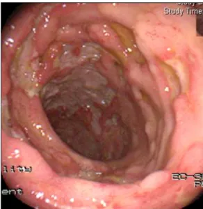
Fig. 1. The colonoscopic finding shows multiple and discrete ul- cers in the ascending colon.
Fig. 2. Microscopic finding of the surgical specimen shows mul- tiple ulcer perforations with diffuse inflammation in the cecum and ascending colon (H&E stain, ×200).
ulcers worldwide.5 Furthermore, gastric involvement is extremely rare, with an incidence lower than that of esophageal BD.6 There have not been any reports on simultaneous esophageal, gastric, ileal, and colon involvement. Here, we report a 55-year-old Korean man with intestinal BD and multiple ileal and colonic ulcerations complicated by perfo- ration, gastric ulcer bleeding followed by perforation, and esophageal ulcers with bleeding.
Case Report
A 55-year-old man was admitted to the emergency department for abdominal pain for 3 months. He had a past history of receiving a coronary angiography for the evaluation of chest pain 8 years prior, but it failed due to severe stenosis of both common iliac arteries and both subclavian arteries. A diagnosis of peripheral arterial occlusive disease was made, then an aorto-left common femoral artery bypass graft and fermoro-fermoral artery bypass graft and stent insertion at both subclavian arteries was performed. He had been taking anti-platelet agent, aspirin 100 mg per oral and anti-hyper- tensive agents since 8 years ago. He also had been suffered from recurrent oral and genital ulcers for the previous 5 years.
Three months before visiting our hospital, he had been admitted to a primary clinic and had undergone esophagogastroduo- denoscopy (EGD) and colonoscopy, which revealed ileocolonic ulcers (Fig. 1). He was referred to our hospital because there had been no improvement in symptom after steroid treatment
with the impression of Crohn’s disease.
On admission, the patient’s vital signs were stable. Although he complained of diffuse abdominal tenderness, there was no rebound tenderness or rigidity. He had well-demarcated, punctuated oral and genital ulcers suggesting BD. Uveitis was not seen on eye exam. Pathergy test was negative. Laboratory values showed a white blood cell count of 32,200/μL, hemoglobin of 13.1 g/dL, a platelet count of 174,000/μL, and an ESR of 14 mm/hr. His liver function test was normal and CRP was 12.7 mg/dL. Anti-cytomegalovirus IgM and anti-amebic IgM antibodies were negative. Although he didn’t have fever on admission day, he developed a fever of 38.1oC on his second day of admission.
His abdominal pain and fever were not improved despite 4 days of antibiotics therapy. Abdominal CT scan showed active inflammation with diffuse wall thickening at the cecum, ascending colon, and transverse colon. Abdominal pain was aggravated and he had newly developed hematochezia. On physical examination, rebound tenderness of whole abdomen became obvious. Accordingly, he underwent emergent right hemicolectomy with ileostomy with suspicion of intestinal perforation. The operation revealed two sites of intestinal perforations from the cecum to the ascending colon. Micro- scopic examination of the specimen showed transluminal inflammation, discrete ulcer margins, and no chronic surroun- ding mucosal change, suggestive of intestinal BD (Fig. 2).
Other types of colitis such as viral or neoplastic diseases were excluded by immunohistochemistric staining. Seven days after the operation, the patient had a large amount of melena. An
108 The Korean Journal of Gastroenterology: Vol. 53, No. 2, 2009
Fig. 3. Esophagogastroduodenoscopic finding shows an active ul- cer with mucosal edema in the lesser curvature of the lower body in the stomach. There is a bleeding stigmata with vessel exposure at the base of the ulcer.
Fig. 5. Esophagoscopic finding shows two esophageal ulcers with current bleeding at 25 cm from the incisors.
Fig. 4. Ileoscopic examination showing the active ulcer at 8 cm from the ileostomy site. There is no evidence of recent bleeding.
emergency EGD showed an oval-shaped active ulcer with a discrete margin in the lesser curvature of the lower body in the stomach. There was a stigmata of recent bleeding (Fig. 3).
Thereafter, melena was spontaneously subsided. On postope- rative day (POD) 25, he was discharged with daily doses of 3 g of mesalazine and 30 mg of prednisolone per oral with the diagnosis of intestinal BD involving stomach and ileocolon.
Twelve days after discharge, the patient was admitted again due to severe abdominal pain. Emergent laparotomy revealed panperitonitis with gastric ulcer perforation. Genital and oral ulcers developed again on PODs 7 and 14, respectively. On POD 19, he had hematochezia. Enteroscopy through the ileostomy site showed no evidence of recent bleeding, but
revealed an ileal ulcer (Fig. 4). However, he had a large amount of melena again on POD 28. Whereas abdominal aortography revealed no bleeding focus, EGD showed two discrete esophageal ulcers with a bleeding vessel at 25-30 cm from upper incisor (Fig. 5). After hemoclipping, the bleeding was stopped. His further postoperative course was uneventful, and the patient was followed-up without any further episode of hemorrhage. After being discharged, he was prescribed 50 mg of azathioprine, 30 mg of prednisolone, and 1.8 mg of colchicine.
Discussion
BD was first described in 1937 by Turkish dermatologist, Huluisi Behcet as a triple complex of chronic, recurrent oral and genital ulcers, and uveitis. The underlying cause of BD is still unknown and the diagnosis is mainly based on the clinical features.1,7 Several sets of criteria had been employed to diagnose and classify BD. In 1990, the international study group (ISG) introduced a new, internationally agreed upon criteria for BD based on a survey of 913 patients recruited from 12 centers in 7 countries. The ISG criteria requires the presence of recurrent oral ulceration plus any two of genital ulceration, typically defined eye lesions, skin lesions, or a positive pathergy test.8 The oral and genital ulcers were found during the initial physical examination of this patient. In addition, the patient’s multiple arterial occlusions and coronary artery occlusive diseases that had developed recurrently for 8 years are thought to be related to the vascular involvement of
Shin DY, et al. Serial Ulcer Perforations in Behcet’s Disease Involving the Whole Gastrointestinal Tract 109
BD. Large vessel involvement is known to occur in one-third of the patients with BD4. Arteries and veins of all sizes can be affected and arterial complications are less common than venous problems in BD, occurring in roughly 1-7% of patients.
Male sex and cigarette smoking are known risk factors.9 His manifestations do not fully satisfy the ISG criteria, but do fulfill two of the main and two of the minor diagnostic criteria for BD proposed by the BD Research Committee of Japan and can be categorized as “incomplete type” BD.10
The frequency of intestinal involvement in patients with BD is known to be 0-60%, with geographic differences.11 Symp- tomatic intestinal involvement is rare in Mediterranean patients with BD, but in East Asian patients, including Koreans, intestinal involvement is more common.12,13 The intestinal lesions are usually resistant to medical treatment and recur frequently (25-78%) after surgical management.14 Intestinal BD with gastrointestinal ulcer is reported to account for only 1% of cases in Western countries. In contrast, Asian patients with BD tend to experience intestinal ulcer more frequently. The main symptoms include anorexia, nausea, abdominal pain, and diarrhea.13 Large, discrete, punched-out-appearing ulcers are characteristics of intestinal BD. Fistula formation, hemorrhage, or perforation occurs in up to 50% of ulcers in BD.15 The most common location of involvement is the ileocecal region (60-75%).16 So far, only nine cases of intestinal Behcet’s disease presenting with both esophageal and ileocolonic ulcers have been reported in the English literature.2 The gastroduodenal mucosa is known to be the least frequently involved area of the gastrointestinal tract, although Ning-Sheng et al. reported a relatively high prevalence of gastric/duodenal ulceration in patients with BD in Taiwan.6 Recently, Isik et al. reported three cases of intestinal perforations in patients with BD. These patients underwent the operations two to four times. The reasons for re-operation were ulcer perforation after primary repair, or postoperative complications like fistula and leakage.17 To our knowledge, there has been no case report of simulta- neous involvement of the esophagus, stomach, ileum, and colon with multiple intestinal perforations in BD. Ulcerogenic medications like aspirin or steroids and the severe stress might have been aggravating factors for the BD associated ulcer rather than direct causes, considering relatively long duration of taking aspirin without ulcer event, refractoriness to conventional anti-ulcer treatment, and normal finding of previous EGD perfomed in other hospital.
In conclusion, we present an exceptionally rare case of
intestinal BD involving the whole gastrointestinal tract and leading to recurrent hemorrhage and ulcer perforation. Although rare, intestinal involvement of BD can lead to very severe complications and can involve multiple sites concurrently. The evaluation of the whole gastrointestinal tract may be necessary in BD patients who develop intestinal symptoms.
References
1. Ghate JV, Jorizzo JL. Behcet's disease and complex aphthosis. J Am Acad Dermatol 1999;40:1-18.
2. Fujiwara S, Shimizu I, Ishikawa M, et al. Intestinal Behcet's disease with esophageal ulcers and colonic longitudinal ulcers.
World J Gastroenterol 2006;12:2622-2624.
3. Sakane T, Takeno M, Suzuki N, Inaba G. Behcet's disease.
N Engl J Med 1999;341:1284-1291.
4. Koc Y, Gullu I, Akpek G, et al. Vascular involvement in Behcet's disease. J Rheumatol 1992;19:402-410.
5. Morimoto Y, Tanaka Y, Itoh T, Yamamoto S, Kurihara Y, Nishikawa K. Esophagobronchial fistula in a patient with Behcet's disease: report of a case. Surg Today 2005;35:
671-676.
6. Ning-Sheng L, Ruay-Sheng L, Kuo-Chih T. High frequency of unusual gastric/duodenal ulcers in patients with Behcet's disease in Taiwan: a possible correlation of MHC molecules with the development of gastric/duodenal ulcers. Clin Rheu- matol 2005;24:516-520.
7. Mutlu S, Scully C. The person behind the eponym: Hulusi Behcet (1889-1948). J Oral Pathol Med 1994;23:289-290.
8. Criteria for diagnosis of Behcet's disease. International Study Group for Behcet's Disease. Lancet 1990;335:1078-1080.
9. Calamia KT, Schirmer M, Melikoglu M. Major vessel in- volvement in Behcet disease. Curr Opin Rheumatol 2005;17:
1-8.
10. Suzuki Kurokawa M, Suzuki N. Behcet's disease. Clin Exp Med 2004;4:10-20.
11. Yurdakul S, Tuzuner N, Yurdakul I, Hamuryudan V, Yazici H. Gastrointestinal involvement in Behcet's syndrome: a con- trolled study. Ann Rheum Dis 1996;55:208-210.
12. Shimizu T, Ehrlich GE, Inaba G, Hayashi K. Behcet disease (Behcet syndrome). Semin Arthritis Rheum 1979;8:223-260.
13. Oshima Y, Shimizu T, Yokohari R, et al. Clinical studies on Behcet's syndrome, with special reference to 100 personal cases. Naika 1962;9:701-714.
14. Iida M, Kobayashi H, Matsumoto T, et al. Postoperative re- currence in patients with intestinal Behcet's disease. Dis Colon Rectum 1994;37:16-21.
110 대한소화기학회지: 제53권 제2호, 2009
15. Jarrahnejad P, Gadepalli S, Zurkovsky E, Tulsyan N, Rolan- delli R. Behcet's disease: a rare cause of lower gastro- intestinal bleeding. Int J Colorectal Dis 2006;21:856-858.
16. Kasahara Y, Tanaka S, Nishino M, Umemura H, Shiraha S, Kuyama T. Intestinal involvement in Behcet's disease: review of 136 surgical cases in the Japanese literature. Dis Colon
Rectum 1981;24:103-106.
17. Isik B, Ara C, Kirimlioglu H, et al. Single or multiple perfo- rations with varying locations as a complication of intestinal Behcet's disease: report of three cases. Scand J Gastroenterol 2005;40:599-603.