Ⅰ.
서 론‘대칭’이라는 용어는 물체의 좌, 우가 거울상(mirror
images)에서 정확하게 일치하는 것으로 정의할 수 있다
1.
그러나 사람의 두개 안면 영역에서 이러한 대칭은 거의 없 다. 그러나 안면 비대칭은 시상면이나 수직적인 악골의 불 균형을 동반한 환자에서도 일반적으로 흔한 주소가 될 수 있다. 반대로, 안면 비대칭은 치열의 이상 배열이나, 연조 직의 보상, 머리 위치의 경사 등에 의해 가려지는 경우 역 시 발생할 수 있다
2.
안면 비대칭에 영향을 주는 요소로는 상악골 자체의 길 이 차이, 하악체 길이의 차이, 하악지 길이의 차이, 두개저 형태의 차이 등이며
3,4, 임상적으로 이러한 요소들은 환자 의 성장 방향이나, 성장량에 영향을 주며, 복합적으로 3차 원적인 부조화를 야기하여 이부의 편위 및 안면 비대칭으 로 표현된다
5,6. 안모의 비대칭은 악골과 연관된 현상일 때 더욱 현저하게 나타나며, 이들은 주로 하악골과 연관되어 나타난다
7.
안면 비대칭 환자의 하악골의 비대칭 요소를 알아보기 위한 여러 연구가 있었으나
3,4,8, 하악골체의 시상 단면에 대 한 연구는 미비한 실정이다. 안면 비대칭의 치료를 위한 악 교정 수술 시 하악골 단독으로 수술을 하는 경우나 양악수 술을 하는 경우 모두에서 하악골체부 및 하악지에 대한 수 술이 시행되며 이 때 비대칭환자에서의 하악 단면에 대한 해부학적 이해가 수술계획 수립 및 술후 합병증의 감소를 위해 필요하다고 생각한다.
이에 본 연구에서는 computed tomography (CT)를 이용하
황 대 석626-770 경남 양산시 물금읍 범어리
부산대학교 치의학전문대학원 구강악안면외과학교실 Dae-Seok Hwang
Department of Oral and Maxillofacial Surgery, Pusan National University Hospital
3-3, Beomeo, Mulgeum, Yangsan, 626-770, Korea TEL: +82-55-360-5103 FAX: +82-55-360-5104 E-mail: [email protected]
안면 비대칭환자의 하악골체부의 시상 단면에 관한 연구
이재열
1∙김용일
2∙황대석
1∙김용덕
1∙신상훈
1∙김욱규
1∙정인교
1부산대학교 치의학전문대학원
1구강악안면외과학교실,
2교정학교실
Cross-sectional study of the mandibular body in patients with facial asymmetry
Jae-Yeol Lee
1, Yong-Il Kim
2, Dae-Seok Hwang
1, Yong-Deok Kim
1, Sang-Hun Shin
1, Uk-Kyu Kim
1, In-Kyo Chung
1Depatments of
1Oral and Maxillofacial Surgery,
2Orthodontics, School of Dentistry, Pusan National University, Yangsan, Korea
Introduction: To correct the facial asymmetry by mandibular jaw surgery, it is important to know the anatomy of the mandible including the mandibular canal positioning of patients with facial asymmetry. This study was performed to evaluate the differences in the cross-sectional surface in the body of the mandible between the deviated side and opposite side in patients with facial asymmetry.
Materials and Methods:The study was conducted on 37 adult patients composed of 2 groups, the asymmetry group (n=20) and non-asymmetry group (n=17). Using the cross-sectional computed tomography (CT) images, the distance from the buccal aspect of the mandibular canal to the outer aspect of the buccal cortex, distance from the buccal aspect of the mandibular canal to the inner aspect of the buccal cortex, distance from the inferior aspect of the mandibular canal to the inferior border of the mandible, thickness of the mandible, and cross-sectional surface area of the mandible were measured in each side of the mandible.
Results:The cross-sectional area of the mandible including the mandibular canal positioning in the deviated side was not statistically different from the opposite side in the asymmetry group. Only the distance from the inferior aspect of the mandibular canal to the inferior border of the mandible in the ramus area of the deviated side was significantly longer than opposite side. On the other hand, the bucco-lingual width of the asymmetry group was thinner than the non-asymmetry group.
Conclusion:The cross-sectional area including the mandibular canal of the mandible did not appear to be modified by the facial asymmetry.
Key words:Facial asymmetry, Cross-sectional anatomy, Mandibular nerve
[paper submitted 2010. 11. 24 / revised 2011. 2. 10 / accepted 2011. 4. 5]
Abstract (J Korean Assoc Oral Maxillofac Surg 2011;37:109-13)
여 안면 비대칭을 보이는 환자의 하악골의 시상 단면에서 하악관의 위치를 포함하여 여러 계측항목의 변위측과 반 대측의 차이가 있는지 알아보고 비대칭이 없는 환자군과 비교분석하였다.
Ⅱ.
연구 대상 및 방법1. 연구 대상
본 연구의 대상은 2009년 2월부터 2010년 7월까지 부산 대학병원 구강악안면외과에 내원하여 CT 검사를 시행한 37명의 환자 중 안면 정중선과 하악 이부 사이의 변위가 3 mm 이상인 환자를 비대칭군으로(asymmetric group), 두개 안면부의 연조직이나 경조직에 기형이 없으며 Angle씨 분 류 Ⅰ급 교합을 가진 사람을 대칭군(non-asymmetry group) 으로 분류하였다. 비대칭군의 경우 모두 악교정수술을 위 해 내원한 환자였으며 교정장치를 부착한 상태에서 CT 검 사를 시행하였고, 평균이부 변위량은 6.2 mm였으며 좌측 으로 변위된 경우가 12명, 우측으로 변위된 경우가 8명이 었다.(Table 1)
2. 연구 방법
1) 삼차원 영상의 재구성
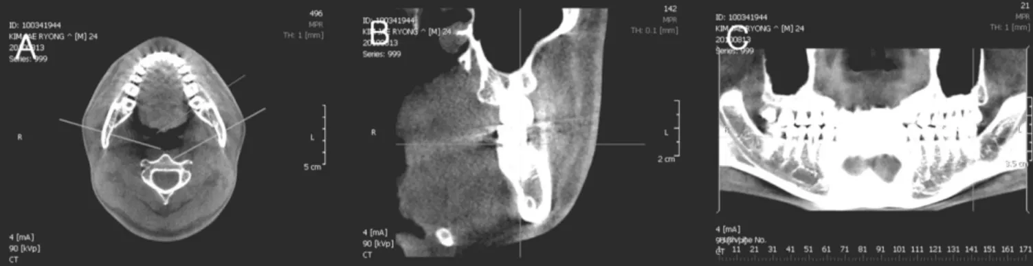
Cone beam CT 촬영은 DCT pro (Vatech Co., Seoul, Korea) 을 이용하여 노출시간 24초, 0.3 mm 간격의 영상을 digital imaging and communications in medicine (DICOM) 3.0 file로 전환하였으며, Ondemand program (Cybermed Inc., Seoul, Korea)을 통해 하악 전치 절단연과 양측 하악 제1대구치 근
심협측 교두정을 지나는 하악 교합평면을 수평면으로 하 고, 악궁의 형태에 수직인 횡단면 영상으로 전환하였 다.(Fig. 1)
2) 연구 항목 및 위치
변환된 횡단면 영상에서 측정지점은 제1대구치 근심면, 제2대구치 원심면, 하악지의 오훼돌기의 최상방점을 지나 는 면으로 하였고 실험군에서는 변위측(이부가 변위된 방 향)과 반대측으로, 대조군에서는 좌측과 우측을 각각 구분 하여 다음과 같은 항목을 측정하였다.(Fig. 2)
Fig. 1.Converted images by using Ondemand program. A: Axial image, B: Cross-sectional image, C: Panoramic image.
Table 1.Patients distribution
Group Male/Female Mean age Chin deviation
Non-asymmetry (n=17) 9/8 23.7±4.28
Asymmetry (n=20) 7/13 22.6±3.55 6.2±2.9 (Rt./Lt.=8/12)
Fig. 2. Measuring points and measurements in the cross sectional images.
(A: distance from the buccal aspect of the mandibular canal to outer aspect of the buccal cortex, B: distance from the buccal aspect of the mandibular canal to inner aspect of the buccal cortex, C: distance from the inferior aspect of the mandibular canal to inferior border of the mandible, D: thickness of the mandible, E: cross-sectional surface area of the mandible)
(1) 하악관의 협측면에서 협측 피질골의 외측까지의 거 리(distance from the buccal aspect of the mandibular canal to buccal aspect of the buccal cortex, A)
(2) 하악관의 협측면에서 협측 피질골의 내측까지의 거 리(distance from the buccal aspect of the mandibular canal to lingual aspect of the buccal cortex, B)
(3) 하악관의 하연에서 하악 하연까지의 거리(distance from the inferior aspect of the mandibular canal to inferi- or border of the mandible, C)
(4) 하악골의 폭경(thickness of the mandible, E)
(5) 하악골의 단면적(cross-sectional surface area of the mandible, F)
3) 통계학적 분석
비대칭군에서 변위측과 반대측, 대칭군에서 좌, 우측의 차이를 비교하기 위해 SPSS version 12 (SPSS Inc., Chicago, IL, USA)를 사용하여 paired t-test를 실시하였고, 실험군과 대조군의 비교 및 성별에 따른 차이를 보기 위해 indepen- dent t-test를 사용하였다. 이때 P value가 0.05 이하일 경우 통계적으로 유의하다고 판정하였다.
Ⅲ.
연구 결과비대칭군에서 변위측과 반대측의 각 측정항목의 계측결 과, 하악관의 협측면에서 협측 피질골까지의 거리는 양측 모두에서 하악지에서 제2대구치로 오면서 커지다가 제1대 구치에서 작아지는 양상을 보였으며, 반대측에서 제1대구 치에서의 값이 더 컸지만 통계학적 유의성은 없었다. 하악 지에서 하악관의 하연에서 하악 하연까지의 거리는 하악 지 측정지점에서의 값이 가장 크고 제2대구치에서 줄어들 다가 제1대구치에서 다시 증가하는 양상을 보이며, 하악지 에서 변위측이 10.3 mm, 반대측이 11.51 mm로 반대측이 더 크게 나왔다.(P<0.05) 하지만 하악 협측면에서 협측 피질골 의 내측까지의 거리, 하악골의 폭경 및 하악골의 단면적은 모든 계측지점에서 통계학적으로 유의한 차이를 나타내지 않았다.(Table 2) 대칭군에서는 모든 측정지점과 측정항목 에서 통계학적인 차이를 나타내지 않았다.(Table 3)
Paired t-test 결과 유의성이 나타나지 않은 계측지점 및 항 목은 비대칭군과 대칭군의 비교를 위해 각 군에서 좌, 우 값을 통합하여 통계적으로 분석하였다. 비대칭군의 경우 거의 모든 측정지점 및 측정항목에서 대칭군에 비해 측정 값이 작게 나타났으며, 특히 하악관의 협측면에서 협측 피
Table 3.Measurements in the normal occlusion group
Right side Left side P value
1st molar 2nd molar Ramus 1st molar 2nd molar Ramus 1st molar 2nd molar Ramus
A 5.43±1.48 7.01±1.83 5.36±1.72 5.37±1.23 7.08±1.81 5.24±1.85 0.868 0.797 0.777
B 3.03±1.39 4.57±1.70 3.14±1.75 3.07±1.09 4.78±1.82 3.06±1.84 0.919 0.484 0.855
C 9.68±2.48 7.87±1.64 11.40±2.42 8.16±2.04 7.97±1.50 10.49±1.64 0.436 0.742 0.195
D 12.46±2.45 13.70±2.00 12.26±2.48 12.30±2.37 13.91±1.77 12.29±1.63 0.668 0.485 0.945
E 368.60±78.38 401.64±80.00 410.00±60.17 369.39±68.37 403.63±68.69 422.66±54.90 0.887 0.859 0.116 (A: distance from the buccal aspect of the mandibular canal to outer aspect of the buccal cortex, B: distance from the buccal aspect of the mandibular canal to inner aspect of the buccal cortex, C: distance from the inferior aspect of the mandibular canal to inferior border of the mandible, D: thickness of the mandible, E: cross-sectional surface area of the mandible)
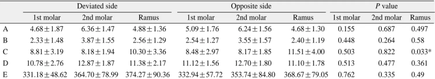
Table 2.Measurements in the asymmetry group
Deviated side Opposite side P value
1st molar 2nd molar Ramus 1st molar 2nd molar Ramus 1st molar 2nd molar Ramus
A 4.68±1.87 6.36±1.47 4.88±1.36 5.09±1.76 6.24±1.56 4.68±1.30 0.155 0.687 0.497
B 2.33±1.48 3.87±1.55 2.56±1.29 2.54±1.27 3.55±1.57 2.40±1.19 0.448 0.264 0.58
C 8.81±3.19 8.18±1.94 10.30±3.36 8.48±2.97 8.17±1.85 11.51±4.00 0.503 0.822 0.033*
D 10.78±2.76 12.87±1.87 11.38±2.17 11.12±1.56 12.70±1.80 11.10±1.78 0.513 0.477 0.361
E 331.18±48.62 364.70±78.99 374.27±90.36 332.94±57.72 353.74±84.80 368.67±79.05 0.762 0.335 0.49 (A: distance from the buccal aspect of the mandibular canal to outer aspect of the buccal cortex, B: distance from the buccal aspect of the mandibular canal to inner aspect of the buccal cortex, C: distance from the inferior aspect of the mandibular canal to inferior border of the mandible, D: thickness of the mandible, E: cross-sectional surface area of the mandible) *P<0.05
질골의 내측까지의 거리, 하악골의 폭경과 하악골의 단면 적은 통계적으로 유의한 차이를 나타냈다.(Table 4)
비대칭군과 대칭군 모두에서 성별에 따른 차이는 모든 계측지점과 계측항목에서 통계학적인 유의성을 나타내지 않았다.
Ⅳ.
총괄 및 고찰안면 비대칭은 그 종류가 매우 다양하고 형태가 복잡하 여 대칭적인 안면의 회복은 술자에게는 항상 도전과제가 되고 있다. 또한 하악 전돌이나 하악 후퇴를 가지는 환자들 중 약 21-67%에 이르는 환자들이 안면 비대칭을 가지고 있 어 안면 비대칭의 문제는 악교정수술에 있어서 중요한 고 려 대상이 되었다
9,10. 안면 비대칭의 원인은 유전적인 요인 뿐만 아니라 태아나 유아기, 성장기의 환경적인 요인과도 관련이 있다
11. 또한 과두의 과성장
12이나 저작근의 기능적 부조화
13, 유아기의 머리 위치
14, 사두증(plagiocephay)
6등 다 양한 원인인자들이 안면 비대칭에 관여한다.
이러한 안면의 비대칭은 악골과 연관된 경우 더욱 현저 하게 나타나는데, 특히 하악골에서 주로 좌, 우의 차이가 발생한다. Baek 등
4은 비대칭환자에서 변위측이 반대측에 비해 하악지의 높이가 더 짧으며, 하악지의 경사가 내측으 로 덜 기울어져 있다고 하였으며, 하악 골체의 길이는 양측 에서 유의할 만한 차이가 없다고 하였다. Choi 등
15은 이부 편위에 가장 영향을 주는 요소는 좌, 우 하악지의 길이의 차이라고 하였으며, 비변위측에서 정중 시상면과 하악지 가 이루는 각이 더 크다고 하였다. 본 연구에서도 하악관의 수직적 위치를 나타내는 하악관의 하연에서 하악 하연까 지의 거리가 하악지의 측정지점에서 변위측에서 평균 10.3 mm로 반대측의 평균 11.51 mm보다 짧게 나타났다. 이는 하악지의 길이 차이에 의한 것으로 생각한다. 하지만,
Maeda 등
3은 안면 비대칭은 상악과 하악지에 비해 하악골
체부의 좌, 우 차이가 더 많이 나타난다고 하였으며, Fong 등
8역시 이부 편위는 수직적 요소의 차이보다 좌, 우의 수 평적 길이 차이와 연관하여 생긴다고 하였다.
안면 비대칭에 대해 2차원적 또는 3차원적 하악골의 좌, 우 차이에 대한 연구는 많이 있었으나 하악골의 시상 단면 에서의 연구는 미비한 실정이다. 본 연구결과, 안면 비대칭 환자에서 하악관의 위치를 포함하여 시상 단면 자체의 좌, 우 차이는 거의 없는 것으로 나타났다. 이는 하악골의 성장 에서 수직적, 전, 후방적 성장 차이에 의해 비대칭이 나타 나며, 하악골체의 내, 외측 성장 차이는 크지 않음을 의미 한다.
술후 나타나는 지각이상은 악교정수술, 특히 하악지 시 상분할 골절단술 시 발생하는 가장 흔한 합병증 중 하나이 며, 약 30%에서 40%의 환자에서 일시적 또는 영구적인 지 각이상을 보인다고 보고되고 있다
16,17. 이러한 지각이상은 하악관의 위치와 관련이 높은데, Yamamoto 등
18은 하악골 의 협측 피질골과 접촉해 있는 하악관의 위치를 가지는 환 자에서 술후 지각이상이 높게 발생한다고 하였으며 협측 피질골과 하악관 사이의 골수강(marrow space)이 0.8 mm 이하인 경우 술후 1년 이후에도 지각이상이 계속될 수 있 다고 하였다. Yoshioka 등
19도 하악골의 협측 피질골에서 하 악관의 협측면까지의 거리가 짧을수록 지각이상의 가능성 이 높다고 하였다. 본 연구에서는 이러한 하악관의 위치가 안면 비대칭환자에서 좌, 우측이 차이가 있는지 알기 위해 하악관의 위치를 하악골의 표면에서의 거리를 측정하여 보았다. 그 결과 비대칭군에서 변위측과 반대측의 차이는 없었으나, 비대칭이 없는 군과 비교해 볼 때 제1대구치와 제2대구치 부위에서 골수강의 크기가 작은 것으로 나타났 으므로(P<0.05) 이러한 환자의 수술 시 신경손상에 대한 고 려가 필요하다.
하악골의 두께에 관해서는 Hallikainen 등
20은 후퇴된 하 악골의 협설측 두께가 전돌된 하악의 두께보다 유의하게 크다고 하였으며, Jung 등
21은 하악 전돌증 환자에서 정상 교합군에 비해 하악골의 협설측 두께가 작다고 하였고 두 군 모두 남자가 여자보다 두께가 크다고 하였다. 본 연구에 서는 비대칭이 있는 환자군에서 하악골의 협설측 두께가 현저하게 작게 나타났는데, 이는 비대칭환자 중 하악 전돌 을 동반한 경우가 많기 때문으로 보인다. 하지만 성별에 따 른 차이는 보이지 않았다.
비대칭환자의 하치조 신경관의 위치 및 하악골의 시상 단면의 좌, 우 차이를 알아보기 위해 이번 연구를 시행하였 으나, 큰 차이를 발견하지는 못하였다. 하지만, 연구대상의 수가 부족하고 시상 단면에서 관찰한 계측항목이 적은 한 계가 있었다. 향후 비대칭 정도에 따른 차이나 동반된 하악 전돌 여부 및 교합관계에 따른 하악골의 좌, 우 차이에 대 한 3차원적 연구가 필요하다고 생각한다.
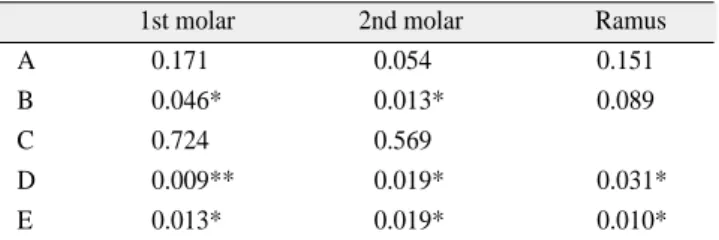
Table 4.Comparison of the measurements according to group
1st molar 2nd molar Ramus
A 0.171 0.054 0.151
B 0.046* 0.013* 0.089
C 0.724 0.569
D 0.009** 0.019* 0.031*
E 0.013* 0.019* 0.010*
(A: distance from the buccal aspect of the mandibular canal to outer aspect of the buccal cortex, B: distance from the buccal aspect of the mandibular canal to inner aspect of the buccal cortex, C: distance from the inferior aspect of the mandibular canal to inferior border of the mandible, D: thickness of the mandible, E: cross-sectional surface area of the mandible) *P<0.05, **P<0.01
Ⅴ. 결 론
본 연구는 안면 비대칭을 보이는 환자에서 하악골의 시 상 단면에서 변위측과 반대측의 차이 및 비대칭이 관찰되 지 않는 환자와의 차이를 알아보기 위해 시행되었다. Cone beam CT를 이용하여 비대칭군과 대칭군에서 제1대구치의 근심면과 제2대구치의 원심면, 하악지의 오훼돌기 최상방 을 지나는 하악골의 시상 단면에서 하악관의 위치와 하악 골의 두께, 하악골의 단면적을 조사하여 다음과 같은 결과 를 얻었다.
1. 비대칭군에서 하악지 측정지점에서 하악관의 하연에 서 하악 하연까지의 거리는 변위측이 반대측에 비해 작게 나타났으며(P<0.05), 나머지 측정지점에서의 측 정항목의 값은 통계적 유의성이 없었다.
2. 대칭군에서는 모든 측정지점에서의 모든 측정값에서 통계적 유의성이 없었다.
3. 비대칭군은 대칭군에 비해 하악관의 협측면에서 협측 피질골의 내측까지의 거리, 하악골의 폭경과 하악골의 단면적이 작았다.(P<0.05)
4. 각 군에서 각 측정값의 성별에 따른 차이는 관찰되지 않았다.
이상의 결과 비대칭이 있는 하악골 단면과 하악관 위치 의 좌,우 차이는 없지만, 비대칭이 없는 환자에 비해 하악 골의 협설폭 및 단면적이 작으므로 이러한 환자의 하악골 수술 시 좁은 하악골 두께에 대한 고려가 필요하다고 생각 한다.
References
1. Kronmiller JE. Development of asymmetries. Semin Orthod 1998;4:134-7.
2. Burstone CJ. Diagnosis and treatment planning of patients with asymmetries. Semin Orthod 1998;4:153-64.
3. Maeda M, Katsumata A, Ariji Y, Muramatsu A, Yoshida K, Goto S, et al. 3D-CT evaluation of facial asymmetry in patients with maxillofacial deformities. Oral Surg Oral Med Oral Pathol Oral Radiol Endod 2006;102:382-90.
4. Baek SH, Cho IS, Chang YI, Kim MJ. Skeletodental factors af- fecting chin point deviation in female patients with class III mal- occlusion and facial asymmetry: a three-dimensional analysis us- ing computed tomography. Oral Surg Oral Med Oral Pathol Oral Radiol Endod 2007;104:628-39.
5. Sakurai A, Hirabayashi S, Sugawara Y, Harii K. Skeletal analysis of craniofacial asymmetries in plagiocephaly (unilateral coronal
synostosis). Scand J Plast Reconstr Surg Hand Surg 1998;32:81-9.
6. St John D, Mulliken JB, Kaban LB, Padwa BL. Anthropometric analysis of mandibular asymmetry in infants with deformational posterior plagiocephaly. J Oral Maxillofac Surg 2002;60:873-7.
Erratum in: J Oral Maxillofac Surg 2005;63:419.
7. Haraguchi S, Takada K, Yasuda Y. Facial asymmetry in subjects with skeletal Class III deformity. Angle Orthod 2002;72:28-35.
8. Fong JH, Wu HT, Huang MC, Chou YW, Chi LY, Fong Y, et al.
Analysis of facial skeletal characteristics in patients with chin de- viation. J Chin Med Assoc 2010;73:29-34.
9. Severt TR, Proffit WR. The prevalence of facial asymmetry in the dentofacial deformities population at the University of North Carolina. Int J Adult Orthodon Orthognath Surg 1997;12:171-6.
10. Tani M, Iketani M, Watanabe M, Suda S, Fujimura N, Miyazawa M, et al. Posterior-anterior cephalometric analysis in patients with dentofacial deformities. J Jpn Stomatol Soc 1989;35:1749- 59.
11. Pirttiniemi PM. Associations of mandibular and facial asymme- tries-a review. Am J Orthod Dentofacial Orthop 1994;106:191- 200.
12. Obwegeser HL, Makek MS. Hemimandibular hyperplasia-hemi- mandibular elongation. J Maxillofac Surg 1986;14:183-208.
13. Poikela A, Kantomaa T, Pirttiniemi P. Craniofacial growth after a period of unilateral masticatory function in young rabbits. Eur J Oral Sci 1997;105:331-7.
14. Persing J, James H, Swanson J, Kattwinkel J; American Academy of Pediatrics Committee on Practice and Ambulatory Medicine, Section on Plastic Surgery and Section on Neurological Surgery. Prevention and management of positional skull deformities in infants. Pediatrics 2003;112:199-202.
15. Choi J, Min S, Oh SH, Kwon KH, Choi MK, Lee J, et al.
Skeletal pattern analysis of facial asymmetry patient using three dimensional computed tomography. J Korean Assoc Oral Maxillofac Surg 2008;34:622-7.
16. Westermark A, Bystedt H, von Konow L. Inferior alveolar nerve function after mandibular osteotomies. Br J Oral Maxillofac Surg 1998;36:425-8.
17. Yoshida T, Nagamine T, Kobayashi T, Michimi N, Nakajima T, Sasakura H, et al. Impairment of the inferior alveolar nerve after sagittal split osteotomy. J Craniomaxillofac Surg 1989;17:271-7.
18. Yamamoto R, Nakamura A, Ohno K, Michi KI. Relationship of the mandibular canal to the lateral cortex of the mandibular ra- mus as a factor in the development of neurosensory disturbance after bilateral sagittal split osteotomy. J Oral Maxillofac Surg 2002;60:490-5.
19. Yoshioka I, Tanaka T, Khanal A, Habu M, Kito S, Kodama M, et al. Relationship between inferior alveolar nerve canal position at mandibular second molar in patients with prognathism and possi- ble occurrence of neurosensory disturbance after sagittal split ra- mus osteotomy. J Oral Maxillofac Surg 2010;68:3022-7.
20. Hallikainen D, Iizuka T, Lindqvist C. Cross-sectional tomogra- phy in evaluation of patients undergoing sagittal split osteotomy.
J Oral Maxillofac Surg 1992;50:1269-73.
21. Jung YH, Nah KS, Cho BH. The location of the mandibular canal in prognathic patients compared to subjects with normal occlu- sion. Korean J Oral Maxillofac Radiol 2007;37:217-20.