plication4.
Anatomical variants of the paranasal sinus and nasal cav- ity are commonly detected, with an estimated prevalence of 68%5. Some anatomic variants on the nasal cavity and ostium of maxillary sinus, such as deviated nasal septum, concha bullosa or paradoxical middle turbinate, bending of the unci- nated process, and Haller cells, are very important because of their contribution to the blockage of ostio-meatal units. These anatomic variants can disturb the drainage and ventilation of the maxillary sinus and, thereby, can affect the risk of sinus mucosal disease6.
With maxillary sinus floor elevation, postoperative swell- ing of the Schneiderian membrane is an unavoidable sequela.
Although the mucosa of the sinus membrane heals rapidly and recovers its homeostasis7, if the ostio-meatal complex is unfavorable due to anatomic variants, its healing can be de- layed, and risk of sinusitis is increased.
In this study, we evaluated the computed tomography (CT) images of patients who had undergone sinus floor elevation and investigated the correlations between anatomic variants of the lateral nasal wall and maxillary sinus and risk of max-
I. Introduction
Maxillary sinus floor elevation is known as a reliable op- tion to enable insertion of dental implants for patients with severe atrophy in the posterior area of the maxilla1. In spite of vague criteria for evaluation and diagnosis, maxillary si- nusitis is the most common complication of this procedure2. This unpleasant complication can occur as a result of con- tamination of the maxillary sinus with oral or nasal pathogen or because of a lack of asepsis during the surgery3. However, obstruction of the ostium caused by postoperative swelling of the maxillary sinus mucosa can also be a source of this com-
Kyung-Hwan Kwon
Department of Oral and Maxillofacial Surgery, College of Dentistry, Wonkwang University, 460 Iksan-daero, Iksan 54538, Korea
TEL: +82-63-859-2921 FAX: +82-63-847-4002 E-mail: denhouse@wonkwang. ac.kr
ORCID: http://orcid.org/0000-0002-5257-8440
This is an open-access article distributed under the terms of the Creative Commons Attribution Non-Commercial License (http://creativecommons.org/licenses/by-nc/4.0/), which permits unrestricted non-commercial use, distribution, and reproduction in any medium, provided the original work is properly cited.
CC
Correlations between anatomic variations of maxillary sinus ostium and postoperative complication after sinus lifting
Jang Won Lee, Ji Yong Yoo, Seung Jae Paek, Won-Jong Park, Eun Joo Choi, Moon-Gi Choi, Kyung-Hwan Kwon Department of Oral and Maxillofacial Surgery, Wonkwang University Dental Hospital,
College of Dentistry, Wonkwang University, Iksan, Korea
Abstract(J Korean Assoc Oral Maxillofac Surg 2016;42:278-283)
Objectives: The maxillary sinus mucosa is reported to recover to preoperative sterility after sinus floor elevation. However, when drainage of maxil- lary sinus is impaired, recovery can be delayed and maxillary sinusitis can occur. Therefore, in this study, we investigated the correlations between anatomic variants that can interrupt the ostium of the maxillary sinus and incidence of complication after sinus lifting.
Materials and Methods: The subjects are 81 patients who underwent sinus lifting in Wonkwang University Dental Hospital (Iksan, Korea). Com- puted tomography (CT) images of the subjects were reviewed for presence of nasal septum deviation, anatomic variants of the middle turbinate, and Haller cells. Correlations between anatomic variations and occurrence of maxillary sinusitis were statistically analyzed.
Results: Patients with anatomic variants of ostio-meatal units, such as deviated nasal septum, concha bullosa or paradoxical curvature of the middle turbinate, or Haller cells, showed a higher rate of complication. However, only presence of Haller cell showed statistically significant.
Conclusion: Before sinus lifting, CT images are recommended to detect anatomic variants of the ostio-meatal complex. If disadvantageous anatomic variants are detected, the use of nasal decongestants should be considered to reduce the risk of postoperative sinusitis.
Key words: Maxillary sinus, Sinus floor augmentation, Maxillary sinusitis, Anatomic variation, Ostio-meatal complex
[paper submitted 2016. 6. 23 / revised 1st 2016. 8. 26, 2nd 2016. 10. 4 / accepted 2016. 10. 4]
Copyright Ⓒ 2016 The Korean Association of Oral and Maxillofacial Surgeons. All rights reserved.
2. Methods
Each CT image was reviewed by the author, investigating the presence of anatomic variants. The radiological assess- ments were used to detect conditions that could jeopardize the physiological maxillary ventilation and drainage functions, impairing the antral homeostasis, such as septal deviations, concha bullosa, paradoxical bending of the middle turbinate, or Haller cells.(Fig. 1-4)
Sinusitis is characterized by a typical triad of symptoms;
nasal congestion or obstruction, pathological secretion, and headache8. However, these symptoms are extremely variable.
Sinusitis is also suspected in patients complaining of pain illary sinusitis after sinus floor elevation.
II. Materials and Methods
1. Subject
At the Department of Oral and Maxillofacial Surgery in Wonkwang Dental Hospital (Iksan, Korea), we reviewed a total of 366 patients who had undergone sinus floor elevation from January 2009 to December 2015. Of these, 267 patients were excluded because of lack of adequate CT images with view of the ostio-meatal complex. Finally, 99 patients were included in this study.
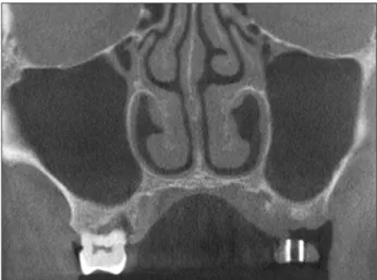
Fig. 2. Concha bullosa of middle turbinate and Haller cell on left.
Jang Won Lee et al: Correlations between anatomic variations of maxillary sinus ostium and postoperative complication after sinus lifting. J Korean Assoc Oral Maxillofac Surg 2016
Fig. 4. Haller cell on left maxillary ostium.
Jang Won Lee et al: Correlations between anatomic variations of maxillary sinus ostium and postoperative complication after sinus lifting. J Korean Assoc Oral Maxillofac Surg 2016
Fig. 3. Paradoxical curvature of left middle turbinate.
Jang Won Lee et al: Correlations between anatomic variations of maxillary sinus ostium and postoperative complication after sinus lifting. J Korean Assoc Oral Maxillofac Surg 2016
Fig. 1. Deviated nasal septum.
Jang Won Lee et al: Correlations between anatomic variations of maxillary sinus ostium and postoperative complication after sinus lifting. J Korean Assoc Oral Maxillofac Surg 2016
composed of 81 cases, comprising 54 males (66.7%) and 27 females (33.3%) with a mean age of 51.48 years.(Table 1)
The operation was performed on the right side in 37 cases (45.7%) and on the left side in 44 cases (54.3%). In addition, 68 cases (84.0%) used the lateral window technique and 13 cases (16.1%) used the crestal approach.(Table 2) Simultane- ous bone graft was conducted in 75 cases.
In our study, 10 cases (12.4%) showed nasal septum de- viation to the operation site. Concha bullosa or paradoxical curvature of the middle concha was discovered in 12 cases (14.8%), and Haller cells were identified in 30 cases (37.0%).
(Table 3)
Among 81 cases, 7 cases that showed consistent pain for longer than 2 weeks or nasal discharge were considered to be demonstrating a complication.
Statistical analysis was performed to evaluate the correla- tions between each anatomical variant and prolonged healing after sinus floor elevation. Correlation of nasal septum devia- tion did not show a significant result (P=0.206), and neither did that of the middle concha (P=0.276). However, although most patients who showed variants of the middle concha showed nasal septum deviation, these cases were excluded from the nasal septum deviation group due to deviation direc- tion. In this regard, we evaluated the relation between com- plication and variants on the nasal septum or middle concha, and its significance level was 0.070. Unlike other anatomical variants, correlations of Haller cells was statistically signifi- cant (P=0.009).
or tenderness in the region of the sinus, in combination with mucopurulent rhinorrhea. A physical examination was per- formed after the sinus floor elevation procedure to assess the presence of maxillary sinusitis. In particular, the patients were assessed for development of any sign or symptom suggesting postoperative maxillary sinusitis, including nasal obstruction or a purulent nasal discharge, facial pain or tenderness, fever, or purulent oral discharge. In this study, the patients who ex- perienced pain for longer than 2 weeks or nasal obstruction were considered to have complication after sinus floor eleva- tion.
The statistical analysis was useful to assess the correlations between anatomic variants and the development of maxillary sinusitis. Correlations were evaluated using Fisher’s exact test. A chi-square test was not appropriate for this data be- cause the expected frequency was less than 5 for more than 25% of the cells in the contingency tables. Analysis was con- ducted using PASW Statistics version 18 (IBM Co., Armonk, NY, USA). Statistical significance was set at P<0.05.
III. Results
Of the 99 cases, 10 patients who underwent functional en- doscopic surgery were excluded from this study. In addition, 8 cases in which the sinus membrane was perforated during the operation were excluded. Finally, the study group was
Table 3. Correlations between anatomic variations and postoperative sinusitis (n=81)
Anatomic variants Patient Complication OR (95% CI) P-value
Deviated nasal septum
Concha bullosa or paradoxical curvature Haller cells
Any anatomic variants
10 (12.4) 12 (14.8) 30 (37.0) 37 (45.7)
2/10 (20.0) 2/12 (16.7) 6/30 (20.0) 6/37 (16.2)
3.30 (0.55-19.89) 2.56 (0.44-15.03) 12.50 (1.42-109.72)
8.32 (0.95-72.66)
0.206 0.276 0.009 0.043 (OR: odds ratio, CI: confidence interval)
Values are presented as number (%) or OR (95% CI).
Jang Won Lee et al: Correlations between anatomic variations of maxillary sinus ostium and postoperative complication after sinus lifting. J Korean Assoc Oral Maxillofac Surg 2016 Table 1. Sex and age distributions (n=81)
Patient No. of patients (%)
Gender Male Female Age (yr) ≤30 31-40 41-50 51-60 61-70
54 (66.7) 27 (33.3) 4 (4.9) 4 (4.9) 23 (28.4) 41 (50.6) 9 (11.1)
Jang Won Lee et al: Correlations between anatomic variations of maxillary sinus ostium and postoperative complication after sinus lifting. J Korean Assoc Oral Maxillofac Surg 2016
Table 2. Operation site and approach (n=81)
Operation No. of patients (%)
Operation site Right Left Approach Lateral window Crestal
37 (45.7) 44 (54.3) 68 (84.0) 13 (16.1)
Jang Won Lee et al: Correlations between anatomic variations of maxillary sinus ostium and postoperative complication after sinus lifting. J Korean Assoc Oral Maxillofac Surg 2016
procedure may impair the physiology of the sinus mucosa and cause unpleasant complications. In spite of the prompt postoperative recovery of the maxillary mucosa with a rapid return to preoperative sterility, it must be pointed out that the sinus compliance, which represents the ability to recover nor- mal maxillary sinus homeostasis after sinus floor elevation, depends on its baseline condition18.
Anatomical variants of the paranasal sinus and nasal cav- ity are commonly detected, with an estimated prevalence of 68%5. Because of their contribution to the blockage of ostio- meatal units, some anatomic variants on the lateral nasal wall, such as deviated nasal septum, concha bullosa or paradoxi- cal middle turbinate, bending of the uncinated process, and Haller cells, are very important. These variants can interfere with drainage and ventilation of the maxillary sinus and, thereby, can affect the risk of sinus mucosal disease6. These variants and their relations with the ostium of the maxillary sinus can be easily detected on computed tomographic im- ages19.
Theoretically, anatomical variants of the lateral nasal wall are likely to shift and alter ostio-meatal complex components, possibly resulting in obstruction of maxillary sinus mucous drainage19,20. Altered anatomic relations in the nasal cavity and ostio-meatal complex might be involved in sinus drain- age impairment. Compromised maxillary sinus drainage is closely associated with a reduction of the maxillary ostium21. Previous studies on the function of this ostium have shown a reduced size of ostium diameter in cases of sinusitis22.
The nasal septum is an osseocartilaginous wall that divides the nasal cavity. In the junction of the nasal cartilage with the vomer, acute angulation can occur23, resulting in deviation of the nasal septum. Severe nasal septal deviation has been assumed to be related with sinusitis by interfering with the ostio-meatal complex19,24,25.
The term concha bullosa was first introduced by Zucker- landl in 1862, as a pneumatization of the middle turbinate.
Paradoxical curvature of the middle turbinate is known as a convexity pointing toward the middle meatus. These com- mon anatomical variants might cause secondary deformity of the turbinate, followed by the risk of obstruction of the middle meatus and mucosal pathologies23,26.
Another common anatomic variant is the presence of infra- orbital ethmoidal cells, also known as Haller cells, which can be found between the maxillary sinus and orbital floor. This is a clinically important anatomical variant because it has been assumed to be an etiologic factor in recurrent maxillary sinusitis due to reduction of the ostio-meatal complex27. In
IV. Discussion
The ostio-meatal unit plays an important role in the devel- opment of maxillary sinusitis by impairment of the muco- ciliary clearance system9. If the patency of the ostio-meatal unit is interrupted, clearance of the maxillary sinus can be delayed, and this unfavorable event can increase the risk for development of sinusitis.
Given the close relationship between the Schneiderian membrane and the ostium of the maxillary sinus and the fact that inflammatory reaction after any surgical procedure is inevitable, surgeons must consider the risk of infectious se- quelae after sinus floor elevation. Because of the temporary interference of ciliary activity caused by the traumatic eleva- tion of the Schneiderian membrane, altered mucous composi- tion and bacterial infection can occur10,11. After sinus floor elevation, the maxillary sinus may be filled with hematoma or seroma, requiring removal. However, a mild inflammatory reaction can occur as a normal physiological activity of the mucosal airway system12, and this swelling of the mucosa can lead to obstruction of the patency of the ostio-meatal unit. As a result, sinus floor elevation might compromise the physi- ological drainage of the maxillary sinus into the middle me- atus by transient inflammatory swelling on the mucosa of the ostium or due to other mechanisms that can predispose the patient to acute maxillary sinusitis13 and possibly lead to bone graft loss.
Although a transient or persisting effect on the ciliated mucosa can be expected as a result of raising the mucosa of the maxillary sinus, the mucosal reaction has been reported to be negligible7. Fortunately, the maxillary sinus mucosa is ca- pable of adapting adequately to the alteration following sinus floor elevation14,15. In particular, in healthy persons, drainage of the maxillary sinus did not seem to be impaired after the procedure12. This can explain why the incidence of maxil- lary sinusitis after sinus floor elevation in the literature is less than 1%16,17. Using generally accepted Ear, Nose, and Throat (ENT) criteria for diagnosing sinusitis, occurrence of maxil- lary sinusitis after sinus floor elevation has been reported at 1.3%13. Despite the low incidence of maxillary sinusitis after this procedure, there is still inherent risk of disturbing the physiology of the sinus. It is generally assumed that altera- tion of the maxillary sinus anatomy, such as elevation of the Schneiderian membrane with bulging or injured sinus mu- cosa, might impair the physiology of the maxillary sinus.
Even though sinus floor elevation is considered an ef- ficacious procedure, surgeons should remember that this
References
1. Raghoebar GM, Brouwer TJ, Reintsema H, Van Oort RP. Aug- mentation of the maxillary sinus floor with autogenous bone for the placement of endosseous implants: a preliminary report. J Oral Maxillofac Surg 1993;51:1198-203; discussion 1203-5.
2. Doud Galli SK, Lebowitz RA, Giacchi RJ, Glickman R, Jacobs JB. Chronic sinusitis complicating sinus lift surgery. Am J Rhinol 2001;15:181-6.
3. Misch CM. The pharmacologic management of maxillary sinus elevation surgery. J Oral Implantol 1992;18:15-23.
4. Drettner B. The permeability of the maxillary ostium. Acta Oto- Laryngologica 1965;60:304-14.
5. Bolger WE, Butzin CA, Parsons DS. Paranasal sinus bony ana- tomic variations and mucosal abnormalities: CT analysis for endo- scopic sinus surgery. Laryngoscope 1991;101:56-64.
6. Bayram M, Sirikci A, Bayazit YA. Important anatomic variations of the sinonasal anatomy in light of endoscopic surgery: a pictorial review. Eur Radiol 2001;11:1991-7.
7. Timmenga NM, Raghoebar GM, Liem RS, van Weissenbruch R, Manson WL, Vissink A. Effects of maxillary sinus floor elevation surgery on maxillary sinus physiology. Eur J Oral Sci 2003;111:
189-97.
8. Yonkers AJ. Sinusitis--inspecting the causes and treatment. Ear Nose Throat J 1992;71:258-62.
9. Bertrand B, Eloy P. Relationship of chronic ethmoidal sinusitis, maxillary sinusitis, and ostial permeability controlled by sinuso- manometry: statistical study. Laryngoscope 1992;102:1281-4.
10. Misch CE. Maxillary sinus augmentation for endosteal implants:
organized alternative treatment plans. Int J Oral Implantol 1987;4:
49-58.
11. Zimbler MS, Lebowitz RA, Glickman R, Brecht LE, Jacobs JB.
Antral augmentation, osseointegration, and sinusitis: the otolaryn- gologist's perspective. Am J Rhinol 1998;12:311-6.
12. Timmenga NM, Raghoebar GM, Boering G, van Weissenbruch R.
Maxillary sinus function after sinus lifts for the insertion of dental implants. J Oral Maxillofac Surg 1997;55:936-9; discussion 940.
13. Timmenga NM, Raghoebar GM, van Weissenbruch R, Vissink A.
Maxillary sinusitis after augmentation of the maxillary sinus floor:
a report of 2 cases. J Oral Maxillofac Surg 2001;59:200-4.
14. Stammberger H. Endoscopic endonasal surgery--concepts in treat- ment of recurring rhinosinusitis. Part II. Surgical technique. Otolar- yngol Head Neck Surg 1986;94:147-56.
15. Jensen OT, Shulman LB, Block MS, Iacono VJ. Report of the si- nus consensus conference of 1996. Int J Oral Maxillofac Implants 1998;13 Suppl:11-45.
16. Bhattacharyya N. Bilateral chronic maxillary sinusitis after the sinus-lift procedure. Am J Otolaryngol 1999;20:133-5.
17. Zinner ID, Small SA. Sinus-lift graft: using the maxillary sinuses to support implants. J Am Dent Assoc 1996;127:51-7.
18. Torretta S, Mantovani M, Testori T, Cappadona M, Pignataro L.
Importance of ENT assessment in stratifying candidates for sinus floor elevation: a prospective clinical study. Clin Oral Implants Res 2013;24:57-62.
19. Laine FJ, Smoker WR. The ostiomeatal unit and endoscopic sur- gery: anatomy, variations, and imaging findings in inflammatory diseases. AJR Am J Roentgenol 1992;159:849-57.
20. Scribano E, Ascenti G, Cascio F, Racchiusa S, Salamone I. Com- puterized tomography in the evaluation of anatomic variations of the ostiomeatal complex. Radiol Med 1993;86:195-9.
21. Aust R, Drettner B. The functional size of the human maxillary ostium in vivo. Acta Otolaryngol 1974;78:432-5.
22. Stierna P, Söderlund K, Hultman E. Chronic maxillary sinusitis.
Energy metabolism in sinus mucosa and secretion. Acta Otolaryn- gol 1991;111:135-43.
23. Arslan H, Aydinlioğlu A, Bozkurt M, Egeli E. Anatomic variations of the paranasal sinuses: CT examination for endoscopic sinus sur-
previous studies, the incidence of Haller cells was variable;
from 6% to 45.1%5,23. A possible reason for this discrepancy is differences in criteria for diagnosing Haller cells or in the technique of CT scanning. In this study, the prevalence of Haller cells was 37.0%.
In this study, correlations of nasal septum deviation and middle concha variants with complications were not sig- nificant (P=0.206 and P=0.276, respectively). On the other hand, the correlation of Haller cells with complications was statistically significant (P=0.009). We concluded that this dif- ference was due to regional proximity of Haller cells. Unlike other variants, Haller cells are located right above the ostium, and this proximity may contribute to reduction of the ostium after the operation. Another reason for this result is the small number of investigated cases. Unlike Haller cells, incidences of anatomic variants of nasal septum and middle concha were 12.4% and 14.8%, respectively. If the number of included cases was sufficient, these variants could have been found to be significant.
V. Conclusion
Considering the influence of anatomic variants of ostio- meatal complex, it is recommended that candidates for maxil- lary sinus floor elevation with any radiological or anamnestic evidence suggesting impaired maxillary sinus conditions should undergo an ENT evaluation that includes CT of the maxillofacial district28. If anatomic variants that might impair the patency of the maxillary sinus are detected, the surgeon should inform the patient of increased risk of postoperative sinusitis and consider the use of nasal decongestants as a pro- phylaxis.
Conflict of Interest
No potential conflict of interest relevant to this article was reported.
ORCID
Jang Won Lee, http://orcid.org/0000-0003-2809-3424 Ji Yong Yoo, http://orcid.org/0000-0002-9390-1494 Seung Jae Paek, http://orcid.org/0000-0002-5095-7543 Won-Jong Park, http://orcid.org/0000-0001-6687-5043 Eun Joo Choi, http://orcid.org/0000-0001-5377-8893 Moon-Gi Choi, http://orcid.org/0000-0003-3502-7652 Kyung-Hwan Kwon, http://orcid.org/0000-0002-5257-8440
gery. Auris Nasus Larynx 1999;26:39-48.
24. Fadda GL, Rosso S, Aversa S, Petrelli A, Ondolo C, Succo G. Mul- tiparametric statistical correlations between paranasal sinus ana- tomic variations and chronic rhinosinusitis. Acta Otorhinolaryngol Ital 2012;32:244-51.
25. Ameri AA, Eslambolchi A, Bakhshandeh H. Anatomic variants of paranasal sinuses and chronic sinusitis. 2005;2:121-4.
26. Nouraei SA, Elisay AR, Dimarco A, Abdi R, Majidi H, Madani SA, et al. Variations in paranasal sinus anatomy: implications for
the pathophysiology of chronic rhinosinusitis and safety of endo- scopic sinus surgery. J Otolaryngol Head Neck Surg 2009;38:32-7.
27. Zinreich SJ, Kennedy DW, Rosenbaum AE, Gayler BW, Kumar AJ, Stammberger H. Paranasal sinuses: CT imaging requirements for endoscopic surgery. Radiology 1987;163:769-75.
28. Pignataro L, Mantovani M, Torretta S, Felisati G, Sambataro G.
ENT assessment in the integrated management of candidate for (maxillary) sinus lift. Acta Otorhinolaryngol Ital 2008;28:110-9.