I. INTRODUCTION
Atherosclerosis is a common and insidious dis- ease, which accounts for most of the morbidity seen in humans with coronary heart diseases and stroke.
According to WHO report, cardiovascular diseases represent 20% of deaths worldwide (14 million annually), with 50% (leading cause) of all deaths in developed countries, and 16% ( the third leading cause) of all deaths in developing countries. In Korea, 21% of death are due to complications asso- ciated with atherosclerotic disease1.
Risk factors for atherosclerosis include: older age, male gender, smoking, body-mass index, levels of serum lipids, diabetes, hypertension, family history, and infection. Periodontal disease, as an infectious disease, has been associated with atherosclerosis in humans. Matilla has shown that dental infections are a major risk factor for adverse coronary events and
overall mortality2, 3. DeStefano et al. has shown that men with severe periodontal disease have a 1.72 rel- ative risk of death due to CHD as compared to men without periodontal disease4. It was concluded that an increased risk of coronary heart disease is associ- ated with dental disease, particularly in young men under the age of 50. Recent evidence in the peri- odontal literature suggests that host inflammatory responses to bacteria, and not just the bacterial pathogens themselves, are responsible for the marked destruction found in severe periodontal dis- ease. However, the molecular and biological mech- anisms of this association are far from being under- stood. Several possibilities exist for the potential influence of chronic periodontal disease on the rate of progression of atherosclerosis. First, periodontal disease may alter host responses in such a way as to accelerate the atherosclerotic process. Second, an underlying alteration in immune function(for
Establishment of a Mouse Model of Infection-Induced Atheroma Formation
Hyun-Ju Chung1, In-Chul Ryu2, Soo-Boo Han2, Jannett H Southerland3, Catherine ME Champagne3, Steven Offenbacher3
1Dept of Periodontology, College of Dentistry, Chonnam National University
2Dept of Periodontology, College of Dentistry, Seoul National University
3Center for Oral and Systemic Diseases and Dept of Periodontology, College of Dentistry, University of North Carolina.
대한치주과학회지 : Vol. 33, No. 1, 2003
This study was supported by a grant of the Korea Health 21 R&D Project, Ministry of Health & Welfare, ROK(HMP-00-CH-10-0009).
Corresponding author: HJ Chung, Dept of Periodontology, College of Dentistry, Chonnam National University, 5 Hakdong, Donngu. Gwangju, 501-190
instance “hyperactive”monocyte/macrophage response5, 6) may increase the host susceptibility to atherosclerosis and periodontal disease, with no influence of the infection on the progression of ath- erosclerosis. Third, the above possibilities exist with the addition of bacterial infection of the atheroma- tous lesion (bacterial tropism to the atheroma) may be responsible for accelerating the progression of the atherosclerotic process with or without modula- tion of immune responses. Genetics is certain to play a significant role. Studies have shown various types of atherosclerotic and severe periodontal dis- eases are passed down through families. Another factor is the bacterial challenge itself7. Thus, it is pro- posed that these three major factors influence a hyperactive immune response: genetics, environ- ment (diet and stress), and bacterial challenge.
Our researches have been focusing on developing an animal model to elucidate this mechanism of association between 2 disease entities. The subcuta- neous chamber model of infection is a well-estab- lished and commonly used model of chronic infec- tion with periodontal pathogens such as Porphyromonas gingivalis(Pg)8, 9. ApoE knockout -/- mice are highly susceptible to atherosclerosis when fed a high-fat diet10. Combined models can be used to evaluate the effects of periodontal infection on
the development of atheroma lesions. By using the mouse as a model, these factors can be varied and outcomes assessed. The ApoE +/-is predisposed to atheroma lesions. This line will manifest atheroscle- rotic lesions in a relatively short period of time, as early as two months using high-fat diets, while C57B6 mice are predisposed to a much lesser degree to atherosclerosis and require six months of high-fat feeding to show mild atheroma formation.
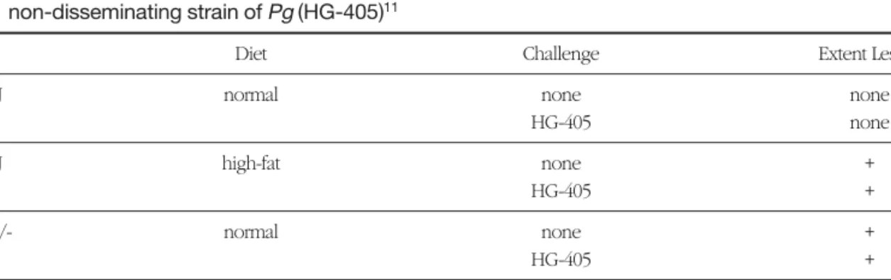
Previously, Gibbs et al11 used ApoE heterozygous (ApoE+/-) mice with a C57B6 background mouse line which were chronically infected with a non- dis- seminating strain of Pg, HG-405. This experiment failed to demonstrate an atherogenic response using Pgstrain HG-405, in mice fed either a normal or a high-fat diet(Table 1).
This present study was aimed to examine the effects of chronic infection with a more toxic, dis- seminating strain of P. gingivalis, in the heterozygous ApoE+/- mice maintained on a normal diet, on local inflammation, and on short-term aortic atheroma for- mation. The hypothesis in this experiment was that disseminating strains of P. gingivalis(such as A7436) could enhance atheroma formation as compared to non-disseminating strains (HG-405) and infection- mediated cytokines could induce hyperlipidemia and thereby enhance atheroma formation.
Table 1. Atherogenic effects of normal and high-fat diet on wild type (C57B6J) and ApoE+/- mice, using a non-disseminating strain of Pg (HG-405)11
Mice Diet Challenge Extent Lesion
C57B6J normal none none
HG-405 none
C57B6J high-fat none +
HG-405 +
ApoE+/- normal none +
HG-405 +
ApoE+/- high-fat none +++
HG-405 ++
II. MATERIALs and METHODs
C57B6J Mice were purchased from Jackson Labs (Barharbor, ME ) at 6 weeks of age and ApoE-/- mice were supplied from Dr. Maeda's lab in UNC Medical School. Heterozygous mice ApoE+/- were bred using female ApoE-/- and male C57B6J repro- ductive pairs. Approximately nine weeks was need- ed to produce the ApoE+/-mice ready for the experi- ment. Female ApoE+/- mice were maintained on normal chow diet(from Purina) after weaning at 4 weeks of age. Genotype was confirmed by PCR using specific primers and genomic DNA extracts prepared from tail clippings.
Mice were immunized at week 8 with an intraperi- toneal injection of either broth (bacterial culture medium) or a preparation of heat-killed P.g.strain A7436 (109 CFU/100㎕). Two subcutaneous cham- bers (0.4 X 1cm, 12 stainless steel coil) were implant- ed bilaterally in the flanks of each mouse at week 10 under general anesthesia by intraperitoneal injection of 0.10~0.15 ml/mouse of mixture of Ketaset (0.062 ml) and Rompum (0.22 ml) in saline (3.16 ml).
Animals were randomized for the four groups: 1) live challenge with PgA7436 and high fat diet (west-
ern type); 2) live challenge with PgA7436 and low fat diet; 3) chamber injection with medium alone and high fat diet; 4) chamber injection with medium alone and low fat diet. The high fat diet was a mix- ture of 1 part of Thomas Hartroft diet (Teklad (TD88051) Test Diets, Madison, WI.) and 3 parts of Purina Breeder chow (resulting in the composition of total fat 15%, cholesterol 1.25%, and cholic acid 0.5%
). At weeks 12 and 15, mice were challenged by injecting into chamber with either 100㎕ broth or a suspension of live P.g. (108CFU/100㎕).
Animals were monitored weekly until sacrifice for clinical signs of cachexia and weight loss and the clinical response of chamber and surrounding tis- sue. The sloughing of coil chamber was monitored as result of inflammation and the day of sloughing documented. Chamber fluid samples were collected at days 0, 7, and 14 after each challenge and processed for evaluation of inflammatory mediator levels. Blood samples were collected by retro-orbital bleeding at weeks 12, 15, and 18, and further processed for evaluation of lipid levels and inflam- matory mediator levels. At weeks 18, the mice were fasted overnight prior to being overdosed with Ketaset /Rompum mixture. The chest was opened Figure 1. Animal protocol timeline.
and the heart and vascular trees perfused with fresh 4%(wt/vol) phosphate-buffered (0.12 mol/L) paraformaldehyde (ph 7.4). The heart and attached aorta and other organ tissues were removed and placed in fresh paraformaldehyde until processed.
1. Evaluation of Chamber Fluid
The syringe was chamber fluid samples were col- lected by aspiration in a 1cc syringe containing loaded with 100㎕ of PBS buffer (to wet and pre- vent sticking of sample to the syringe) and weighed, and volume of collected fluid was estimated by weight (1 g = 1 ml). Collected fluid samples were stored frozen (-80℃) until analysis.
For chamber Pgverification, The 10 ㎕ aliquots of chamber fluid, already diluted into 500 ㎕ of RIA buffer, was plated on a Wilkins-Chalgren plate and grown in under anaerobic conditions at 37℃ for 4-5 days. Pg scores were determined by counting colonies as follows: Pg-score=0 for no colonies, Pg- score=1 for 1-10 colonies, Pg-score=2 for 10-100, Pg-score=3 for non-confluent but greater than 100 colonies and Pg-score=4 for confluent colonies11.
Levels of inflammatory mediators were measured by ELISA using commercially available kits (Cytoscreen mouse IL-1β, mouse IL-6, mouse TNF- α, BioSource International, Camarillo CA; PGE2, Cayman Chemical Company, Ann Arbor, MI).
Chamber fluids were diluted (1:10 for IL-1β, 1:50 for IL-6; 1:200 for PGE2, and 1:50 for TNF-α) and mea- sured quantities were expressed in pg/ml.
Finally, measured amounts of mediators were divided by chamber fluid weight collected and expressed in ㎍/ml.
2. Evaluation of Serum Lipid Levels
Blood samples were allowed to coagulate and
plasma was collected after centrifugation and stored at -80℃ until analysis. Serum levels of total choles- terol and triglycerides were determined using diag- nostic kits (Wako Pure Chemical Industries, Ltd., Osaka, Japan, and SigmaDiagnostics, Inc., St. Louis, MO, respectively) by colorimetric enzymatic assay adapted to a microplate reader. Results were expressed as mean±SE of the average values obtained at weeks 15 and 18 for each animal and for each group of animals .
3. Evaluation of Atheroma Lesion
Mice were sacrificed by transcardial perfusion with 0.1M phosphate buffer containing 1 unit/ml Heparin sodium for 5 minutes, followed by 0.1M phosphate buffer containing 4 % paraformaldehyde for 10 minutes. The heart with the surrounding tis- sues was dissected and incubated in the same fixa- tive formaldehyde solution for 48 hours at 4oC.
Heart and aorta were cleaned from surrounding tis- sues, leaving the ascending aorta intact according to the method described by Paigen12. The upper part of the heart, containing the aorta was embedded in 25% gelatin, and sectioned on a line according to the tips of the atria, moving from the heart towards the aorta. This is at an angle corresponding to that which the aorta exits from the heart. Five mm-thick sections were mounted on slides and dried. The aortic sinus became apparent, as evidenced by the non-round shape of the aorta, with the aortic valves also being seen. When a round aorta is present again, and the valves are still clearly seen, sections are made every 10㎛ on a cryostat. Alternate 10㎛
sections were fixed on gel-coated slides. Histological sections were stained with Oil red O, and counter- stained with hematoxylin.
For scoring of atherosclerotic lesions, five sections at 80㎛ intervals were evaluated for the cross-sec-
tional area of lesion, beginning where the aorta was rounded and valves appeared distinctly through to the endpoint where the valves disappeared, a dis- tance of approximately 350㎛. These sections were evaluated on the basis of total foam cell deposit area divided by the aortic luminal area. Quantitative eval- uation of the atherosclerotic lesion was performed by direct light microscopy and digital analysis of captured video images with the Scion Image soft- ware downloaded from the NIH. This software cal- culates the atheroma lesion score as the ratio of the outlined area of lesion by the area of lumen.
4. Statistical Analysis
Time and group effects on chamber cytokine lev- els and atheroma scores was determined by 1 way analysis of variance. Significant differences, if pre- sent, were compared using paired or non-paired T- test. Exploratory regression modeling for atheroma scores using cytokines for predictive variables may be considered.
III. RESULTs
1. Chamber Infection OutcomeWhen an infection has established in the chamber space, an immune response produced the cardinal signs of inflammation, erythema and edema. An established infection by intrachamber innoculation led to sloughing of the chamber. A striking difference was seen in chamber slough between high-fat and low-fat diet groups . The high-fat group rejected both chambers in 5 out of 8 animals (52.5%) and the low fat rejected 100 % out of 7 animals. The 1st chamber sloughed in 15 days in 6 among 8 animals(75%). and the second chamber in 10 days in 7 among 8 animals (87%) in high fat group and in low fat group in 12
days and 9 days in all animals(100%) after challenges.
This slough of 1st chambers occurred earlier in low- fat fed group than high fat group.
As for Pg-scores, different patterns were seen between the various groups and strains. For both strains of mice the Pg-scores of the high-fat slough group were mostly in the 3 and 4 category indicat- ing that the Pg levels remained very high until the chambers were sloughed. A different pattern was seen in the non-sloughing groups (high-fat and low- fat diets). The Pg-scores for both high-fat and low- fat animals with non-sloughing chamber were less than 2 or 0 categories. There was no difference in Pgscore between high-fat and low-fat groups. Also, the APOE+/- strain tended to have more mice in the higher P.g.-Score categories as compared to the C57B6J strain.
2. Chamber Fluid Cytokine level
Chamber fluid levels of inflammatory mediators IL-1βand PGE2were significantly increased in P.
gingivalis-challenged animals as compared to the non-challenged animals at p < 0.05. IL-6 was detect- ed in high level in P. gchallenged mice, compared to non-dectable level in the control animals. TNF-α level showed large individual variations and no sig- nificant difference between the challenged and non- challenged groups.
On low fat diet, the post-challenge chamber levels of IL-1βand TNF αwere significantly higher in Pg challenged animals compared to non-challenged animals (p<0.05), while IL-6 and PGE2levels were not. On high fat diet, the post-challenge chamber levels of PGE2were significantly higher in Pgchal- lenged animals compared to non-challenged ani- mals(p<0.05), while the other three cytokines had only the trend to increase in Pg challenged ani- mals(p>0.05). IL-1β level in challenged animals was
significantly higher in low fat group than in high fat group(p<0.05).
3. Plasma lipid profile
Serum levels of both total cholesterol and triglyc-
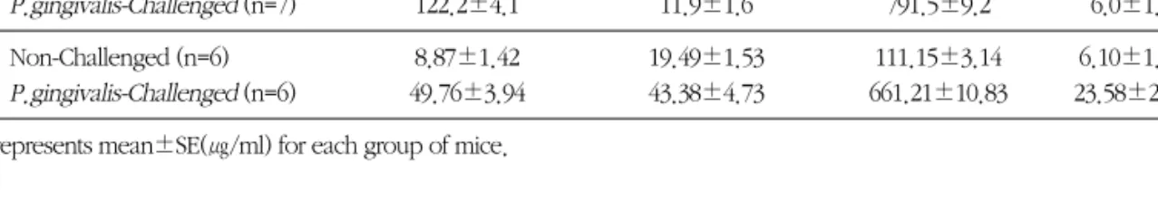
erides were increased in P. gingivalis-challenged animals compared to the non-challenged animals. In low- fat diet group, P. g challenge resulted in signifi- cant increase of cholesterol level and triglyceride both(P< 0.05, non-paired ttest). Blood HDL level and HDL/total cholesterol ratio were significantly Table 2. Effect of chronic infection with a disseminating strain of Pg (A7436) on chamber fluid levels of inflam-
matory mediators in ApoE+/- mice maintained on a normal (low fat) and high fat diet
IL-1β IL-6 PGE2 TNF-α
LFD Non-Challenged (n=6) 1.3±0.6 0 309.9±10.0 0
P.gingivalis-Challenged (n=7) 122.2±4.1 11.9±1.6 791.5±9.2 6.0±1.1
HFD Non-Challenged (n=6) 8.87±1.42 19.49±1.53 111.15±3.14 6.10±1.29
P.gingivalis-Challenged (n=6) 49.76±3.94 43.38±4.73 661.21±10.83 23.58±2.18 Values represents mean±SE(㎍/ml) for each group of mice.
Table 3. Effects of P. gingivalis infection on lipid profiles in mice
cholesterol(mg/dl) triglycerides(mg/dl) HDL(mg/dl) HDL/cholesterol
LFD Non-Challenged (n=6) 50.88±0.68 55.91±0.61 23.82±1.11 48.0±1.60
P.gingivalis-Challenged (n=7) 60.55±0.34 92.48±2.26 24.65±1.27 40.39±1.67
HFD Non-Challenged (n=6) 223.42±2.01 38.46±1.19 14.11±0.82 5.31±0.55
P.gingivalis-Challenged (n=6) 278.48±2.64 38.81±1.09 15.66±0.90 7.55±0.69 Values represents mean±SE(mg/dl) for each group of mice.
Figure 2. Effects of chronic P. gingivalis (A7436) infection on serum lipid levels in ApoE+/- mice maintained on a regular (low-fat) diet or high- fat diet. (* means the differences between 2 groups at P value< 0.05).
higher in low- fat diet group compared to the high- fat group, and showed no signigicant difference between challenged and non-challenged groups.
4. Aorta Atheroma Score
The aorta atheroma formation was characterized as follows: non-challenged high-fat diet group(64775±89㎛2)> challenged, high-fat diet
group (48104±35㎛2) >>>challenged, low-fat diet group >nonchallenged, low-fat diet group. Overall, lesion size was much greater for the APOE+/- high- fat group than the APOE+/- low-fat group. The mean total area and extent of atherosclerotic lesions were greater in P. gingivalis- challenged animals, as com- pared to non-challenged animals (162 ± 8 vs 89 ± 8㎛2), respectively in low- fat diet.
In ApoE+/-mice maintained on a high fat diet, a Figure 3. Effect of chronic infection with a disseminating strain of Pg (A7436) on atheroma lesion scores in ApoE+/- mice maintained on a high fat(A) and low fat normal diet(B).Plotted values represent calcu- lated means, and error bars represent SEM, for each group of mice. Number of animals per group were: 8 challenged and 6 unchallenged high fat diet; 7 challenged and 6 unchallenged low fat diet.
Comparisons between challenged and unchallenged mice were not statistically significant (P = 0.6 for high fat diet and 0.3 for low fat diet groups (t test).
A. ApoE+/-mice maintained on a high fat diet.
B. ApoE+/-mice maintained on a normal (low fat) diet
chronic infection with the disseminating strain of Pg (A7436) did not increase the atheroma lesion size at any of the section levels examined (Figure 3A).
However, in ApoE+/-mice maintained on a normal diet, a chronic infection with the disseminating strain of Pg(A7436) induced an increase in the size of atheroma lesion, especially at the section levels closest to the heart (Figure 3B).
When the chamber fluid cytokine and blood lipid levels were plotted versus the aorta atheroma lesion, atheroma lesion size was correlated to serum cho- lesterol levels (R2=0.58, P=0.038), and chamber fluid levels of TNF-α(R2=0.38, P=0.199) and serum cho-
lesterol levels were correlated to chamber fluid lev- els of TNF-α(R2=0.60, P=0.029)(Figure 4.)
IV. DISCUSSION
Growing evidence form case-control and popula- tion-based studies supports an association between periodontal disease and atherosclerosis2-5. but does no define whether the relation is casual or other- wise. Recent evidence in the periodontal literature suggests that host inflammatory responses to bacte- ria, and not just the bacterial pathogens themselves, are responsible for the marked destruction found in Figure 4. Associations between atheroma lesion, serum cholesterol levels, and chamber TNFa levels in ApoE+/- mice maintained on a low fat diet, infected or not with the disseminating strain of Pg A7436. The chamber fluid cytokine and blood lipid levels were plotted versus the aorta atheroma lesion to examine the data for a correlation between these outcomes. Atheroma lesion size was sig- nificantly correlated to serum cholesterol levels (R2=0.58), and chamber fluid levels of TNF-α (R2=0.38) and serum cholesterol levels were correlated to chamber fluid levels of TNF-α(R2=0.60 ).
severe periodontal disease. That is, the metabolic machinery used to destroy the periodontal tissues and bone is derived mostly from the host.6 Vascular pathology caused by immunologic reaction to bacte- ria appears to be similar to that caused by atheroma formation. In addition to dietary and genetic factors, some authors suggest that bacterial infections of unknown origin may contribute to acceleration of cardiovascular disease13-15.
The present study was designed to test whether innoculation of an established periodontal pathogen into subcutaneous chamber imitating periodontal infection contributes to the development and pro- gression of atherosclerosis in a susceptible animal model. The ApoE+/-mice used in this study have on 1 functional ApoE gene and are more susceptible to atherosclerosis than are normal mice (C57BL/6)-10, unlike ApoE-/-mice, in which atherosclerosis spon- taneously develops. This decreased ability to pro- duce Apo-E protein negatively alters serum lipid profiles when the animals are subjected to a high-fat diet. By using these mice as model, these factors can be varied and outcomes assessed. We utilized surgical stainless steel wire coil implanted subcuta- neously8,9.Using this chamber model, it's known that this sequestered space permits a minimal infec- tious dose that is reduced by more than 100x over the non- chamber subcutaneous challenge model.
This line would manifest atherosclerotic lesions in a relatively short period of time, as early as two months using high-fat diets11, while C57BL/6 mice are predisposed to a much lesser degree to athero- sclerosis and require six months of high-fat feeding to show mild atheroma formation. Being challenged with noninvasive strain of Porphyromonas gingivalis (Pg HG405), the ApoE+/-mice produced more pro- nounced lesion and much higher PGE2in chamber fluid and compared to C57BL/6 mice regardless of diets. The chamber cytokine response to infection of
increased PGE2 secretion was severe in C57BL/6 mice on a high fat diet than mice on a low fat diet and similar in ApoE+/- mice between high and low fat diet. However, chronic exposure to noninvasive strain of P. gingivalis led to neither accelerated pro- gression of atheroma formation in the aorta nor increased chamber PGE2 production in susceptible mice. In this previous study, Pg challenge did not increase atheroma lesion in high-fat diet group, in contrast to increasing lesion in low-fat diet group. It was reported that lipoproteins bind and inactivate bacterial endotoxin and lipoprotein-bound endotox- in is less able to elicit TNF release by macrophages16-18. This protective effect of high serum lipoprotein in ApoE+/-mice maintained high fat-diet could explain why bacterial challenge didn't show any significant effect on the lesional progres- sion and PGE2 production.
In this study, interval of challenge was determined as 2~3 weeks, based on the Genco et al7,8 that viable isolates were recovered from the chamber fluid to 14 days in immunized Balb/c mice. We have con- firmed this by taking culture of the chamber fluid aliquot Until chambers sloughed off 7~10 day after Pg inoculation, chamber fluid aliquots (10㎕) result- ed in more than 100 colonies. This finding means presence of 50x100 viable cells in whole chamber fluid( total 500㎕ after being diluted in PBS) and compared to the original bacterial cells (109) inocu- lated, the recovery rate was above the level of 1/200000. A striking difference was seen in chamber slough between high-fat (52.5%) and low-fat diet groups (100 %) . This sloughing of 1st chambers occurred earlier in low-fat fed group than high fat group. This result may indicate that dietary factors modulate the inflammatory response (proinflamma- tory cytokine) to the Gram negative bacteria P. gin- givalis. Especially increased blood fat level could neutralize the endotoxicity. Interestingly, no differ-
ence was detected between the high-fat and low-fat diet groups for the APOE+/- strain. Chamber fluid levels of inflammatory mediators IL-1β and PGE2 were increased in P. gingivalis-challenged animals as compared to the non-challenged animals. On low- fat diet, the post-challenge levels of IL-1β and TNFαwere significantly higher in Pgchallenged ani- mals compared to non-challenged animals, while IL- 6 and PGE2 levels were not. On high fat diet, the post-challenge chamber levels of PGE2were signifi- cantly higher in Pgchallenged animals compared to non-challenged animals, while the other three cytokines had only the trend to increase in Pgchal- lenged animals.. IL-1β level in challenged animals was significantly higher in low fat group than in high fat group
Blood levels of both total cholesterol and triglyc- erides were increased in P. gingivalis-challenged animals. In low- fat diet group, Pg challenge result- ed in significant increase of cholesterol level. Blood HDL level and HDL/total cholesterol ratio were sig- nificantly higher in low-fat diet group compared to the high-fat group, and showed no significant differ- ence between challenged and non-challenged groups. This result implicate that gram(-) bacterial infection could affect the lipid metabolism and result in hyperlipidemia depending on the fat contents in diet. These results provide evidence that this process could be mediated by cytokine production under the influence of bacterial infection and result in increase in the level of neutral fat and contribute to the atheroma formation. In this process, intraoral periodontal infection, as one very common entity of chronic localized infection, could be the etiologic or contributing factor. Therefore, further researches are need to elucidate the mechanism and the predispos- ing factors.
The aorta atheroma formation was different according to the diet fed and Pg challenge, as fol-
lows: non-challenged high-fat diet group> chal- lenged, high-fat diet group >>>challenged, low-fat diet group >nonchallenged, low-fat diet group.
Overall, lesion size was much greater in high-fat group than low-fat group. The lesion was confined to aorta area adjacent to heart in low-fat diet group whereas it was located extensively in high-fat diet group. In low-fat diet, the mean total area and extent of atherosclerotic lesions were increased in Pg challenged animals, compared to non-chal- lenged animals. These data suggest that P. gingivalis infectious challenge enhances atheroma lesion for- mation ion low-fat diet group, associated with an increase in local inflammatory mediator levels.
However, the protective effect of high serum lipoprotein in ApoE+/-mice maintained high fat-diet could explain why bacterial challenge didn't show any significant effect on the Il-1β and TNF-α level and lesional progression.
The atheromatous lesion in low-fat diet group was so small even though the bacteria was chal- lenged. ApoE+/- mice which were frequently chal- lenged with disseminating Pg strain for long periods (longer than 3 months) would be expected to devel- op atherosclerotic plaques earlier and to a greater degree than non-challenged mice. Li et al19investi- gated the effect of repeated systemic inoculations with Pg, a putative periodontal pathogen, on the progression of atherosclerosis in ApoE+/- mice. In their experiment, 10-week-old, male ApoE+/- mice fed either a high-fat diet or regular chow were inoc- ulated intravenously with live Pg (107 CFU) or vehi- cle once per week for 10, 14, or 24 consecutive weeks(relative long period). Atherosclerotic lesions of the proximal aortas and aortic trees were more advanced in Pg-challenged animals than in vehicle control animals and occurred earlier (at 14 weeks) when no lesions were apparent in control animals.
At 24 weeks after inoculation, proximal aortic lesion
size quantified by histomorphometry was 9-fold greater in chow-fed mice inoculated with Pg than in noninoculated mice and was 2-fold greater in Pg- inoculated versus noninoculated high-fat diet fed mice; all atherosclerotic lesions were macrophage- rich. Pg ribosomal DNA was found in the aortas, liv- ers, and hearts 24 weeks after inoculation. These results provide evidence that long-term systemic challenge with Porphyromonas gingivalis, an oral pathogen, can accelerate atherogenic plaque pro- gression. Li et al's study did not prime the mice and used less viable bacteria cells.
As for the relation between bacterial infection and atherosclerosis, other lines of evidence exist that implicate non-dental local infections as risk factors for heart disease20-22. Controlled clinical studies have suggested that "milder bacterial infections" like res- piratory and dental infections are risk factors for CHD. Valtonen's review of the literature on this sub- ject concludes that a growing amount of evidence is present to link infection and atherosclerosis.15 Chronic Chlamydia pneumonia infections have been well known to be associated with coronary heart disease26, 21. Certainly, more severe infections are seen to be major risk factors for acute myocar- diac infarction and stroke. In the more acute infec- tions, thrombosis formation may be mostly related to hypercoagulable states. Lipid and prostaglandin metabolisms are significantly altered by acute infec- tions15.
Multiple metabolic effects can be seen in response to LPS or infection. Triglyceride clearance may be reduced in response to LPS challenge due to reduc- tions in lipoprotein lipase activity. Increases in VLDL levels may be seen due to increased de novo syn- thesis of fatty acids or re-esterification of fatty acids23,
24.Bacterial challenge induces the production of sev- eral inflammatory mediators known to change the metabolism of fatty acids. In the rat model, TNF is
thought to mediate lipogenesis through stimulation of interleukin-6 production which stimulates hepatic lipogenesis by increasing hepatic citrate concentra- tions25. Thus, inflammatory mediators may have a role in the induction of hyperlipidemic states, espe- cially in individuals which overproduce these cytokines. It appears that inflammation can lead to hyperlipidemic states, which may then increase inflammatory responses, presenting a possible vicious cycle26.
In conclusion, the disseminating strain of Pg A7436 appears to be atherogenic, although mild, in ApoE+/- mice maintained on low fat diet.
Chronic and repetitive infection with the dissemi- nating strain of PgA7436 triggers a local inflammato- ry response that is associated with an elevation of serum cholesterol levels and with the magnitude of the atheroma score.
V. CONCLUSION
In this particular mouse model of the effect of dis- seminating strain of PgA7436 in ApoE+/- mice main- tained on low fat diet Pginfection leads to increased local inflammatory mediator levels, some of which were significantly associated with enhanced athero- ma formation.
VI. REFERENCES
1. WHO, World health statistics annual electronic file 2001(July)
2. Mattila KJ, Valtonen VV, Nieminen M, Fluttunen JK. Dental infection and the risk of new coro- nary events: prospective study of patients with documented coronary artery disease. Clinical Infectious Diseases 1995; 20:588-92.
3. Mattila KJ, Valle MS, Nieminen MS, Valtonen VV, Hietaniemi KL. Dental infections and coro-
nary atherosclerosis. Atherosclerosis 1993;
103:205-l1.
4. DeStefano F, Anda RF, Kahn HS, Wilhamson DF, Russell CM. Dental disease and risk of coro- nary heart disease and mortality [see comments).
BMJ 993; 306:688-91.
5. Beck JD, Garcia RI, Heiss G, Vokonas PS, Offenbacher S. Periodontal disease and cardio- vascular disease. J Periodontol 1996 ;67:1123-37 6. Offenbacher S, Collins JG, Yalda B, Haraden G.
Role of prostaglandins in high risk periodontitis patients. In: Genco RJ, Hamada S, Lehner T, Mergenhagen S. eds. Molecular Pathogenesis of Periodontal Disease. Washington DC:ASM Press;
1994:203-214
7. Collins JG, Windley HW III, Arnold RR, Offenbacher S. Effects of Porphyromonas gingi- valis infection on inflammatory mediator response and pregnancy outcome in hamsters.
Inf Imm 1994; 62:4356-61.
8. Genco CA, Cutler CW, Kapczynski D, Maloney K, Arnold RR. A novel mouse model to study the virulence of and host response to Porphyromonas gingivalis. Inf Imm 1991,59:1255-63
9. Genco CA, Kapczynski D, Cutler CW, Arko RJ, Arnold RR. Influence of immunization of Porphyromonas gingivalis colonization and inva- sion in the mouse chamber model. Inf Imm 1992, 60:1447-54
10. Zhang SH, Reddick RL, Burkey B, Maeda Nl.
Diet induced atherosclerosis in mice heterozy- gous and homozygous for apolipoprotein E gene disruption. J Clin Invest. 1994:94:937-945 11. Gibbs P, Offenbacher S, Arnold R, Maeda N .P.
gingivalis Infection in Murine Atherosclerosis Model Infectious challenge and atherosclerosis.
Thesis of UNC, 1998
12. Paigen B, Morrow A, Holmes PA, Mitchell D,
Williams RA. Quantitative assessment of athero- sclerotic lesions in mice. Atherosclerosis 1987;68:231-40
13. Heinle H, KIng D, Betz E. Metabolism of fibro- muscular and atheromatous plaques in an experimental model: causal mechanisms for the development of intimal necrosis. [Review].
Current Topics in Pathology 1993; 87:193-221.
14. Lopes-Virella MF, Virella G. Immunological and microbiological factors in the pathogenesis of atherosclerosis. Clinical Immunology &
Immunopathology 1985; 37:377-86.
15. Valtonen VV. Infection as a risk factor for infarc- tion and atherosclerosis. [Review]. Annals of Medicine 1991; 23:539-43.
16. Read et al. The protective effect of serum lipoprotein against bacterial LPS, Eur Heart J 1993;14(supp K):125-9
17. Harris HW, Grunfeld C, Feingold KR et al.
Chylomicrons alter the fate of endotoxin, decreasing TNF release and preventing death. J Clin Inves. 1993;91:1028-34
18. Flegel WA, Wolpl A, Mannel DN, Northoff H.
Inhibition of endotoxin-induced activation og human monocytes by human lipoproteins. Inf Imm, 1989;57:2237-45)
19. Li L, Messas E, Batista EL Jr, Levine RA., Amar S.
Porphyromonas gingivalis Infection Accelerates the Progression of Atherosclerosis in a Heterozygous Apolipoprotein E deficient Murine Model Circulation. 2002;105:861-867
20. Saikku P, Leinonen M, Tenkanen L, et al.
Chronic Chlamydia pneumoniae infection as a risk factor for coronary heart disease in the Helsinki Heart Study [see comments]. Annals of Internal Medicine 1992; 116:273-8.
21. Thorn DH, Grayston JT, Siscovick DS, Wang SP, Weiss NS, Daling JR. Association of prior infec- tion with Chlarnydia pneumoniae and angio-
graphically demonstrated coronary artery dis- ease. JAMA 1992; 268:68-72.
22. Moazed TC, Kuo C-c,Grayston JT, Campbell LA.
Murine models of Chlamydia pneumoniae infec- tion and atherosclerosis. J Inf Dis 1997;175:883- 90
23. Adi S, Pollock AS, Shigenaga JK, Moser AH, Feingold KR, Grunfeld C. Role for monokines in the metabolic effects of endotoxin. Interferon- gamma restores responsiveness of C3HIHeJ mice in vivo. Journal of Clinical Investigation 1992; 89:1603-9.
24. Lopes-Virella MF. Interactions between bacterial lipopolysaccharides and serum lipoproteins and their possible role in coronary heart disease. Eur Heart J 1993;14(Supp K): 118-24
25. Grunfeld C, Adi S, Soued M, Moser A, Fiers W, Feingold KR. Search for mediators of the lipogenic effects of tumor necrosis factor: poten- tial role for interleukin- 6. Cancer Research 1990;50:4233-8.
26. Bates EJ, Ferrante A, Harvey DP, Poulos A.
Polyunsaturated fatty acids increase neutrophil adherence and integrin receptor expression.
Journal of Leukocyte Biology 1993; 53:420-6.