대한소화기학회지 2006;47:1-4 □IMAGE OF THE MONTH □
증례: 34세 남자 환자가 혈변을 주소로 내원하였다. 환자 는 내원 6일 전 혈변이 처음 발생하여 개인병원에서 위내시 경검사를 받았으며, 검사 결과 이상소견이 없어 대장내시경 검사를 할 예정이었으나 내원 당일 2차례 혈변이 더 발생하 여 본원 응급실을 경유하여 입원하였다. 응급실 내원 시 활 력징후는 정상이었고, 신체검사에서 결막이 창백해 보이는 것 이외에 이상소견은 없었다. 말초혈액검사에서 백혈구 5,100/mm3, 혈색소 7.1 g/dL, 혈소판 269,000/mm3로 빈혈이 있었으며 생화학검사, 흉부방사선검사, 단순복부촬영은 정 상이었다. 내원 2일째 시행한 대장내시경검사에서 다량의 선혈과 혈변이 있었으나 출혈병소로 의심할 만한 병변은 관 찰되지 않았다. 내원 3일째 복부 CT 검사 결과 원위부 회장 부위에서 조영제가 관강에 흘러나온 것이 관찰되어 활동성 출혈이 있음을 추정할 수 있었으나(Fig. 1) 종괴나 소장벽이 두꺼워져 있는 등의 이상소견은 관찰되지 않았다. 그러나 CT 검사 당일 오후 9시경 갑자기 쇼크가 발생하여 응급으 로 혈관조영술을 하였으며, 역시 원위부 회장에서 활동성 출혈이 있음을 확인한 후 영양혈관에 코일을 이용하여 색전 치료를 하였다(Fig. 2).
내원 4일째 시술 후 출혈 여부를 확인하기 위하여 시행한 RBC 스캔검사 결과 24시간 영상에서 회장대장 연결부위에 방사능이 관찰되었으며 추가로 얻은 26시간 영상에서 상행 결장으로 방사능이 이동하는 양상을 나타냈다(Fig. 3). 이에 소장병변을 진단하기 위해 시행한 소장조영술 검사에서 이
상소견이 발견되지 않았으나 이중풍선 소장내시경검사 결 과 회맹판에서 60-70 cm 떨어진 부위에 게실이 있었고 게실 안쪽에서 혈전이 흘러나오는 것을 관찰할 수 있었다(Fig. 4).
Meckel 게실 의심하에 개복수술하여 게실을 제거한 후(Fig.
5) 1주일 뒤 퇴원하였다.
진단: Meckel 게실에서 발생한 위장관 출혈
위장관 출혈 원인 중 90% 이상은 기존의 상부, 하부 위 장관 검사법으로 진단이 가능하나 2-10%는 원인을 알 수 없으며, 이러한 경우의 대부분은 소장이 그 출혈부위로 추 정된다. 소장 출혈의 원인은 혈관이형성증, 소장종양이 가 장 흔하며, 그 다음으로 Meckel 게실, 크론병, 비스테로이 드 소염제로 인한 궤양이다. 그러나 소장출혈이 의심될 경 우 기존의 진단법은 낮은 진단율로 한계를 갖고 있다. 최 근 캡슐내시경의 개발로 전체 소장을 육안으로 관찰할 수 있게 되었으며, 실제 여러 연구 결과 소장에서 출혈이 발 생하였을 때 기존의 방사선 검사, 즉 소장조영술과 핵의학 검사의 진단율은 약 15%, push형 소장내시경의 진단율은 21-38%인 데 비하여 캡슐내시경 진단율은 48-76%로 상당 히 우수한 검사임이 입증되었다.1,2 그러나 캡슐내시경검사 역시 몇 가지 단점이 있는데 검사 중 통기(air insufflation) 가 안 되어 점막을 자세히 관찰하기 어렵고, 캡슐 전면에 이물질이 붙어도 제거할 방법이 없어 제한된 화면을 얻는 경우가 많으며 마지막으로 진단에 중요한 조직검사나 치
이중풍선 소장내시경으로 진단한 Meckel 게실
성균관대학교 의과대학 내과학교실
박정호․손정일
A Case of Meckel's Diverticulum Diagnosed by Double Balloon Enteroscopy
Jung Ho Park, M.D., and Chong Il Sohn, M.D.
Department of Internal Medicine, Kangbuk Samsung Hospital, Sungkyunkwan University College of Medicine, Seoul, Korea
ꠏꠏꠏꠏꠏꠏꠏꠏꠏꠏꠏꠏꠏꠏꠏꠏꠏꠏꠏꠏꠏꠏꠏꠏꠏꠏꠏꠏꠏꠏꠏꠏꠏꠏ
연락처: 박정호, 110-746, 서울시 종로구 평동 108번지 강북삼성병원 소화기내과
Tel: (02) 2001-2494, Fax: (02) 2001-2049 E-mail: [email protected]
ꠏꠏꠏꠏꠏꠏꠏꠏꠏꠏꠏꠏꠏꠏꠏꠏꠏꠏꠏꠏꠏꠏꠏꠏꠏꠏꠏꠏꠏꠏꠏꠏꠏꠏ Correspondence to: Jung Ho Park, M.D.
Department of Internal Medicine, Kangbuk Samsung Hospital Sungkyunkwan University College of Medicine, 108 Pyeong-dong Jongno-gu, Seoul 110-746, Korea
Tel: +82-2-2001-2494, Fax: +82-2-2001-2049 E-mail: [email protected]
2 대한소화기학회지: 제47권 제1호, 2006
료 등의 시술을 할 수 없다는 점이다.3,4 이에 반해 이중풍 선 소장내시경은 전체 소장을 자유롭게 육안으로 관찰할 수 있고 조작하기 쉬우며, 또한 병변이 발견되면 조직검사 와 치료 시술을 할 수 있다는 장점이 있다.5
Meckel 게실은 위장관에 발생하는 가장 흔한 선천 기형 이며, 대부분 증상 없이 지내나 5% 미만에서 출혈, 장폐 쇄, 염증, 천공 등의 합병증을 동반한다. 이 중 출혈은 대 부분 게실 내 이소 점막인 위점막에서 분비한 위산에 의 해 인접한 회장점막의 궤양에서 발생한다.6 진단법으로
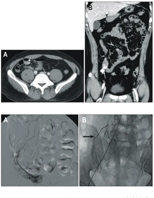
99mTc pertechnetate를 이용한 스캔은 소아에서는 비교적 민 감도와 특이도가 높지만 성인에서 발생한 Meckel 게실에 는 기여도가 적고,7 전산화단층촬영도 진단할 수 있다고 하나 개복술 후 진단이 가능한 경우가 대부분이다. 최근 Meckel 게실을 새로운 검사법으로 진단하려는 시도가 계 속되고 있는데, 캡술내시경으로 진단한 보고8,9가 2예, 이중풍 Fig. 1. The abdominal computed tomogram shows focal spillage of contrast material in the lumen of distal ileum. This finding suggested the possibility of active bleeding in the small intestine. (A) Transverse view and (B) sagittal view.
A
B
Fig. 2. (A) Emergency angio- graphy does not demonstrate active bleeding focus. (B) However, a feeding vessel in the isolated bowel loop from a branch of SMA is detected and it is treated by embo- lization with coil.
A B
Fig. 3. Tc-99m labeled RBC bleeding scan showed active bleeding on ileocecal area.
24 h delay 24 h delay
26 h 26 h
박정호 외 1인. 이중풍선 소장내시경으로 진단한 Meckel 게실 3
선 소장내시경으로 진단한 경우가 1예10 보고되었다. Meckel 게실은 대부분 회장 원위부에 발생하므로 이중풍선 소장 내시경을 이용하여 진단하는 데 무리가 없으며, 앞으로 Meckel 게실 진단에 중요한 역할을 할 것으로 기대한다.
참고문헌
1. Ell C, Remke S, May A, Helou L, Henrich R, Mayer G. The first prospective controlled trial comparing wireless capsule endoscopy with push enteroscopy in chronic gastrointestinal bleeding. Endoscopy 2002;34:685-689.
2. Hartmann D, Schilling D, Bolz G, et al. Capsule endoscopy versus push enteroscopy in patients with occult gastroin- testinal bleeding. Z Gastroenterol 2003;41:377-382.
3. Costamagna G, Shah SK, Riccioni ME, et al. A prospective trial comparing small bowel radiographs and video capsule
endoscopy for suspected small bowel disease. Gastroentero- logy 2002;123:999-1005.
4. Lewis BS, Swain P. Capsule endoscopy in the evaluation of patients with suspected small intestinal bleeding: results of a pilot study. Gastrointest Endosc 2002;56:349-353.
5. May A, Nachbar L, Wardak A, Yamamoto H, Ell C. Double- balloon enteroscopy: preliminary experience in patients with obscure gastrointestinal bleeding or chronic abdominal pain.
Endoscopy 2003;35:985-991.
6. Bemelman WA, Hugenholtz E, Heij HA, Wiersma PH, Obertop H. Meckel's diverticulum in Amsterdam: experience in 136 patients. World J Surg 1995;19:734-736.
7. Schwartz MJ, Lewis JH. Meckel's diverticulum: pitfalls in scintigraphic detection in the adult. Am J Gastroenterol 1984;
79:611-618.
8. Golder S, Schmidt J, Kolmsee P, et al. Identification of a Fig. 4. (A) Double-balloon enteros- cope shows Meckel’s diverti- culum at 80 cm proximal to the ileocecal valve (above). (B) En- teroscopic view when the scope is further advanced into the bowel.
A B
Fig. 5. (A) Meckel’s diverticulum in 10 cm size is attached to the distal ileum. (B) Histological examination shows ectopic gastric mucosa inside the diverticulum (hematoxylin and eosin, 1:250).
A B
4 The Korean Journal of Gastroenterology: Vol. 47, No. 1, 2006
Meckel's diverticulum by wireless capsule endoscopy. Endo- scopy 2005;37:608.
9. Park SM, Chun HJ, Jeen YT, Yoon I. A case of chronic gas- trointestinal bleeding from a Meckel's diverticulum detected by wireless capsule endoscopy. Korean J Gastroenterol 2004;
43:125-128.
10. Gasbarrini A, Di Caro S, Mutignani M, et al. Double-balloon enteroscopy for diagnosis of a Meckel's diverticulum in a patient with GI bleeding of obscure origin. Gastrointest Endosc 2005;61:779-781.