Comparison of Cage and Allograft Mixed Bone
Marrow for Monosegmental Instrumented Posterior Lumbar Interbody Fusion
Chae Hyun Lim, M.D., Dae Hee Kim, M.D., Sang Ho Ahn, M.D. Yong Soo Choi, M.D.
Department of Orthopedic Surgery, Kwangju Christian Hospital, Gwangju, Korea
Study Design: A retrospective study.
Objectives: To compare the radiological and clinical results between cage and cancellous allograft mixed with bone marrow for monosegmental instrumented posterior lumbar interbody fusion (PLIF).
Summary of the Literature Review: Allograft has potential problems, such as delayed union. Autologous bone marrow provides for improving the capability of bone induction with allograft. There are rare reports on PLIF using allograft mixed with autologous bone marrow.
Materials and Methods: Monosegmental instrumented PLIF was performed on 51 patients who had lumbar degenerative disease, cage for 28 patients (cage group) and allograft mixed with bone marrow for 23 patients (allograft group). The clinical and radiological results in each group were compared.
Results: The mean follow-up was 45 (30 - 111) months. At the final follow up, there was no significant difference between the cage group and the allograft group in the Korean Version Oswestry Disability Index (p=0.72) and Visual Analogue Score for back pain (p=0.54) and radiating pain to the leg (p=0.26). The radiological fusion rate was 92.8% in the cage group, and 82.6% in the allograft group (p=0.02).
At the last follow up, disc height was decreased to 1.5±0.8 mm of the cage group, and 3.0±1.5 mm of the allograft group (p=0.0001).
Conclusions: PLIF using cancellous allograft mixed bone marrow has low fusion rate contrast to good clinical results. It is necessary to take a careful selection of the allograft mixed bone marrow for PLIF.
Key Words: Lumbar spine, Posterior lumbar interbody fusion, Cage, Cancellous allograft
Received: March 13, 2012 Revised: August 16, 2012 Accepted: November 15, 2012 Published Online: December 31, 2012 Corresponding author: Yong Soo Choi, M.D.
Department of Orthopedic Surgery, 264 Yangrim-dong Nam-gu Gwangju, Kwangju Christian Hospital, Kwangju, Korea
TEL: 82-62-650-5064, FAX: 82-62-650-5066 E-mail: [email protected]
“This is an Open Access article distributed under the terms of the Creative Commons Attribution Non-Commercial License (http://
creativecommons.org/licenses/by-nc/3.0/) which permits unrestricted non-commercial use, distribution, and reproduction in any medium, provided the original work is properly cited.”
서 론
퇴행성 요추 질환의 치료 방법으로 후방 추체간 유합술에 사 용되는 추체간 이식물의 종류는 다양하며 자가 장골, 국소 자가 골, 금속이나 비금속 cage와 같은 인공 구조물, 동종골 등이 있 다. 그중에서 자가 장골을 이용하는 방법이 골 유합 등에서 우수 한 결과를 보이나 이식 공여부 동통과 출혈 등 단점이 있어,1-3) 자가골의 대체물로 동종골, 인공 삽입물에 이식골을 채워 넣는 방법 등 많은 방법들이 여러 저자들에 의해 소개되었다.4) 공여 부 문제를 해결하기 위해 후방 감압시 획득한 국소 조각골을 추 체간과 cage 내부에 충전하여 이식하는 방법은 성공적인 결과를 보여주고 있으며5,6) cage를 이용한 후방 추체간 유합술과 척추경 나사못 고정술은 퇴행성 요추 질환 치료에 널리 사용되고 있다.
동종골 효과에 대해서는 많은 저자들이 다양한 견해를 보고하 고 있다. 동종골의 부정적인 결과 보고,7-9) 와 긍정적인 결과들이
있다.10-13) Malloy와 Hilibrand14)은 동종골이 유합율은 낮으나 공
여부 장애가 적기 때문에 이식골의 선택에 있어서 이런 문제를 함께 고려해야 한다고 하였다. 종합해 보면 동종골의 근본적인 문제는 유합 기간이 지연되고 역학적인 안정성이 약화 될 수 있
다는 것이다.
자가 골수 이식은 임상적으로 신생골 형성에 우수한 것으로 알려져 있고15-17) 채취로 인한 합병증이 없으나 골수 그 자체로 는 골 전도 능력이 제한되고, 골수 내의 골형성 세포의 수 또한 제한된 양만 존재하여 단독으로 적용하기에는 한계가 있다.18,19)
저자들은 후방요추체간 유합술을 위한 방법 중 해면 동종골의 단점을 보완하기 위해 상용화한 해면 동종골에 자가골수를 혼합 하였고, 해면 동종골의 역학적 강화를 위해 수술 수기상 고관절 인공관절의 골 결손부를 재건할 때 적용하는 압박 골이식 수기 를 적용하여 후방 요추체간 유합술을 시행하였다.
본 연구의 목적은 퇴행성 요추 질환에서 단분절 추체간 유합 술을 위해 자가 골수 혼합 해면 동종골 삽입술의 방사선학적 및 임상 결과를 cage 내고정술과 비교 분석하여 해면 동종골의 유 용성을 알아보고자 하였다.
대상 및 방법
1. 연구 대상
2002년 9월부터 2008년 6월까지 본원에서 퇴행성 요추 질환 으로 단분절 추체간 유합술을 시행하였던 51예를 대상으로 하 였다. Cage를 이용하여 후방 추체간 유합술 및 감압술, 척추경 나사못 고정술을 시행한 환자 중 2년 이상 추시 가능한 28명을 cage 군으로 분류하였고, cage 없이 파쇄 해면 동종골과 자가 골 수를 혼합하여 후방 요추체간 유합술을 시행한 23명을 동종골 군으로 분류하였다.
Cage 군의 평균 연령은 59.5세 였으며, 남자 7명, 여자 21명 이 었다. 척추관 협착증이 11명, 척추 전방 전위증이 17명 이었다.
L3-4가 2명, L4-5가 23명, L5-S1이 3명 이었다. 동종골 군의 평균 연령은 61.3세 였으며, 남자 7명, 여자 16명 이었다. 척추 관 협착증이 19명, 척추 전방 전위증이 4명 이었다. L3-4가 3명, L4-5가 20명 이었다. 추시 기간은 평균 45(30 ~ 111)개월이었 다.
2. 수술 방법
Cage 군에서는 후방 감압술 후 절제된 추궁판과 내측 후방 관절에서 획득한 국소골을 2~3mm로 조각내어 cage(Ogival interbody cage, Stryker)에 충진하고, 나머지 국소 조각골은 척추 체 사이에 충진한 후 cage를 삽입하고 척추경 나사못과 강봉을 사용하여 단단히 압박 고정을 하였다(Fig. 1).
동종골 군에서는 후방 추체간 유합술을 위해 추간판을 철저히 제거하고 종판을 노출 시킨 후 상용화된 해면 동종골을 자가 장 골 골수와 혼합하여 준비된 깔대기를 통해 압박 골 이식술을 양
측에서 시행하여 추간판 공간을 충분히 채운 후 내측 후관절 골 편을 케이지 모양으로 만들어 입구에 삽입하여 파쇄 해면 동종 골의 탈출을 막고 추체간 지지를 유지하고자 하였다(Fig. 2).
3. 연구 방법
임상적 결과는 한국어판 장애지수(Korean Version Oswestry Disability Index; KODI)20)와 요통에 대한 시각통증점수(Visual Analogue Score)를 이용하여 평가하였고, 측면 방사선 사진을 이용하여 유합율, 추체간 간격의 변화를 측정하여 각각 비교하 였다.
유합의 판정은 Brantigan & Steffee 분류법21)을 이용하여 A, B, C 단계를 불유합으로, D, E 단계를 유합으로 판정하였으며
Fig. 1. Case of cage group. A 63 year old female patient who had spon- dylolisthesis L4 on L5 was undergone posterior lumbar interbody fusion with cage and local bone.
Fig. 2. Case of allograft group. Photograph of the allograft and bone block (A), this bone block (arrow) that was harvested from facet joint could be provided not only mechanical stability of the disc space as a cage but also prevention from extrusion of mosellized graft. Postoperative lateral radiograph (B).
(Table 1), 특히 케이지군의 유합 판정은 전후면 방사선 사진에 서 케이지 내부 이식골과 추체간 그리고 케이지 주위 추체간 골 성 연결을 준용하여 적용하였다. 추체간 간격은 상위 척추체 종 판의 중심점에서 수직선을 그어 하위 척추체의 종판과 만나는 거리로 정하였다(Fig. 3).
한국어판 장애지수와 VAS를 이용한 요통과 하지방사통은 각 각 Fisher’s exact 검정으로 비교 분석하였고, 방사선학적 골 유합 은 X2 검정으로 분석하였으며, 추체간 높이의 변화는 T 검정을
이용하여 분석하였다. 자료의 통계학적인 분석을 위하여 SPSS 12.0 for Windows(SPSS, Chicago, IL, USA) 프로그램을 사용하 였고, 통계학적인 유의 수준은 0.05로 하였다.
결 과
1. 임상적 결과
최종 추시상 한국어판 장애지수는 Cage 군에서 수술 전 33.3
±6.7에서 수술 후 최종 추시상 16.1±4.3으로 호전되었고, 동종 골 군에서 수술전 32.2±8.5에서 수술 후 최종 추시상 15.7±5.8 로 호전되었다. 요통의 시각통증 점수는 Cage 군에서 수술 전 6.6±2.7에서 수술 후 최종 추시상 2.7±1.9로 호전 되었고, 동종 골 군에서 수술 전 4.9±2.5에서 수술 후 최종 추시상 2.4±1.6으 로 호전되었다. 하지 방사통의 시각통증 점수는 Cage 군에서 수 술 전 5.0±3.3에서 수술 후 1.8±1.8로 호전되었고 동종골 군에 서 수술 전 7.0±1.9에서 수술 후 최종 추시상 2.4±2.2로 호전되 었다. 한국어판 장애지수(p=0.72), 요통(p=0.54)과 하지방사통 (p=0.26)의 시각통증 점수에서 각각 두 군 간에 유의한 차이는 없었다(Table 2).
Fig. 3. The disc height was measured at the preoperative, postoperative and follow-up lateral radiograph.
Table 1. Brantigan and Steffee classification for radiologic union
Classification Description
A: obvious Pseudoarthrosis, collapse of construct, loss of disc height, vertebral slip, broken screw, displacement of the carbon cage, resorption of bone graft
B: probable pseudoarthrosis Significant resorption of the bone graft, major lucency or gap visible in fusion area(2>mm around the entire periphery of graft)
C: uncertain(here nonunion) Bone graft visible in the fusion area at approximately the density originally achieved at surgery; a small lucency or gap may be visible involving just a portion of the fusion area at least half of graft area showing no lucency between graft bone & vertebral bone
D: probable fusion Bone bridge entire fusion area at surgery; there should be no lucency beween the donor bone & vertebral bone E: fusion Bone in the fusion area is radiographically adhesive at surgery; optimally, there is no interface between the donor bone
& the vertebral bone, although a sclerotic line between graft & vertebral bone indicates fusion; other sign of solid fu- sion include the fusion area, resorption of anterior traction spur, anterior progression of the graft within disc space, fusion of facet joint
Table 2. The Clinical Results and Radiological Fusion Rate between two Groups
KODI score VAS score
(back pain) VAS score
(radiating pain) Radiological fusion rate (B-S classification)
Cage group 16.1±4.3 2.7±1.9 1.8±1.8 92.8%
Allograft group 15.7±5.8 2.4±1.6 2.4±2.2 82.6%
P value 0.72 0.54 0.26 0.02
KODI, Korean Version Oswestry Disability Index, VAS, Visual Analogue Score, B-S classification, Brantigan and Steffee classification
Fig. 4. Revision for one case of allograft group. At 6-months follow-up, radiograph shows loss of disc height and absorption of the allograft (B). Histological finding reveals inflammation around dead allograft (C). After radical curettages, two cages filled
2. 방사선학적 결과
방사선학적으로 추체간 유합은 Cage 군 28 예 중에서 26예 (92.8%)에서 유합, 동종골 군에서 23 예 중에서 19 예 (82.6%) 의 유합을 얻었다. 동종골군의 유합률이 낮았다(p=0.02). 추간 판 간격의 변화는 Cage 군에서 평균 수술 전 8.6±2.4mm에서 수술 직후 13.3±1.3mm로 회복되어 최종 추시상 11.8±1.2mm 로 감소하였다. 동종골 군에서 평균 수술전 9.3±2.4mm에서 수 술 직후 14.2±2.4mm로 회복되어, 최종 추시상 11.2±2.1mm로 감소하였다. 수술 직후 추간판 높이의 회복정도는 Cage 군에서 평균 4.6±2.6mm, 동종골 군에서 5.1±2.4mm로 양군간 차이가 없었으나(p=0.47) 최종 추시 상 Cage 군 평균 1.5±0.8mm, 동 종골 군 평균 3.0±1.5mm 소실되어 Cage 군이 추체간 높이 유 지에 양호한 결과를 얻었다(p=0.0001)(Table 3). 방사선학적으 로 Cage 군 2 예, 동종골 군 4 예에서 불유합의 결과를 얻었으나 Cage 군 2 예와 동종골 군 3예는 임상 증상이 양호하여 추적 관 찰하였으며, 동종골 군 1예에서 이식골의 흡수 및 추체 간격 감 소와 요통으로 수술 후 6개월째 cage 고정 및 척추경 나사못의 재삽입술을 시행하였다(Fig. 4).
3. 합병증
수술의 합병증으로 감압술 중에 발생한 경막 손상이 cage 군 에서 2예 있었으며 신경근 증상의 합병증 없이 치료되었다.
고 찰
요추의 추체간 유합술을 위한 이식골로 자가 장골은 잘 알려 진 바와 같이 숙련된 수술자가 시행하더라도 이식 공여부 동통, 심부감염, 이식공여부의 골절 등의 단점이 있고,1-3) 다분절 유합 이나 불유합에 따른 재 수술시 제한된 양으로 인한 문제가 있어 현재 다양한 골이식 대체물에 대한 연구 및 개발이 진행되고 있 다.
Cage를 이용한 추체간 유합술과 후방 기기 고정술은 최근 가 장 널리 쓰이는 수술 방법으로 cage 내에 수술시 제거된 국소골 을 cage 내부에 삽입하여 사용함으로써 이식골의 역학적 기능을 cage가 수행하여 높은 안정성과 요추 전만도 회복에 유용한 술 식이다. Okuyama 등5)의 보고에 의하면 93.5% 높은 유합율과 비교적 높은 임상적 만족도를 보여주고 있다. 저자들의 cage 군 도 유합율 92.8%, 임상적 결과가 양호하여 유사한 결과를 얻었 다.
자가골의 대체물로 동종골은 자가골 사용의 단점인 채취에 따 른 문제점이 없고, 인공 삽입물과 달리 생물학적인 유합이 가능 하며 그리고 생역학적으로도 추체와 물성이 비슷한 장점이 있 다. 하지만 동종골 이식술은 몇가지 약점이 있다.
먼저 골유합율 면에서 An 등9)은 자가골과 동종골을 이용하여 척추의 후외측 유합율을 시행함에 있어서 자가골은 80%의 유 합율을 보이나 동종골은 45%의 유합율을 얻었다고 보고하였고, Brantigan 등21)은 동종골을 이용한 요추 추체간 유합술후 56%
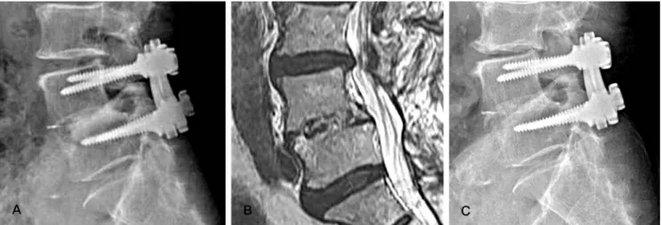
Fig. 5. At 36 months follow-up, radiograph (A) and MRI (B) didn’t achieved bony fusion of the allograft exactly. But at 54 months follow-up, radiograph revealed well bony fusion and maintenance of the disc height.
Table 3. The Changes of Disc Height on Lateral Radiograph
Preoperative Postoperative Last follow up
Cage group 8.6±2.4mm 13.3±1.3mm 11.8±1.2mm
Allograft group 9.3±2.4mm 14.2±2.4mm 11.2±2.1mm
P value 0.47 0.0001
골유합 성공율을 보고하여 동종골의 골유합율의 성적이 자가골 과 비교할 때 좋지 않았다. 최근 골유합을 향상시키기 위해 동종 골에 BMP를 혼합하여 우수한 유합 결과가 보고되고 있으나,22) Vaidya 등23)은 동종골의 골유도능을 위해 재조합 BMP를 혼합 하여 골유합율은 크게 향상 시켰으나 이식골의 침강이 50%이상 에서 발생하여 더 이상 혼합사용을 권하지 않는다 보고하였다.
자가 골수는 1986년 Connolly와 Shindell24)이 발표한 경골의 불 유합을 치료하기 위해 자가 골수의 경피적 주입으로 치료한 결 과를 보고한 이후 골 형성 능력이 있는 세포를 쉽게 얻을 수 있 는 방법으로 임상에서 사용되어지고 있다. 그러나 자가 골수는 그 자체로는 골 전도 능력이 제한되어 파쇄 동종골과 혼합 사용 한 경우 우수한 임상 결과의 보고가 점차 늘고 있다.17,25) Curylo 등26)은 후외방 유합술 토끼 모델에서 탈무기질 동종골과 자가 골수 혼합의 결과가 방사선학적 유합율 76.5%, 탈무기질 단독 사용의 52.3% 보다 높은 결과로 자가 골수의 척추 후외방 유합 술 모델에서 자가 골수의 역할을 보여주었다. 따라서 자가 골수 와 해면 동종골 혼합 이식은 이론적으로 동종골이 가지고 있는 골 전도성과 제한적이지만 자가 골수의 골 유도성과 골형성 세 포를 활용할 수 있는 좋은 방법으로 사료된다. 저자들의 결과에 서 82.6%의 방사선학적 골유합의 결과를 얻어 이전의 동종골을 이용한 유합술의 연구보다 유합율이 향상된 결과를 얻었으나 케 이지 고정보다 낮은 유합의 결과를 얻었다. 또한 저자들이 초기 불유합으로 판정한 해면 동종골 이식 환자의 36개월 추시 방사 선 및 자기공명검사에서 해면 동종골이 자가골로 완전 치환되지 않았으나 54개월 추적 방사선 사진에서 완전 골유합을 이루어 (Fig. 5), 해면 동종골의 추적 관찰에서 골유합을 얻을 가능성을 시사하였다.
또 하나의 약점으로 역학적으로 안정성이 뛰어난 특성을 가진 자가 피질골과 달리 해면 동종골은 안정성이 떨어져 동종골 이 식 후 이식골의 추체내로 침강 또는 붕괴가 일어날 수 있다는 것 이다.27) 해면 동종골의 압박 골이식 수기가 인공 고관절 치환술 의 골 결손을 재건이나 대퇴골두 무혈성 괴사에서 핵심감압술 후 압박골이식술의 임상적 적용의 양호한 결과가 보고되고 있 어,28) 해면 동종골의 역학적 취약점을 보완하는 방법으로 저자 들은 파쇄 해면 동종골의 압박 골이식술을 시행하였고 추가적으 로 감압으로 채취된 내측 후관절 골편을 케이지처럼 지지골로 사용하였다. 그러나 압박 골이식술과 내측 후관절 골편의 지지 대 사용에도 추체간 높이 유지에 한계가 있었다.
본 논문이 환자의 흡연이나 질환 등 특성 조사가 되지 않았고, 대상 환자 수가 적을 뿐 아니라 대상 환자의 적응증의 차이, 후 향적 조사의 제한점이 있으나, 요추 후방 추체간 유합술을 위한 자가 골수 혼합 해면 동종골의 압박 골이식 수기의 적용은 기존
에 보고된 동종골 이식술보다는 골 유합을 향상시킨 결과를 고 려할 때 자가골 이식의 대체방법의 하나로 선택될 수 있으리라 사료되며, 향후 많은 환자를 대상으로 불유합 위험군 연구 등 동 종골 이식의 금기증 연구가 필요하리라 사료된다.
결 론
요추 후방 추체간 유합술에서 자가골수 혼합 해면 동종골은 임상적으로 양호한 결과를 얻었으나 방사선학적으로 추체간 유 합에 불량한 결과로 자가 골수 혼합 해면 동종골 이식술의 요추 후방 추체간 유합술 적용에 신중한 선택이 필요하리라 사료된 다.
REFERENCES
1. Enker P, Steffee AD. Interbody fusion and instrumentation.
Clin Orthop Relat Res. 1994;300:90-101.
2. Lin PM. Posterior lumbar interbody fusion technique: com- plications and pitfalls. Clin Orthop Relat Res. 1985;193:90- 102.
3. Verlooy J, De Smedt K, Selosse P. Failure of a modified posterior lumbar interbody fusion technique to produce ad- equate pain relief in isthmic spondylolytic grade 1 spondy- lolisthesis patients: A prospective study of 20 patients. Spine (Phila Pa 1976). 1993;18:1491-5.
4. Brantigan JW, Steffe AD, Geiger JM. A carbon fiber implant to aid interbody lumbar fusion. Mechanical testing. Spine (Phila Pa 1976). 1991;16(6 Suppl):S277-82.
5. Okuyama K, Kido T, Unoki E, Chiba M. PLIF with a ti- tanium cage and excised facet joint bone for degenerative spondylolisthesis in augmentation with a pedicle screw. J Spinal Disord Tech. 2007;20:53-9.
6. Miura Y, Imagama S, Yoda M, Mitsuguchi H, Kachi H. Is local bone viable as a source of bone graft in posterior lum- bar interbody fusion? Spine (Phila Pa 1976). 2003;28:2386- 9.
7. Jorgenson SS, Lowe TG, France J, Sabin J. A prospective analysis of autograft versus allograft in posterolateral lumbar fusion in the same patients: A minimum of 1-year follow- up in 144patients. Spine (Phila Pa 1976). 1994;19:2048-53.
8. Aurori BF, Weierman RJ, Lowell HA, Nadell Cl, Parsons JR. Pseudoarthrosis after spinal fusion for scoliosis: A com-
parison of autogenous and allogenic bone grafts. Clin Or- thop Relat Res. 1985;199:153-8.
9. An HS, Lynch K, Toth J. Prospective Comparison of au- tograft vs. allograft for adult posterolateral lumbar spine fusion: differences among freeze-dried, frozen, and mixed grafts. J Spinal Disord. 1995;8:131-5.
10. Dodd CA, Fergusson CM, Freedman L, Houghton GR, Thomas D. Allograft versus autograft bone in scoliosis sur- gery. J Bone Joint Surg Br. 1988;70:431-4.
11. Bridwell KH, O’Brien MF, Lenke LG, Baldus C, Blanke K.
Posterior spinal fusion supplement with only allograft bone in paralytic scoliosis. Does it work? Spine (Phila Pa 1976).
1994;19:2658-66.
12. Fabry G. Allograft versus autograft bone in idiopathic sco- liosis surgery: a multivariate statistical analysis. J Pediatr Orthop. 1991;11:465-8.
13. Gibson S, McLeod I, Wardlaw D, Urbaniak S. Allograft versus autograft in instrumented posterolateral lumbar spinal fusion: a randomized control trial. Spine (Phila Pa 1976). 2002;27:1599-603.
14. Malloy KM, Hilibrand AS. Autograft versus allograft in degenerative cervical disease. Clin Orthop Relat Res.
2002;394:27-38.
15. Connolly JF, Guse R, Tiedeman J, Dehne R. Autologous marrow injection as a substitute for operative grafting of tibial nonunions. Clin Orthop Relat Res. 1991;266:259-70.
16. Connolly JF. Injectable bone marrow preparations to stim- ulate osteogenic repair. Clin Orthop Relat Res. 1995;313:8- 18.
17. Seitz WH Jr, Froimson AI, Leb RB. Autogenous bone mar- row and allograft replacement of bone defects in the hand and upper extremities. J Orthop Trauma. 1992;6:36-42.
18. Lane JM, Yasko AW, Tomin E, et al. Bone marrow and
recombinant human bone morphogenetic protein-2 in os- seous repair. Clin Orthop Relat Res. 1999;361:216-27.
19. Vaccaro AR, Chiba K, Heller JG, et al. Bone grafting alter- natives in spinal surgery. Spine J. 2002;2:206-15.
20. Jeon CH, Kim DJ, Lee HM, et al. Cross-cultural adaptation of the korean version of the oswewtry disability index(ODI).
J Korean Soc Spine Surg. 2005;12:146-52.
21. Brantigan JW. Pseudarthrosis rate after allograft posterior lumbar interbody fusion with pedicle screw and plate fixa- tion. Spine (Phila Pa 1976). 1994;19:1271-9.
22. Slosar PJ, Josey R, Reynolds J. Accelerating lumbar fusions by combining rhBMP-2 with allograft bone: a prospec- tive analysis of interbody fusion rates and clinical outcomes.
Spine (Phila Pa 1976). 2007;7:301-7.
23. Vaidya R, Weir R, Sethi A, Meisterling S, Hakeos W, Wybo CD. Interbody fusion with allograft and rhBMP-2 leads to consistent fusion but early subsidence. J Bone Joint Surg Br.
2007;89:342-5.
24. Connolly JF, Shindell R. Percutaneous marrow injection for an ununited tibia. Nebr Med J. 1986;71:105-7.
25. Scoff HD. Bone marrow/allograft component therapy: A clinical trial. Am J Orthop (Belle Mead NJ). 1995;24:40-7.
26. Curylo LJ, Johnstone B, Petersilge CA, Janicki JA, Yoo JU.
Augmentation of spinal arthrodesis with autologous bone marrow in a rabbit posterolateral spine fusion model. Spine (Phila Pa 1976). 1999;24:434-8.
27. Kumar A, Kozak JA, Doherty BJ, Dickson JH. Interspace distraction and graft subsidence after anterior lumbar fu- sion with femoral strut allograft. Spine (Phila Pa 1976).
1993;18:2393-400.
28. Jung KH, Jang JS, Choi YR. Core decompression and Im- paction bone graft in osteonecrosis of the femoral head. J Korean Hip Soc. 2005;17:223-9.
단분절 후방 요추체간 유합술에서 Cage 내고정군과 자가 골수 혼합 동종골군의 방사선학적 및 임상 결과 비교
임채현 • 김대희 • 안상호 • 최용수 광주기독병원 정형외과
연구계획: 후향적 연구
목적: 퇴행성 요추 질환에서 단분절 추체간 유합술을 위해 cage 내고정술과 자가 골수 혼합 해면 동종골 삽입술의 방사선학적 및 임상 결과를 비교 하 였다.
선행문헌의 요약: 동종골의 문제로 유합 기간 지연이 지적되고 있다. 자가 골수 이식은 신생골 형성에 우수한 것으로 알려져 있어 요추 질환에서 추체간 유합술을 위해 자가 골수 혼합 동종골 삽입술 결과 보고는 드물다.
대상 및 방법: 퇴행성 요추 질환으로 단분절 추체간 유합술을 시행하였던 51예를 대상으로 하였다. Cage를 이용한 28예를 Cage 군으로, cage없이 해 면 동종골과 자가 골수를 혼합하여 후방 요추체간 유합술을 시행 받은 23예를 동종골 군으로 분류하였다. 두 군간의 임상적 및 방사선학적 결과를 비교 분석하였다.
결과: 45(30-111)개월 추적 조사되었다. 최종 추시상 한국어판 장애지수 (p=0.72), 요통 (p=0.54)과 하지 방사통 (p=0.26)의 시각통증지수에서 Cage 군과 동종골 군간 차이를 보이지 않았다. 방사선학적 추체간 유합율은 Cage군 92.8%, 동종골군이 82.6%로 동종골군의 유합률이 낮았다(p=0.02).
최종 추시 상 추체간 높이가 Cage군 평균1.5±0.8mm, 동종골군 평균 3.0±1.5mm 소실되어 Cage군이 추체간 높이 유지에 양호한 결과를 얻었다 (p=0.0001).
결론: 요추 후방 추체간 유합술에서 자가골수 혼합 해면 동종골은 임상적으로 양호한 결과를 얻었으나 방사선학적으로 추체간 유합에 불량한 결과로 자가 골수 혼합 해면 동종골 이식술의 요추 후방 추체간 유합술 적용에 신중한 선택이 필요하리라 사료된다.
색인 단어: 요추, 후방 추체간 유합술, Cage, 해면 동종골 약칭 제목: 케이지 또는 해면 동종골을 이용한 요추 유합술