pISSN 1598-9992 eISSN 2233-6869
CASE REPORT
괴사성 췌장염의 합병증으로 발생한 췌장-직장루의 치료: 순차적 치료 전략의 유용성
임성균, 김성훈, 서승영, 양희찬, 이승옥
전북대학교병원 의생명연구원 소화기내과
Feasibility of Adopting the "Step-up Approach" in Managing Necrotizing Pancreatitis-induced Pancreatic-colonic Fistula
Sung Kyun Yim, Seong Hun Kim, Seung Young Seo, Hee Chan Yang and Seung Ok Lee
Division of Gastroenterology, Department of Internal Medicine, Biomedical Research Institute, Chonbuk National University Hospital, Jeonju, Korea
Managing acute pancreatitis is clinically challenging because of the generally poor patient condition, the variety of treatment options depending on the severity and complications, and the uncertainty of outcomes. Recently, the step-up approach, which involves less invasive initial treatment and more invasive subsequent treatment, where necessary, has been proposed as the mainstay of managing pancreatitis. This paper presents a case of a 57-year-old man with severe acute pancreatitis, who developed an unexpected fistula in the rectum, which was treated successfully using the step-up approach. In managing this case, the authors faced clinical challenges in determining the infection of necrotic tissue in the early phase of the disease, the optimal timing and method of drainage, and the fistula closure or repair technique. Successful management of this case using the step-up approach validated current recom- mendations and suggests that it is a reasonable treatment strategy for pancreatic-colonic fistulas. This case also highlights the im- portance of an awareness that pancreatitis-associated complications can develop in an unexpected manner. (Korean J Gastroenterol 2019;73:365-369)
Key Words: Pancreatitis, acute necrotizing; Colon; Rectum; Fistula
Received January 1, 2019. Revised February 18, 2019. Accepted March 10, 2019.
CC This is an open access article distributed under the terms of the Creative Commons Attribution Non-Commercial License (http://creativecommons.org/licenses/
by-nc/4.0) which permits unrestricted non-commercial use, distribution, and reproduction in any medium, provided the original work is properly cited.
Copyright © 2019. Korean Society of Gastroenterology.
교신저자: 이승옥, 54907, 전주시 덕진구 건지로 20, 전북대학교병원 소화기내과
Correspondence to: Seung Ok Lee, Division of Gastroenterology, Department of Internal Medicine, Chonbuk National University Hospital, 20 Geonji-ro, Deokjin-gu, Jeonju 54907, Korea. Tel: +82-63-250-1289, Fax: +82-63-254-1609, E-mail: [email protected], ORCID: https://orcid.org/0000-0003-0243-215X
Financial support: None. Conflict of interest: None.
INTRODUCTION
Severe acute pancreatitis is a critical condition caused by the development of pancreatic and peri-pancreatic necrosis, organ failure and the possibility of subsequent infection.1-3 The infection of necrotic tissue, which is a major cause of mortality, is difficult to detect in the early phase of the dis- ease, but requires prompt removal.1-4 In the past, aggressive surgery with debridement was the treatment of choice, but
it resulted in high mortality.1 In recent years, however, man- agement has tended to be more noninvasive. In this regard, the “step-up” approach has been proposed as the mainstay in the management of pancreatitis and its complications, in- volving less invasive modalities initially, and then progressing stepwise to more invasive options.3,4 Nevertheless, treatment is still a clinical challenge owing to the generally poor patient condition, various treatment options depending on the se- verity or complications, and the uncertain outcome.
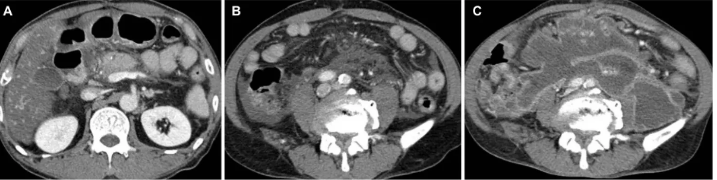
Fig. 1. Computed tomography (CT) scans of the patient. Initial CT scan reveals edematous change of the pancreatic head with acute peripancreatic fluid collection (A) extending to the pelvic cavity (B). Follow-up scan at day 20 shows a massive increase in necrotic collection and wall formation at the same level (C).
This paper presents a case of severe acute pancreatitis with fistula formation in the rectum, an unexpected and un- usual complication site. The present report documents the challenges in critical decision-making in this case and empha- sizes the importance of a multidisciplinary step-up approach in the management of severe acute pancreatitis and its complications.
CASE REPORT
A 57-year-old man was admitted due to severe epigastric pain. The patient had been diagnosed with diabetes mellitus, hypertension, and alcoholic cirrhosis of the liver. Upon admis- sion, his blood pressure, heart rate, and body temperature was 110/70 mmHg, 84 beats/min, and 38.0℃, respectively.
The patient showed tenderness on the epigastrium. The labo- ratory examination revealed the following: white blood cells 7,290/μL (neutrophils, 76.9%); hemoglobin 11.2 g/dL; plate- lets 53×103/μL; AST 136 IU/L; ALT 21 IU/L; ALP 76 IU/L;
GGT 376 IU/L; total bilirubin 2.30 mg/dL; amylase 1,776 IU/L;
lipase 2,271 IU/L; BUN 43.5 mg/dL; creatinine 0.67 mg/dL;
lactate dehydrogenase 835 IU/L; and CRP 68.18 mg/L.
Abdominal CT revealed edema of the pancreatic head with slightly decreased enhancement. In addition, fluid collections were present with extension to the pelvic cavity, which was consistent with acute peripancreatic fluid collection (Fig. 1A, B).
Intravenous (IV) fluid replacement was started as soon as the pancreatitis was detected on the laboratory and CT results. Normal saline and Hartmann’s solution was supplied at a rate of 5 mL/kg/hr and then adjusted where necessary.
Given the patient had fever at presentation, IV antibiotics were administered upon admission.
On day 3, his mental status became drowsy, and the pa- tient showed hemodynamic instability (tachycardia 148 beats/min; blood pressure 69/35 mmHg) and he was trans- ferred to the intensive care unit. His blood pH was 7.286, lactate level was elevated to 7.8 mmol/L, and partial pressure arterial oxygen was 78.5 mmHg with a nasal oxygen supply for 5 L/min. His white blood cell count was 12,630/μL (neutrophil, 75%), hemoglobin 9.5 g/dL. His renal function deteriorated with BUN and creatinine levels of 92.5 and 1.37 mg/dL, respectively. A blood culture at the time of admission showed no microorganisms. Mechanical ventilation was start- ed given his drowsy mental status and unstable respiration.
Although fluid replacement was continued, the hypotension persisted and an IV inotropic was needed for blood pressure control. At this point, continuing supportive care with IV anti- biotics versus immediate drainage was discussed. For the management of pancreatitis, delaying drainage until complete walling-off of the lesion, which takes an average of four weeks after onset, is a widely accepted strategy. In addition, continue supportive treatment was decided because most pancreatic infections occur after the second week and that a white blood count elevation with organ failure can occur in sterile pancreatitis.4,5 On hospital day five, the hemodynamic in- stability improved and the IV inotropic was discontinued.
Ventilator weaning was also possible on day 5 and extubation was performed on day 6. In the revised Atlanta classification, organ failure (two or more points in the modified Marshall score) for more than 48 hours is defined as severe acute pancreatitis and the current patient required an inotropic for blood pressure control for more than two days (two points in cardiovascular system: systolic blood pressure <90, not flu- id responsive), resulting in a diagnosis of severe acute
A B C
Fig. 2. Sigmoidoscopy image of the fistula opening. Sigmoidoscopy at day 25 reveals a fistula opening in the rectum (A) with pus discharge (B).
Fig. 3. Radiologic confirmation of the fistula tract. Contrast media filled rectum and sigmoid colon after contrast media injection to the percutaneous drainage tube (A) and computed tomography scan showing the fistula tract (B, white arrow).
pancreatitis.6 The patient's condition became stable under supportive care and antibiotic therapy, and he was transferred back to the ward.
The patient showed clinical improvement, but on day 20, he suddenly developed a high fever (39.2℃) and abdominal pain with leukocytosis (14,200/μL) (neutrophils, 88%) and a CRP level of 169.5 mg/L. His general condition deteriorated, and a follow-up CT revealed massive peripancreatic and pelvic necrotic collections with wall formation (Fig. 1C). Although the antibiotic and fluid therapies were continued, his symptoms persisted, and an infection of the necrotic tissue was
suspected. The need for drainage was inevitable but the opti- mal modality (endoscopic, percutaneous, or surgical) was uncertain. On CT, the necrotic collection revealed a deep ex- tension into the pelvic cavity, making endoscopic drainage infeasible. Considering the patient’s poor general condition and current trend of non-invasive management, a decision was made to perform percutaneous drainage, and drainage tubes were inserted into the pelvic cavity and retroperitoneum under fluoroscopy.
Although clinical improvement was noted after tube drain- age, fecal matter was observed in the drainage tube on day
A B
A B
25, which was accompanied by an aggravation of abdominal pain. Sigmoidoscopy revealed a fistula opening in the rectum, 8 cm from the anal verge (Fig. 2). Radiography with contrast media injected via the drainage tube confirmed the communi- cation between the walled-off necrosis and rectum (Fig. 3A).
A follow-up CT also revealed a fistula tract (Fig. 3B). A diag- nosis of pancreatic-rectal fistula was made, which is one of the most serious complications of pancreatitis and is asso- ciated with high mortality. Traditionally, pancreatic-colonic fis- tulas are treated surgically, but in recent years, there have been several reports on successful endoscopic closure of pan- creatic-colonic fistulas.7-9 The patient’s general condition was poor, and it was decided to perform endoscopic fistula closure to block further fecal contamination and maintain percuta- neous drainage. Fistula closure with endoscopic clipping was performed, but the patient showed persistent signs of in- fection, abdominal pain, and drainage of fecal matter continued. The decision for surgery was then made, but a less invasive transient-diverting sigmoid colostomy was per- formed instead of a wide necrosectomy and fistula repair.
After colostomy formation, the tube drainage no longer showed fecal matter and the patient’s condition stabilized.
At 4 weeks after the colostomy, the patient was discharged from hospital with a prescription for oral antibiotics. A fol- low-up CT performed 2 months later revealed a decreased amount of necrotic tissue, and the percutaneous drainage tube was removed 4 months after the colostomy. Sigmoidoscopy also revealed complete closure of the fistula opening.
Colostomy repair was then performed. The patient is currently undergoing outpatient follow-up with a good clinical course.
DISCUSSION
In recent years, the management of acute pancreatitis has changed in favor of a more noninvasive strategy.1-4 In the past, the standard treatment for infected necrotizing pancreatitis was an open necrosectomy with complete removal of the in- fected necrotic tissue, which carries a high risk of complications. As a result, alternative techniques, such as minimally invasive surgery, percutaneous drainage, and endo- scopic drainage, have been proposed and shown promising results.1-4 Therefore, the concept of a “step-up” approach, de- signed and performed by a multidisciplinary team, has been proposed using recent positive multicenter randomized-con-
trol trial data.3 In this regard, the treatment guidelines for acute pancreatitis also recommend percutaneous or endo- scopic drainage as an initial intervention for infected pancreatitis.4 On the other hand, data on the best drainage modality for specific patient subgroups are lacking and further investigations are needed. In the present case, the patient was treated based on the step-up approach, starting with anti- biotics and then moving to percutaneous drainage.
Pancreatic-colonic fistula is a rare critical complication of acute pancreatitis, with a mortality rate of 17-67%.10,11 The precise pathophysiological mechanism of pancreatic-enteric fistula formation is unknown, but several mechanisms or fac- tors have been suggested, as reviewed by Shatney and Sosin.12 The combinations of the proposed factors are likely to be surgical in most cases. One proposed mechanism is the continuous secretion of pancreatic enzymes into the wal- led-off necrosis via communication with the pancreatic duct.
This secretion might result in progressive digestion of the ne- crosis and its neighboring organs, which might in turn result in fistula formation. The increased intracystic pressure caused by fluid accumulation in the walled-off necrosis has also been suggested to compromise the weakest point of the wall and its vasculature, with subsequent necrosis and fistula formation. Other reported factors are the incidentally in- creased intra-abdominal pressure and abdominal trauma, such as needle aspiration or drainage. In the present case, the large size of the necrosis supports the idea of increased pressure. On the other hand, the possibility of iatrogenic fistu- la due to percutaneous drainage cannot be excluded. A pan- creatic-enteric fistula usually arises near the pancreas, such as in the stomach, duodenum, or colon.13 Therefore, anatomi- cal proximity might be an important factor related to the fre- quency with which an organ is involved. Thus, the splenic flexure and transverse colon are the most common sites in the colorectum.7 To the best of the authors’ knowledge, this is the first report of pancreatic-colonic fistula formation in the rectum as a complication of acute pancreatitis. Given its distant location from the pancreas, the rectum is usually spared in colonic complications of pancreatitis. In the present case, the walled-off necrosis extended to the pelvic cavity near the rectum, producing an anatomical proximity.
In most cases of pancreatitis-associated upper gastro- intestinal fistula formation, only supportive care is needed, and spontaneous closure is likely.13,14 The fistula can even
act as a good drainage route. In contrast, spontaneous clo- sure is much less likely in colonic fistulas and they have a poor prognosis compared to fistula formation in other sites of the gastrointestinal tract.11,13,14 Hence, pancreatic-colonic fistula is potentially a continuous infection source, rather than a drainage route, and prompt blocking of fecal contamination is crucial in its management. Traditionally, surgical debride- ment with fistula repair or colostomy formation has been per- formed, but there are several reports of successful endo- scopic fistula closures by endoscopic clipping, endoloop, fibrin glue, and over-the-scope clipping.7-9 For non-severe cases, there have been case reports of successful treatment using only conservative measures, such as continuous lavage through a drainage tube or bowel cleansing alone.7,15 In the present patient, considering the potentially higher perioper- ative morbidity or mortality due to the patient’s poor clinical status and the recently reported success of less invasive man- agement, sigmoidoscopic fistula closure was performed and percutaneous drainage was maintained, but this approach was relatively ineffective. A more aggressive treatment option was considered and a simple transient diverting colostomy was performed to minimize the invasiveness of the operation.
After the colostomy, the patient ultimately recovered without the need for further invasive surgery. This suggests that the step-up approach is a reasonable choice for pancreatic-colon- ic fistulas and supports the general utility of the approach in the management of pancreatitis and its complications.
This case of severe acute pancreatitis was complicated and severe, with unexpected rectal fistula formation. Although the patient’s medical condition deteriorated and many clinical challenges arose, he was treated successfully based on the step-up approach. His recovery validates the value of this ap- proach in the management of pancreatitis and suggests that it can be extended to the management of pancreatic-colonic fistulas. Nevertheless, physicians should be aware that ne- crotizing pancreatitis-associated complications can manifest in unexpected ways.
REFERENCES
1. Zerem E. Treatment of severe acute pancreatitis and its complications. World J Gastroenterol 2014;20:13879-13892.
2. Zerem E, Imamović G, Sušić A, Haračić B. Step-up approach to infected necrotising pancreatitis: a 20-year experience of percu- taneous drainage in a single centre. Dig Liver Dis 2011;43:
478-483.
3. van Santvoort HC, Besselink MG, Bakker OJ, et al. A step-up ap- proach or open necrosectomy for necrotizing pancreatitis. N Engl J Med 2010;362:1491-1502.
4. Working Group IAP/APA Acute Pancreatitis Guidelines. IAP/APA evidence-based guidelines for the management of acute pancreatitis. Pancreatology 2013;13(4 Suppl 2):e1-e15.
5. Gerzof SG, Banks PA, Robbins AH, et al. Early diagnosis of pancre- atic infection by computed tomography-guided aspiration.
Gastroenterology 1987;93:1315-1320.
6. Banks PA, Bollen TL, Dervenis C, et al. Classification of acute pan- creatitis--2012: revision of the Atlanta classification and defi- nitions by international consensus. Gut 2013;62:102-111.
7. Oana S, Shibata S, Matsumoto T. Pancreatico-colonic fistula af- ter endoscopic ultrasound-guided cyst drainage for pancreatic pseudocyst: report of three cases. JOP 2016;17:334-339.
8. Will U, Meyer F, Hartmeier S, Schramm H, Bosseckert H.
Endoscopic treatment of a pseudocystocolonic fistula by band li- gation and endoloop application: case report. Gastrointest Endosc 2004;59:581-583.
9. Karvonen J, Gullichsen R, Salminen P, Grönroos JM. Endoscopic treatment of pseudocystocolonic fistula with fibrin glue.
Gastrointest Endosc 2010;72:664-665.
10. Wille-Jørgensen P, Frederiksen HJ. Colonic necrosis or fistula fol- lowing pancreatitis or gastric surgery. Eur J Surg 1991;157:
137-139.
11. Suzuki A, Suzuki S, Sakaguchi T, et al. Colonic fistula associated with severe acute pancreatitis: report of two cases. Surg Today 2008;38:178-183.
12. Shatney CH, Sosin H. Spontaneous perforation of a pancreatic pseudocyst into the colon and duodenum. Am J Surg 1973;
126:433-438.
13. Clements JL Jr, Bradley EL 3rd, Eaton SB Jr. Spontaneous internal drainage of pancreatic pseudocysts. AJR Am J Roentgenol 1976;
126:985-991.
14. Jiang W, Tong Z, Yang D, et al. Gastrointestinal fistulas in acute pancreatitis with infected pancreatic or peripancreatic necrosis:
a 4-year single-center experience. Medicine (Baltimore) 2016;
95:e3318.
15. Kwon JC, Kim BY, Kim AL, et al. Pancreatic pseudocystocolonic fistula treated without surgical or endoscopic intervention.
World J Gastroenterol 2014;20:1882-1886.