• 검색 결과가 없습니다.

Acute Type A Aortic Dissection in a Patient with Situs Inversus Totalis

N/A
N/A
Protected

Academic year: 2021

Share "Acute Type A Aortic Dissection in a Patient with Situs Inversus Totalis"

Copied!
3
0
0

로드 중.... (전체 텍스트 보기)

전체 글

(1)

321 www.kjtcvs.org

KJTCVS

The Korean Journal of Thoracic and Cardiovascular Surgery

Case Report

Acute Type A Aortic Dissection in a Patient with Situs Inversus Totalis

Dong Kyu Kim, M.D., Ji Min Lee, M.D., Seon Yeong Heo, M.D., Jong Pil Jung, M.D., Chang Ryul Park, M.D., Yong Jik Lee, M.D., Sang Cjeol Lee, M.D., Su Kyung Hwang, M.D., Gwan Sic Kim, M.D.

Department of Thoracic and Cardiovascular Surgery, Ulsan University Hospital, Ulsan University Medical School, Ulsan, Korea

ARTICLE INFO

Received February 25, 2020 Revised April 6, 2020 Accepted April 12, 2020 Corresponding author Gwan Sic Kim

Tel 82-52-250-8726 Fax 82-52-250-8558 E-mail [email protected] ORCID

https://orcid.org/0000-0002-0648-0178

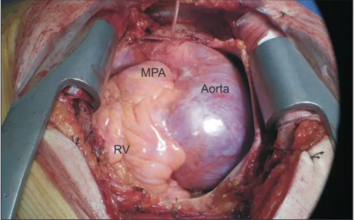
We describe the occurrence of acute type A aortic dissection in a patient with situs inver- sus totalis. A 37-year-old man presented to the emergency department with acute chest pain. Initial chest X-ray findings showed a right-sided heart and a left-sided liver. Con- trast-enhanced computed tomography revealed a Stanford type A acute aortic dissection, aortic root dilatation, and situs inversus totalis. All of the thoracic structures were mirror-im- age reversed and an abnormal coronary artery was observed. The Bentall operation was performed. This report demonstrates that computed tomography and echocardiography were useful for understanding the anatomy and the presence or absence of concurrent anomalies in a patient with situs inversus totalis. The patient’s postoperative course was uneventful.

Keywords: Aortic dissection, Dextrocardia, Situs inversus totalis

Copyright

© The Korean Society for Thoracic and Cardiovascular Surgery. 2020. All right reserved.

This is an Open Access article distributed under the terms of the Creative Commons Attribution Non-Commercial License (http://creativecommons.org/licenses/

by-nc/4.0) which permits unrestricted non-commercial use, distribution, and reproduction in any medium, provided the original work is properly cited.

Case report

Situs inversus totalis is a congenital anomaly character- ized by a mirror image of the normal visceral organs. Un- like dextrocardia, which only involves placement of the cardiac apex in the right thorax, situs inversus totalis in- volves a left-sided right atrium with the venae cavae and a right-sided left ventricle with the aorta. Aortic dissection in patients with this condition is very rare and may be dif- ficult to assess and manage. Here, we describe a 37-year- old man who presented with situs inversus totalis and type A aortic dissection.

A 37-year-old man presented to the emergency depart- ment with acute chest and back pain that had begun 12 hours before his visit. His vital signs upon arrival were blood pressure of 120/65 mm Hg, a regular heart rate of 86 beats/min, respiratory rate of 20 cycles/min, and body tem- perature of 36.5°C. Laboratory testing revealed a white blood cell count of 17,100/mm

3

and a C-reactive protein level of 0.88 mg/dL; other parameters were within normal limits. Initial chest radiography showed mediastinal wid- ening, mild cardiomegaly, and dextrocardia. The patient was aware of this dextrocardia, but did not receive regular

follow-up. Electrocardiography showed a normal sinus rhythm. Portable transthoracic echocardiography revealed an enlarged aortic root and an ascending aorta with an in- timal flap, as well as severe aortic valve regurgitation. The left and right ventricular sizes and systolic function were normal. Contrast-enhanced computed tomography re- vealed a Stanford type A acute aortic dissection and situs inversus totalis (Fig. 1). Emergency surgery was performed with a diagnosis of acute type A aortic dissection with an aortic root aneurysm and severe aortic valve regurgitation.

The operation was performed through a median sternoto- my incision with the patient in the supine position. The right common femoral artery, superior vena cava, and infe- rior vena cava were used as cannulation sites for cardiopul- monary bypass. A vent was inserted through the left supe- rior pulmonary vein. Under moderate hypothermia (26°C), retrograde cold blood cardioplegia solution was infused through the coronary sinus after right atriotomy. After aortic cross-clamping, transverse aortotomy was per- formed. For neurocerebral protection during circulatory arrest, the left innominate artery and right common carot- id artery were cannulated endoluminally with a bal- loon-tipped catheter. Until median sternotomy, the sur-

https://doi.org/10.5090/kjtcs.20.006 pISSN: 2233-601X eISSN: 2093-6516

Korean J Thorac Cardiovasc Surg. 2020;53(5):321-323

(2)

322 www.kjtcvs.org

KJTCVS https://doi.org/10.5090/kjtcs.20.006

geon operated on the right side of the patient. Thereafter, during the main procedure, the surgeon operated on the left side of the patient. In the operative findings, the 3 arch vessels were found to be completely reversed in a mirror image. The ventricular apex was forced to the right side, and the superior and inferior venae cavae were located on the left side. All operative findings of the arteries were completely reversed, compared with normal arteries (Fig.

2). Transverse aortotomy revealed that the intimal tear ex- tended from the sinotubular junction to the ascending aor- ta. The aortic valve had 3 cusps and leaflets. Exploration of the aortic root revealed that the origins of the left and right coronary ostia were within the left coronary cusp (Fig. 3).

Severe annuloaortic ectasia was observed. Thus, the Ben- tall operation was performed using a 23-mm mechanical valved conduit. Both the left and right coronary ostia were cut into a single button and anastomosed to the conduit graft. Pathologic findings of the aortic wall demonstrated hemorrhagic changes with dissection, and the aortic valve demonstrated hyalinization. The postoperative course was uneventful and the clinical findings were normal, without any abnormal findings on follow-up transthoracic echocar- diography or computed tomography. The patient provided written informed consent for publication of clinical details and images.

Discussion

Situs inversus totalis refers to a mirror image of situs sol- itus or the normal arrangement of organs. The incidence of the condition is approximately 1 in 10,000 live births [1].

The male-to-female ratio is 1:1, and the condition has no

ethnic predilection [2]. Acute type A aortic dissection is very rare in patients with this condition. So far, 3 cases have been reported [3-5], of which 1 was traumatic [4].

Congenital cardiac anomalies occur in 3%–5% of indi- viduals with situs inversus [6], and the most common anomaly is a double outlet right ventricle [7]. Nearly all pa- tients with solitary levocardia have an anomaly [2]. Our patient had a coronary anomaly in which both the left cor- onary ostium and the right coronary ostium originated from the left coronary cusp. The proportion of coronary anomalies among all cardiac anomalies is unclear, as few cases have been reported. In addition, the prevalence and types of late complications cannot be estimated due to the rarity of this condition. Our patient exhibited aortic root

A B

Fig. 1. Computed tomography revealed Stanford type A acute aortic dissection and situs inversus totalis. (A) Coronal view. (B) Three-dimensional reconstructed view.

MPA

Aorta

RV

Fig. 2. Intraoperative images. The ventricular apex was forced to the right side. A left-sided aorta was present with a right-sided main pulmonary artery. MPA, main pulmonary artery; RV, right ventricle.

Left coronary ostium

Right coronary ostium

LCC

RCC

Fig. 3. The aortic valve had 3 cusps and leaflets. Exploration of the

aortic root revealed that the origins of the left (arrow) and right

coronary ostia (arrow) were within the left coronary cusp. LCC,

left coronary cusp; RCC, right coronary cusp.

(3)

323

Dong Kyu Kim, et al. Acute Type A Aortic Dissection in a Patient with Situs Inversus Totalis

www.kjtcvs.org

KJTCVS

dilatation and aortic dissection after over 30 years in the context of situs inversus totalis. Therefore, long-term mon- itoring is required.

In regard to histologic findings, 3 similar cases have been reported [3-5]. Niino et al. [3] reported in 2003 that the pathologic findings of the aortic walls included arterio- sclerotic changes. In the present case, the histologic find- ings of the aortic wall were hemorrhage with dissection, and the aortic valve demonstrated hyalinization.

The surgical technique to correct the anomaly is not very technically difficult. However, because the patient’s anato- my may include anomalies and does not match conven- tional anatomical configurations, the operator may become confused. Therefore, it is important to evaluate possible anomalies and anatomical features through preoperative computed tomography and echocardiography. Standing on the left side of the patient can ease the difficulty of the sur- gery, as the anatomy of the patient is inversed.

Conflict of interest

No potential conflict of interest relevant to this article was reported.

ORCID

Dong Kyu Kim: https://orcid.org/0000-0002-0047-2732 Ji Min Lee: https://orcid.org/0000-0001-9317-0313 Seon Yeong Heo: https://orcid.org/0000-0003-4857-7100

Jong Pil Jung: https://orcid.org/0000-0002-2992-7729 Chang Ryul Park: https://orcid.org/0000-0003-2841-7046 Yong Jik Lee: https://orcid.org/0000-0002-0837-4336 Sang Cjeol Lee: https://orcid.org/0000-0003-0502-1022 Su Kyung Hwang: https://orcid.org/0000-0003-2759-2774 Gwan Sic Kim: https://orcid.org/0000-0002-0648-0178

References

1. Lin AE, Krikov S, Riehle-Colarusso T, et al. Laterality defects in the national birth defects prevention study (1998-2007): birth prevalence and descriptive epidemiology. Am J Med Genet A 2014;164A:2581- 91.

2. Fulcher AS, Turner MA. Abdominal manifestations of situs anoma- lies in adults. Radiographics 2002;22:1439-56.

3. Niino T, Shiono M, Inoue T, Hata M, Sezai A, Negishi N. A case of acute aortic dissection type A in a patient with situs inversus. Ann Thorac Surg 2003;75:1963-5.

4. Magishi K, Izumi Y, Ishikawa N, Kimura F. Stanford type A acute aortic dissection caused by blunt trauma in a patient with situs inver- sus. Ann Thorac Surg 2006;81:2294-6.

5. Pecha S, Schirmer J, Yildirim Y, et al. Type A aortic dissection with situs inversus. J Card Surg 2013;28:747-8.

6. Winer-Muram HT, Tonkin IL. The spectrum of heterotaxic syn- dromes. Radiol Clin North Am 1989;27:1147-70.

7. Tripathi S, Ajit Kumar VK. Comparison of morphologic findings in

patients with dextrocardia with situs solitus vs situs inversus: a retro-

spective study. Pediatr Cardiol 2019;40:302-9.

수치

Fig. 1. Computed tomography revealed Stanford type A acute  aortic dissection and situs inversus totalis

참조

관련 문서

Wald-Type Tests for Detecting Breaks in The Trend Function of a Dynamic

3. Specially, Va type these a lot of changes were seen. Va type and Ⅶ type were shown big change after 4 months. Depilation number increases if depilation is gone or it is

We investigated the effects of oligomer (urethane acrylate or polyester acrylate) type, monomer (bisphenol A glycerolate dimethacrylate or bisphenol A ethoxylate

Frequency and clinical significance of the expression of the multidrug resistance proteins MDR1/P-glycoprotein, MRP1, and LRP in acute myeloid leukemia: a

 store value (read from the Register File during decode) written to the Data Memory.  load value, read from the Data Memory, written to

 The disk arm starts at the first I/O request on the disk, and moves toward the last I/O request on the other end, servicing requests until it gets to the other extreme

(Result) The reports of nausea, vomiting, abdominal discomfort, jaundice and hepatomegaly in acute hepatitis A patients and febrile sensation, chillness,

The feed is commonly a solution in a solvent like ethanol or t-butanol, and the nonsolvent is water..