Immediate Effects of Strain-Counterstrain Technique on Pressure Pain Threshold and Muscle Activity in Male Adults
With Upper Trapezius Latent Trigger Point
Ji-hee Jung, B.H.Sc., P.T.
Nam-gi Lee, M.Sc., P.T.
Dept. of Rehabilitation Therapy, The Graduate School, Yonsei University Sung-hyun You, Ph.D., P.T.
Dept. of Physical Therapy, College of Health Science, Yonsei University
Dept. of Ergonomic Therapy, The Graduate School of Health and Environment, Yonsei University
Abstract
1)The aim of this study was to determine the immediate effects of single treatment of strain-counter strain (SCS) on pressure pain threshold (PPT) and muscle activity during scapular plane abduction with 3% body weight load. Fifteen asymptomatic male adults with upper trapezius latent trigger point (LTrP) (PPT<2.9 ㎏/㎠) participated in this study. Pressure algometer was used to measure PPT and surface electromyography was used to record upper, middle and lower trapezius, serratus anterior, infraspinatus and middle deltoid muscle activity and relative ratio during scapular plane abduction between pre- and post-intervention. There was a significant increase in upper trapezius PPT after a 90-second SCS (p<.05). The activity of the upper trapezius and middle deltoid was significantly decreased (p=.014, p=.001), coupled with a decreased muscle activity ratio between the upper and lower trapezius (p<.05).
These results indicate that the SCS may effectively deactivate upper trapezius activity, thereby alleviating muscle balance and reducing pain sensitivity.
Key Words: Eletromyography; Latent trigger point; Muscle activity ratio; Pressure pain threshold;
Strain-counterstrain; Upper trapezius.
Introduction
Upper trapezius (UT) trigger points are common soft tissue impairments that often affect neuromotor control in glenohumeral and scapulothoracic move- ment (Fischer, 1987; Sciotti et al, 2001).
Approximately 90% of the healthy population have latent trigger points (LTrP) combined with muscle shortening and decreased pressure pain threshold (PPT) (Fischer, 1987; Simons, 2002; Simons et al, 1999). These trigger point pain syndrome are caused by postural alignment impairments, muscle im- balance, and repetitive overload or cumulative trau- matic disorders (Huguenin, 2004). This is often manifested with chronic pain and abnormal motor
control patterns, leading to functional movement im- pairments in the shoulder complex (Lucas et al, 2004; Nagrale, 2010). Previous studies (Lucas et al, 2004; Lucas et al, 2009) have shown that individuals with the UT LTrP demonstrated increased muscle activity and altered activation sequence. Prolonged and dominant spontaneous muscle activity in LTrP compromises normal muscle-tension relationship and in turn changes kinetic chain reaction associated with force coupling mechanism between the lower trapezius (LT), serratus anterior (SA), and deltoid muscles during scapulohumeral movements (Hubbard and Berkoff, 1993; Page et al, 2010). These altered kinesiological movement characteristics predispose to kinesiopathological movement impairments including
Corresponding author: Sung-hyun You [email protected]
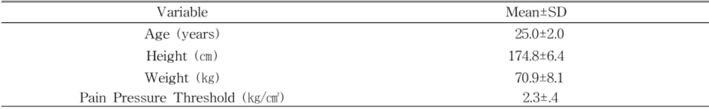
Variable Mean±SD
Age (years) 25.0±2.0
Height (㎝) 174.8±6.4
Weight (㎏) 70.9±8.1
Pain Pressure Threshold (㎏/㎠) 2.3±.4
Table 1. General characteristics of subjects (N=15)
shoulder impingement and rotator cuff tendinitis (Cools et al, 2003; Kibler and Sciascia, 2010; Lucas et al, 2004; Page et al, 2010; Sahrmann, 2002)
Strain-counterstrain (SCS) is a commonly used soft-tissue mobilization technique to alleviate trigger point pain and associated with musculoskeletal dysfunction (Jones, 1981; Lewis, 2001). This technique encompasses two steps: (1) passively holding an involved body segment at a relaxed position for 90 seconds until the desirable level of muscle tension and associated pain reduction are achieved and (2) returning to neutral position slowly. The majority of clinical studies investigating the effect of SCS have specifically focused on pain (Eisenhart et al, 2003;
Lewis, 2001; Pedowitz, 2005), which is often only a secon- dary source of the syndrome, rather than the primary cause-neuromuscular dysfunction. For example, recent studies using PPT and pain scale have shown that SCS is effective in relieving pain hypersensitivity (Lewis, 2010;
Meseguer et al, 2006; Nagrale, 2010). However, SCS can be used in managing muscle imbalance and associated pain because this technique restores optimal movement control by inhibiting overactive muscles and facilitating inhibited muscles (Liebenson, 2007). Nevertheless, whether or not SCS has an inhibitory effect on the muscle of the involved muscle fibers is unknown.
Furthermore, there is a dearth of information about the mechanisms underpinning SCS on clinical management of trigger point pain syndrome. Hence, this study aims to determine the immediate effects of SCS at the UT LTrP on PPT and muscle activ- ity patterns during scapular plane abduction with load. Our hypotheses were that (1) the UT pain threshold would be improved and (2) the SCS technique would decrease the UT activity and de-
crease the associated muscle activity UT/LT ratios during shoulder scapular plane abduction.
Methods Subjects
Fifteen healthy, young male subjects were recruited from Yonsei University. All subjects signed informed consent form prior to the participation of this study. Inclusion cri- teria included: (1) a LTrP in the UT muscle and (2) shoulder abduction at least 160° without compensatory motion. In this study, the LTrP was defined as tender point within a palpable taut band and less than 2.9 ㎏/㎠
PPT. This cutoff pressure was selected because the lowest pressure threshold in the UT of normal male adults was previously identified (Fischer, 1987). Exclusion criteria in- cluded: (1) specific neck and shoulder pain such as radicul- opathy, (2) systematic pathology, and (3) past or present neurologic or musculoskeletal diseases. The subjects’ demo- graphic and anthropometric data are presented in Table 1.
Instruments
Pressure algometer2)
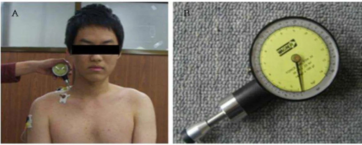
The pressure algometer1) comprising a force gauge with a 1 ㎠ rubber disk, was used to measure PPT.
The rubber disk was placed vertically to the UT LTrP. Pressure was then gradually applied on the rubber disk (representing ㎏/㎠) and recorded when the subjects started to feel pain or discomfort.
Measurement was repeated three times before and after SCS. Mean recorded pressure threshold values were averaged and used for further analysis.
1) FPK 60, Wagner Instruments Inc, Greenwich, CT, U.S.A.
Figure 1. Measuring pressure pain threshold on upper trapezius latent trigger point. A:
Measuring pressure pain threshold, B: Pressure algometer.
Electromyography3)
A surface electromyography (EMG)2) was used to determine shoulder muscle activity and associated ratio during scapular plane abduction. The EMG data were collected at a sampling frequency of 1024 ㎐ with a 60 ㎐ notch filter.4)
Prior to the attachment of electrode, the skin was carefully prepared to reduce skin impedance by shav- ing hair and disinfecting with alcohol. Bipolar Ag/AgCl electrode was placed parallel to the six right shoulder muscles (UT, middle trapezius (MT), LT, infraspinatus (IS), SA, and middle deltoid (MD)) with a 25 ㎜ in- ter-electrode distance. Figure 2 depicts the ex- perimental setup for EMG measurement and electrode placements. For the UT (scapula upward rotator), electrode was placed along the ridge of the shoulder, slightly lateral to and one half the distances between the C7 and acromion. For the MT, electrode was placed horizontally, 2 ㎝ apart, next to the scapula root. For the LT (scapula upward rotator), electrode was placed on a 55° oblique angle, approximately 5 ㎝ down from the scapular spine next to the medial edge of the scapula. For the IS (glenohumeral stabilizer and abductor), electrode was placed approximately 4 ㎝ be- low the spine of the scapula, on the lateral aspect, over the infrascapular fossa of the scapula. For the
SA (scapula upward rotator), electrode was placed just below the axillary area at the level of the inferior tip of the scapula, and just medial of the latissimus dorsi.
For the MD (primary abductor), electrode was placed on the lateral aspect of the upper arm, 2 ㎝ apart, and approximately 3 ㎝ below the acromion (Criswell and Cram, 2011). A reference electrode was placed over the clavicle.
Raw EMG signal was analyzed using Telescan 2.993 software3) and was band-pass filtered at 10-500 ㎐. The EMG data were processed into the root mean square (RMS) using window for 100 ㎳.
The maximal voluntary isometric contraction (MVIC) was used to normalize EMG data for each tested muscle and maximal voluntary muscle contraction test was performed according to Kendall’s manual muscle testing position (Kendall, 2005). EMG data were recorded during a 5-second reference con- traction and repeated three times and stored for data analysis. 30~50 seconds of resting interval was pro- vided between the test trials. The mean EMG ampli- tude data obtained during the middle 3-second of each trial was used for statistical analysis.
Procedures
Subjects were asked to abduct their dominant arm
2) WEMG-8, Laxtha Inc, Daejeon, Korea.
3) Telescan 2.993, Laxtha Inc, Daejeon, Korea.
Figure 2. Experimental setup for EMG muscle activity measurement.
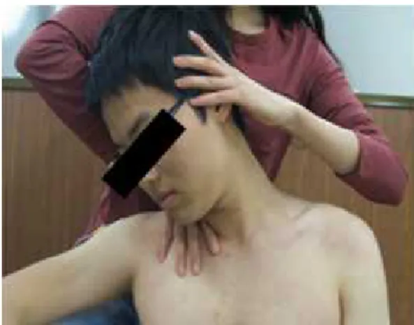
Figure 3. Strain-counterstrain technique in the upper trapezius muscles.
(right side) with 3% of their body weight (Mean=2.1
㎏, SD=.28) (Thigpen et al, 2010) in standing position (Figure 2). The target abduction was set 160° to fo- cus the action of the IS as a stabilizer rather than an external rotator. Abduction movement was guided at 30° scapular plane using a vertical pole, which was fixed from the ground to the ceiling. The pos- tural sway was restricted by using a mirror approx- imately 2 m in front of the subjects at eye level (Lucas et al, 2010). The movement velocity was controlled by auditory cue using beep sound with verbal command. Scapular plane abduction was car- ried out using the 5-4-5-4-5 steps. 5-4-5-4-5 steps comprise five subsets: 5-second for rest, 4-second for raising the arm, 5-second for maintaining abduc- tion posture, 4-second for lowering the arm, and 5-second for rest. Before the test, the subjects were given enough time to familiarize themselves with the 5-4-5-4-5 steps. Three trials were performed with a 4-second rest between trials.
Intervention
Subjects were neutrally sitting positioned for the SCS intervention. The subject’s right shoulder was passively abducted. The examiner found the UT LTrP by palpation. After the detection of trigger point, the examiner applied a gentle pres- sure until subjects felt pain from pressure
sensation. Subjects were passively positioned to reduce the pain level by approximately 70%
(Jones, 1981). To determine whether the pain level decreased, the examiner asked the subjects their perception level compared with the initial condition. In this study, the most common posi- tions that relieve pain are cervical flexion, ipsi- lateral side-bending and contralateral rotation.
Manual pressure was maintained equally for 90 seconds. After the intervention, the subject’s posi- tion was passively returned to the starting posi- tion (Meseguer et al, 2006).
Statistical Analysis
The descriptive data were expressed as mean and standard deviation. A paired t-test was used to de- termine SCS related changes on EMG activity and ratio and PPT. Statistical significance was assigned at p<.05. All statistical analysis was performed using the Statistical Package for the Social Sciences (SPSS) version 18.0 software.
Results Pressure pain threshold
The mean PPT of the UT showed significant dif- ference between pre- and post- intervention (p<.05).
Figure 4. Comparison of the pressure pain threshold between the pre- and post-intervention (*p<.05).
Activity Ratio Pre Post p
Upper trapezius/Middle trapezius 2.64±1.40a 2.34±1.45 .068
Upper trapezius/Lower trapezius 1.38±.88 1.23±.70 .030
Upper trapezius/Serratus anterior .68±.31 .65±.29 .081
aMean±SD, Significant difference (p<.05).
Table 3. Muscle activity ratio during scapular plane abduction at pre- and post-intervention
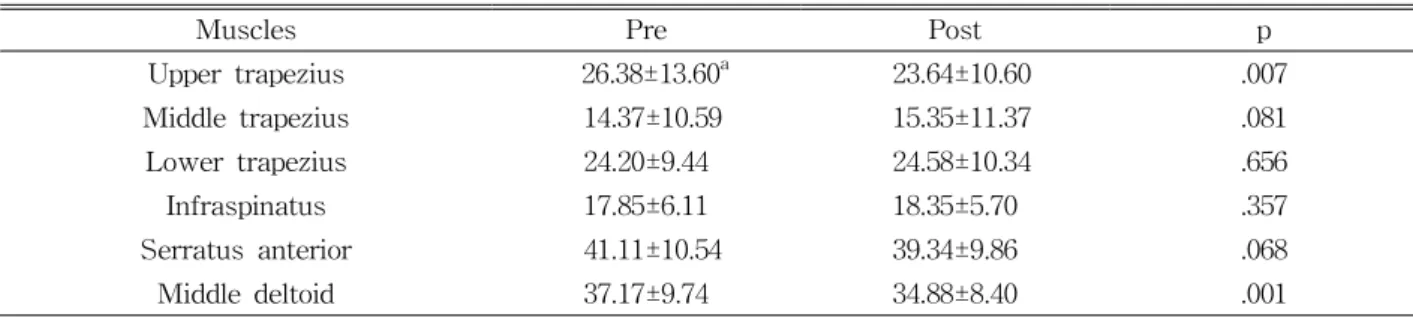
Muscles Pre Post p
Upper trapezius 26.38±13.60a 23.64±10.60 .007
Middle trapezius 14.37±10.59 15.35±11.37 .081
Lower trapezius 24.20±9.44 24.58±10.34 .656
Infraspinatus 17.85±6.11 18.35±5.70 .357
Serratus anterior 41.11±10.54 39.34±9.86 .068
Middle deltoid 37.17±9.74 34.88±8.40 .001
aMean±SD, Significant difference (p<.05).
Table 2. The mean EMG amplitude during scapular plne abduction at pre- and post-intervention (Unit: %MVIC) The PPT was increased (33.04%) from 2.33 ㎏/㎠
(SD=0.35) to 3.10 ㎏/㎠ (SD=.45) following SCS in- tervention, indicating that pain sensitivity of the UT effectively relieved (Figure 4).
EMG muscle activity and ratio
Paired t-test showed significant difference in UT and MD normalized EMG activity during scapular plane abduction (p<.05) (Table 2). The UT and MD muscle activities were decreased 2.74% and 2.29%
respectively. The muscle activity ratio was de- termined: dividing normalized mean UT EMG ampli- tude by normalized mean EMG amplitude of MT, LT, and SA (Cools et al, 2007). The muscle activity ratio between UT and LT was significantly de- creased (p=.030)(Table 3).
Discussion
The present study demonstrated the immediate effects of SCS on EMG muscle activity, relative changes in muscle activity ratio and associated PPT between pre- and post-intervention during the scap- ular plane abduction in male adults with UT LTrP.
As anticipated, the application of SCS on UT LTrP effectively decreased UT EMG activity and relative changes in muscle activity ratios and reduced pain sensitivity as reflected in PPT. Especially, UT and MD muscle activity and UT/LT ratio were de- creased by 2.74%, 2.29%, and 12.20% whereas PPT was reduced by 33.04%.
Certainly, our findings are in agreement with re- cent clinical studies that examined the effects of SCS (Meseguer et al, 2006; Nagrale, 2010) on pain
control. Meseguer et al. (2006) found that neck pain as measured by visual analogue scale (VAS) in in- dividuals with mechanical neck pain was sig- nificantly reduced by 44% following the single appli- cation of SCS (p<.05). Similarly, Nagrale et al.
(2010) demonstrated that the SCS combined with muscle energy technique decreased neck pain (as evident in VAS measure) when compared to the isolated muscle energy technique alone in patients with non-specific neck pain.
One important mechanism for SCS induced modu- lation of pain threshold may be accounted by mechan- ical and neurophysiological components. Mechanically, SCS, which utilizes the localized mechanical pressure on the fibrotic tissue (taut band or shortened sarco- mere), can produce a positional release that restores resting sarcomere length or normalizes the sarcomere length-tension relationship (Simons, 2002). This nor- malized sarcomere length-tension relationship allows optimal actin and myosin cross-bridging to occur, which increases tissue extensibility of the taut band or the fibrotic tissue. In fact, individuals with the up- per trapezius LTrP showed increased muscle activity and altered activation sequence in scapular rotators during scapular plane abduction (Lucas et al, 2004;
Lucas et al, 2009). It was also noted that UT muscle activity in the LTrP area of individuals with LTrP or in the trigger point areas of patients with tension headache or fibromyalgia was more prolonged (up to 50 minutes) and dominant (2 times in the LTrP sub- jects; 9-10 times in patients) than the other non-trig- ger point area when probed with needle EMG meas- urement (Hubbard and Berkoff, 1993). This sustained increased muscle activity together with altered muscle activation sequence can change normal muscle-tension relationship and in turn changes kinetic chain reaction associated with force coupling mechanism between the LT, SA, and deltoid muscles during scapulohumeral movements (Page et al, 2010). This mechanism sug- gests that the trigger point pain syndrome is primar- ily associated with motor control dysfunction rather than pain itself (Simons, 2002).
The other neurophysiological mechanism may be as- sociated with excessive and continuous action potentials cause the release of Ca++ from the sarcoplasmic reticulum. Subsequently, this results in spontaneous and prolonged muscle contraction unless AChE degrades ACh into acetate and choline, thus terminating the re- lease of Ca++. SCS may suppress this excessive release of ACh, which leads to the inhibition of the continuous contraction of sarcomere representing motor endplate noise (spontaneous EMG activity) (Hong and Simons, 1998; Hubbard and Berkoff, 1993; Huguenin, 2004).
Albeit that our preliminary investigation demonstrated novel effects of SCS on pain sensitivity and EMG activ- ity during scapular plane abduction, several shortcomings should be taken into consideration and addressed in fu- ture studies. First, asymptomatic subjects were recruited in the present study; hence, it is difficult to generalize our findings to people with shoulder disorders. This in- vites a future study to examine if similar therapeutic ef- fects exist in individuals with active trigger point pain syndrome. Second, the subjects in this study were small number of male adults threatening external validity.
Third, a case-control study would have provided a more robust design to elucidate the differential effect of SCS on motor control and associated pain modulation in pa- tient populations. Lastly, the long-term effect of SCS should also be explored in future studies.
Conclusion
This study examined the effects of SCS on pain threshold and muscle activity during scapular plane abduction in male adults with UT LTrP. The SCS effectively decreased UT muscle activity and con- currently improved pain threshold. Our results pro- vide empirical evidence that SCS can be an effective soft tissue manipulation technique to reduce pain threshold in individuals with LTrP. Future studies are needed to examine the long-term effect of SCS on neuromuscular control and pain modulation in pathological population with myofascial impairments.
References
Cools AM, Declercq GA, Cambier DC, et al. Trapezius activity and intramuscular balance during iso- kinetic exercise in overhead athletes with im- pingement symptoms. Scand J Med Sci Sports.
2007;17(1):25-33.
Cools AM, Witvrouw EE, Declercq GA, et al. Scapular muscle recruitment patterns: Trapezius muscle latency with and without impingement symptoms.
Am J Sport Med. 2003;31(4):542-549.
Criswell E, Cram JR. Cram's Introduction to Surface Electromyography. 2nd ed. Sudbury, MA, Jones and Bartlett Publishers, 2011.
Eisenhart AW, Gaeta TJ, Yens DP. Osteopathic ma- nipulative treatment in the emergency department for patients with acute ankle injuries. J Am Osteopath Assoc. 2003;103(9):417-421.
Fischer AA. Pressure algometry over normal muscles.
Standard values, validity and reproducibility of pressure threshold. Pain. 1987;30(1):115-126.
Hong CZ, Simons DG. Pathophysiologic and electro- physiologic mechanisms of myofascial trigger points. Arch Phys Med Rehabil. 1998;79(7):863-872.
Hubbard DR, Berkoff GM. Myofascial trigger points show spontaneous needle EMG activity. Spine.
1993;18(13):1803-1807.
Huguenin LK. Myofascial trigger points: The current evidence. Phys Ther Sport. 2004;5(1):2-12.
Jones LH. Strain and counterstrain. Colorado Springs, CO, American Academy of Osteopathy, 1981.
Kendall FP. Muscles: Testing and function with pos- ture and pain. 5th ed. Baltimore, MD, Lippincott Williams & Wilkins, 2005.
Kibler WB, Sciascia A. Current concepts: Scapular dyskinesis. Br J Sports Med. 2010;44(5):300-305.
Lewis C. The use of strain-counterstrain in the treatment of patients with low back pain. J Man Manip Ther. 2001;9(2):92-98.
Lewis C. A randomised controlled study examining the short-term effects of strain-counterstrain treatment on quantitative sensory measures at
digitally tender points in the low back. Man Ther. 2010;15(6):536-541.
Liebenson C. Rehabilitation of the Spine: A practition- er's manual. 2nd ed. Philadelphia, PA, Lippincott Williams & Wilkins, 2007.
Lucas KR, Polus BI, Rich PAPA. Latent myofascial trigger points: Their effects on muscle activation and movement efficiency. J Bodyw Mov Ther.
2004;8(3):160-166.
Lucas KR, Rich PA, Polus BI. Muscle activation pat- terns in the scapular positioning muscles during loaded scapular plane elevation: The effects of la- tent myofascial trigger points. Clin Biomech.
2010;25(8):765-770.
Lucas N, Macaskill P, Irwig L, et al. Reliability of phys- ical examination for diagnosis of myofascial trigger points: A systematic review of the literature. Clin J Pain. 2009;25(1):80-89.
Meseguer AA, Fernández-de-las-Peñas C, Navarro-Poza JL, et al. Immediate effects of the strain/counter- strain technique in local pain evoked by tender points in the upper trapezius muscle. Clin Chiropr.
2006;9(3):112-118.
Nagrale AV, Glynn P, Joshi A, et al. The efficacy of an integrated neuromuscular inhibition technique on upper trapezius trigg er points in subjects with non-specific neck pain: A randomized con- trolled trial. J Man Manip Ther. 2010;18(1):37-43.
Page P, Frank CC, Lardner R. Assessment and Treatment of Muscle Imbalance: The Janda approach. Champaign, IL, Human Kinetics, 2010.
Pedowitz RN. Use of osteopathic manipulative treatment for iliotibial band friction syndrome.
J Am Osteopath Assoc. 2005;105(12):563-567.
Sahrmann S. Diagnosis and Treatment of Movement Impairment Syndromes. Mosby, St.
Louis, Mo., 2002.
Sciotti VM, Mittak VL, DiMarco L, et al.
Clinical precision of myofascial trigger point location in the trapezius muscle. Pain.
2001;93(3):259-266.
Simons DG. Understanding effective treatments of
This article was received February 24, 2011, and was accepted March 31, 2011.
myofascial trigger points. J Bodyw Mov Ther. 2002;6(2):81.
Simons DG, Travell JG, Simons LS. Travell &
Simons' Myofascial Pain and Dysfunction: The trigger point manual. 2nd ed. Baltimore, MD, Williams & Wilkins, 1999.
Thigpen CA, Padua DA, Michener LA, et al. Head and shoulder posture affect scapular mechanics
and muscle activity in overhead tasks. J Electromyogr Kinesiol. 2010;20(4):701-709.