Paper
광학 현미경을 이용한 산화 그래핀 이미지 분석 조건에 관한 연구
이유진* · 김나리* · 윤상수* · 오영석* · 이제욱* · 이원오* †
A Study on Image Analysis of Graphene Oxide Using Optical Microscopy
Yu-Jin Lee *, Na-Ri Kim*, Sang-Su Yoon*, Youngsuk Oh*, Jea Uk Lee*, Wonoh Lee* †
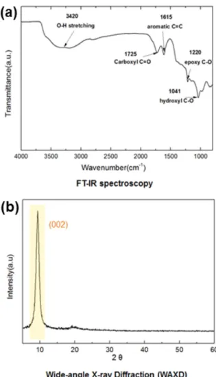
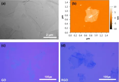
ABSTRACT: Experimental considerations have been performed to obtain the clear optical microscopic images of graphene oxide which are useful to probe its quality and morphological information such as a shape, a size, and a thickness. In this study, we investigated the contrast enhancement of the optical images of graphene oxide after hydrazine vapor reduction on a Si substrate coated with a 300 nm-thick SiO
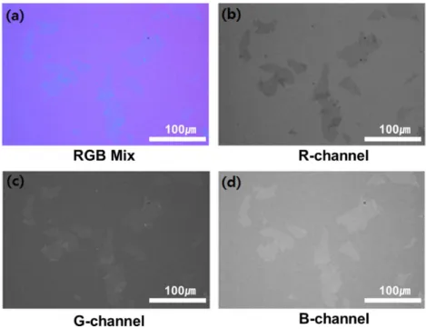
2dielectric layer. Also, a green-filtered light source gave higher contrast images comparing to optical images under standard white light. Furthermore, it was found that a image channel separation technique can be an alternative to simply identify the morphological information of graphene oxide, where red, green, and blue color values are separated at each pixels of the optical image. The approaches performed in this study can be helpful to set up a simple and easy protocol for the morphological identification of graphene oxide using a conventional optical microscope instead of a scanning electron microscopy or an atomic force microscopy.
초 록: