http://e-jbm.org/
45
Copyright © 2016 The Korean Society for Bone and Mineral Research
This is an Open Access article distributed under the terms of the Creative Commons Attribution Non-Commercial Li- cense (http://creativecommons.org/licenses/by-nc/3.0/) which permits unrestricted non-commercial use, distribu- tion, and reproduction in any medium, provided the original work is properly cited.
J Bone Metab 2016;23:45-48
http://dx.doi.org/10.11005/jbm.2016.23.1.45 pISSN 2287-6375 eISSN 2287-7029
Calcaneal Insufficiency Fracture after Ipsilateral Total Knee Arthroplasty
Min Jeong, Jin Woo Jin, Sung Jin Shin, Byoung Youl Kang
Department of Orthopaedic Surgery, Samsung Changwon Hospital, Sungkyunkwan University School of Medicine, Changwon, Korea
Insufficiency fracture of the calcaneus is a rare entity. In the absence of trauma, evaluat- ing a painful ankle in an elderly patient can be difficult and also it might be overlook the insufficiency fracture. We experienced a case of insufficiency calcaneus fracture that oc- curred after ipsilateral total knee arthroplasty. Here, we report our case with a review of literatures.
Key Words: Arthroplasty replacement knee, Calcaneus, Fractures stress
INTRODUCTION
Insufficiency fracture is occurred by repetitive normal stress without significant abnormal force in the area of elastic resistance deficiency.[1] It is known to occur mainly in elderly female patients with osteoporosis.[2] In particular, following hip or knee joint arthroplasty, it occurs very rarely.[3] Asymptomatic cases of insuffi- ciency fracture may be easily overlooked.[1] We describe one case of calcaneal in- sufficiency fracture following ipsilateral total knee arthroplasty with a review of literatures.
CASE
An 80-year-old woman underwent total knee joint arthroplasty due to the de- generative arthritis of the left knee joint. She did not have any history of trauma or coexisting medical illnesses. Her body weight was 45 kg, height was 153 cm and body mass index (BMI) was 19.34. She had been prescribed sodium alendro- nate and cholecalciferol composite agent (Fosamax plus D®; Merck & CO, Inc., West Port, PA, USA) for 1 year at local medical clinic. At a preoperative work-up, the pa- tient received dual energy X-ray absorptiometry (DXA); this showed that the T- score of the femoral neck and lumbar spine were -3.6 and -3.1, respectively. He- matological investigations were within normal limits. A 5 days after operation, the crutch walking was started. The patient was discharged on 1 week after operation without significant complication. On postoperative month 3, the patient visited the outpatient clinic of the clinical department due to the presence of pain in the left heel. The patient told us that there was no trauma. On examination, there were Corresponding author
Jin Woo Jin
Department of Orthopaedic Surgery, Samsung Changwon Hospital, 158 Paryong-ro, Masanhoewon-gu, Changwon 51353, Korea Tel: +82-55-290-1170
Fax: +82-55-290-6888 E-mail: [email protected] Received: January 25, 2016 Revised: February 5, 2016 Accepted: February 7, 2016
No potential conflict of interest relevant to this article was reported.
Case Report
Min Jeong, et al.
46
http://e-jbm.org/ http://dx.doi.org/10.11005/jbm.2016.23.1.45Fig. 1. Lateral and axial plain radiographs of the left calcaneus demonstrating radio-opaque irregular line (white arrow) on calcaneal body.
Fig. 2. Lateral plain radiographs of the left calcaneus showing clearer radio-opaque line, being parallel to posterior facet of the subtalar joint compared to previous radiograph.
edema and tenderness on the lateral and superior side of the left calcaneus. A compression test was also performed for the calcaneus; this also showed positive findings. There were no findings that are suggestive of burning sensation or erythema. On lateral and axial view of a plain radiogra- phy of the posterior region of the calcaneus, radiopaque lesions were shown as an irregular line that is parallel with the posterior joint of the subtalar joint (Fig. 1). The patient was informed of the possibility of insufficiency fracture and then prescribed with anti-inflammatory drugs. In ad- dition, the patient was advised to limit a weight bearing during a gait. One month later, the patient visited the out- patient clinic again, presenting with a persistent presence of edema and pain again. A compression test was also per- formed for the calcaneus; this also showed positive find- ings. Radiopaque lesions that had been seen on lateral view of the diaphysis of calcaneus on plain radiography were found to be a more clear line (Fig. 2). Therefore, the patient underwent magnetic resonance imaging (MRI) scanning.
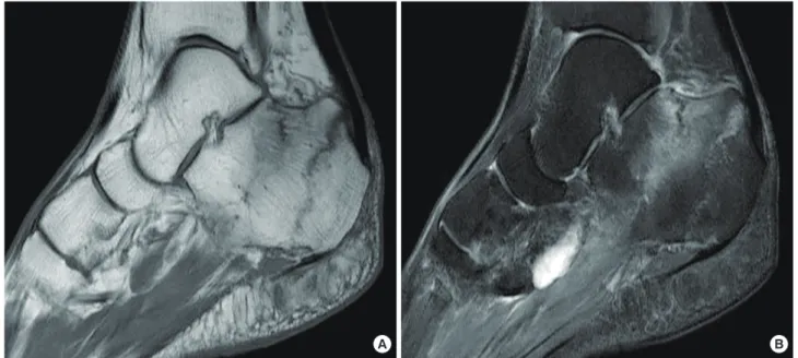
On T2-weighted MRI scans, two linear lesions in the diaph- ysis of the calcaneus, which had not been seen on radiog- raphy, and they were perpendicular to the bone trabecu- lae. Thus, low-intensity signals were found. In the adjacent areas, there were high-signal intensity lesions that are sug- gestive of bone edema (Fig. 3). The patient was diagnosed
with insufficiency fracture of calcaneus. To limit a weight- bearing effect, the patient underwent ankle-foot cast for approximately three months. Following the manifestation of symptoms, the patient achieved an improvement in them at a 2-month final follow-up. On plain radiography, the pa- tient had a loss of radiopaque lesions. Moreover, the pa- tient could also perform a normal gait.
Calcaneal Insufficiency Fracture after Ipsilateral TKA
http://dx.doi.org/10.11005/jbm.2016.23.1.45 http://e-jbm.org/
47
DISCUSSION
Stress fracture is a term that is referred to as a fatigue one or an insufficiency one. It is a microfracture that occurs as a result of the abnormal stress to the normal bone, but an insufficiency one does when the normal loading of the dai- ly living activity to the deficient bony elastic resistance.[1,4]
Insufficiency fracture is known to occur in mainly elderly female patients in association with osteoporosis, rheuma- toid arthritis, hyperparathyroidism, diabetes mellitus, ra- diotherapy or steroid use.[5] It mainly affects pelvic ring, sacrum, tibia and the neck or head of the femur. Although rare, it may also occur in the talus, fibula and calcaneus.[3]
There are also reports that insufficiency fracture occurred after total knee arthroplasty. Most cases in this series occur around the knee joint. Although extremely rare, however, it may also occur in the femoral neck or calcaneus.[6,7]
Still, little is known about the pathophysiologic mecha- nisms underlying the occurrence of insufficiency fracture in the ipsilateral calcaneus after total knee arthroplasty.
Miki et al.[6] reported that insufficiency fracture would not have a relationship with disuse osteoporosis that may oc- cur following total knee arthroplasty. Cakmak et al.[7] not- ed that insufficiency fracture of the calcaneus after ipsilat- eral total knee arthroplasty would occur due to an increas- ed weight bearing after postoperative improvement of pain
Fig. 3. Sagittal T1-weighted image demonstrating two parallel lines with a low signal intensity (A), and T2-weighted image showing high signal intensity in the corresponding lesions (B).
A B
in lower limbs that could not tolerate it prior to surgery.
The current case is an 80-year-old woman undergoing to- tal knee arthroplasty who was diagnosed with insufficien- cy fracture. There were no risk factors other than a diagno- sis of osteoporosis at a pre-operative work-up. Following knee joint arthroplasty, due to the decreased pain, the ac- tivity and the weight bearing were increased as compared with preoperatively. Therefore, it occurred in our patient.
In cases of stress fracture occurring in the calcaneus, sa- crum or pubic ramus, few complications occur. Therefore, it is classified into a low-risk fracture. In cases of stress frac- ture occurring in the scaphoid bone, talus or femur, how- ever, delayed union or malunion may occur at a higher in- cidence. Therefore, such cases are classified into a high-risk fracture.[8] Patients with insufficiency fracture of the calca- neus may present with mild symptoms, which may be mis- diagnosed as bursitis, plantar nerve entrapment syndrome, peroneal tendonitis or plantar fasciitis. This may frequently delay an accurate diagnosis of it.[4,8] If there is a delay in the diagnosis of it, this would limit a gait due to the defor- mity of the calcaneus arising from an intraarticular inva- sion or a malunion of the fracture although it belongs to a low-risk fracture.[4] Therefore, following total knee arthro- plasty, insufficiency fracture of the calcaneus occurs very rarely. If patients undergoing total knee arthroplasty com- plains of pain around the ipsilateral calcaneus, surgeons
Min Jeong, et al.
48
http://e-jbm.org/ http://dx.doi.org/10.11005/jbm.2016.23.1.45should consider the possibility of insufficiency fracture of the calcaneus and then make a differential diagnosis from other diseases. In particular, because elderly patients with osteoporosis are at increased of developing it, clinicians should consider the possibility of it.
REFERENCES
1. Ito K, Hori K, Terashima Y, et al. Insufficiency fracture of the body of the calcaneus in elderly patients with osteoporo- sis: a report of two cases. Clin Orthop Relat Res 2004:190-4.
2. Alonso-Bartolome P, Blanco R, Canga A, et al. Insufficiency fractures of the calcaneus: a diagnostic pitfall for ankle ar- thritis. J Rheumatol 2006;33:1140-2.
3. Soubrier M, Dubost JJ, Boisgard S, et al. Insufficiency frac- ture. A survey of 60 cases and review of the literature. Joint
Bone Spine 2003;70:209-18.
4. Lui TH. Insufficiency fracture of the body of the calcaneus.
Foot (Edinb) 2013;23:93-5.
5. Tsiridis E, Upadhyay N, Giannoudis PV. Sacral insufficiency fractures: current concepts of management. Osteoporos Int 2006;17:1716-25.
6. Miki T, Miki T, Nishiyama A. Calcaneal stress fracture: an adverse event following total hip and total knee arthro- plasty: a report of five cases. J Bone Joint Surg Am 2014;
96:e9.
7. Cakmak S, Mahiroğullari M, Kürklü M, et al. Bilateral femo- ral neck stress fracture following bilateral total knee arthro- plasty: a case report. Acta Orthop Traumatol Turc 2012;46:
312-5.
8. Gehrmann RM, Renard RL. Current concepts review: stress fractures of the foot. Foot Ankle Int 2006;27:750-7.