Case Report
J Gynecol Oncol Vol. 21, No. 4:276-278, December 2010 DOI:10.3802/jgo.2010.21.4.276
276
Combined open surgical and endovascular management of ruptured femoral artery from recurrent vulvar cancer
Vasileios Trompetas1, Andrew JP Sandison1, Hugh J Anderson2
Departments of 1Surgery and 2Radiology, Eastbourne District General Hospital, Eastbourne, UK
We report on the case of a 50-year-old woman with exsanguinating haemorrhage from the common femoral artery as a complication of recurrent vulvar cancer in the groin which was managed successfully with combined open surgical and endovascular intervention. She survived another three months and died from progressive disease without further episodes of bleeding. This complication is rare, presents dramatically, and is usually a terminal event. For those cases where intervention is considered appropriate, the option of combined open surgical and endovascular repair should be kept in mind.
Key Words: Vulvar cancer, Femoral artery rupture, Endovascular repair
Received January 18, 2010, Accepted March 24, 2010 Correspondence to Vasileios Trompetas
Department of Surgery, Eastbourne District General Hospital, Kings Drive, Eastbourne, BN21 2UD, UK
E-mail: [email protected]
INTRODUCTION
Haemorrhage from the femoral vessels as a complication of recurrent vulvar cancer is rarely described.1-3 We report the first such case where the severe bleeding was managed suc- cessfully with immediate surgery followed by endovascular stent graft placement.
CASE REPORT
A 50-year-old lady presented to the emergency department at 23.00 on a Saturday night with exsanguinating haemor- rhage from a defect in the right groin secondary to tumour in- vasion and necrosis. She had a previous history of a T1b N1 moderately differentiated squamous cell carcinoma of the vulva. She had undergone radical vulvectomy and bilateral in- guinal lymphadenectomy with 3/17 positive lymph nodes in the right groin and extracapsular invasion 10 months earlier.
This was followed by adjuvant radiotherapy to the right groin and hemipelvis, 45 Gy in 25 fractions. She was diagnosed with right groin recurrence, four months prior to the urgent admis- sion, and treated with palliative chemotherapy, 3 cycles of Carboplatin and Gemcitabine. An MRI scan a few days before the emergency admission had showed progressive disease but this investigation had been done elsewhere and the result was
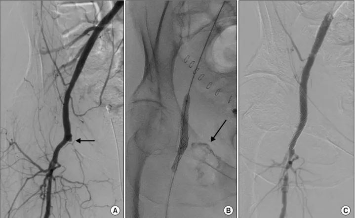
not available to the admitting team, nor were the patient or her partner aware of the result. Upon presentation to the emergency department she was unstable with blood pressure of 100/80 mmHg and heart rate of 145/minute. She had pro- fuse arterial bleeding from the right groin which was partly controlled by direct pressure. There was no interventional ra- diology service immediately available at that time. She was taken to theatre where control of the external iliac artery was achieved via an intraperitoneal approach. Exploration of the right groin showed a large cavity with necrotic tissue. The femoral vein was absent, completely destroyed by the re- current cancer. There was bleeding from the medial aspect of the common femoral artery secondary to destruction by the tumour. The bleeding area was controlled with 4-0 prolene sutures. The cavity was packed and dressed. The patient was taken to intensive care unit for further resuscitation, but a de- cision was made not to reintervene surgically if there was fur- ther haemorrhage. She was transferred to a ward the next day after she became stable. As the femoral artery was still exposed and the tissues were very frail, it was felt that the surgical re- pair was not adequate and the likelihood of rebleeding high. A right femoral arteriogram with left femoral access was there- fore carried out. It showed occlusion of the superficial femoral artery at its origin (Fig. 1A). There was irregularity of the dis- tal common femoral artery, but no extravasation of contrast was demonstrated. Two 8 mm by 20 mm covered stents (WallgraftTM Endoprosthesis, Boston Scientific, Natick, MA, USA) were deployed across the bifurcation of the common femoral artery into the profunda femoris artery. On balloon expansion of the second stent there was seen to be some ex- travasation of contrast (Fig. 1B) but a completion angiogram
Femoral artery rupture from recurrent vulvar cancer
277
Fig. 1. (A) Occlusion of the superficial femoral artery at its origin (arrow). (B) Two 8 mm by 20 mm covered stents (WallgraftTM Endoprosthesis) deployed across the bifurcation of the common femoral artery into the profunda femoris artery, extravasation of contrast (arrow). (C) Completion angiogram, stented segment is widely patent with no extravasation of contrast.
showed the stented segment to be widely patent with no ex- travasation of contrast (Fig. 1C). After five days of overall stay in hospital she was discharged to a hospice. The oncological decision was for palliative management. The patient died from progressive disease without further bleeding three months later.
DISCUSSION
Recurrent vulvar cancer in the groin particularly after pre- vious lymphadenectomy and radiotherapy is very difficult to manage. It often involves the femoral vessels and it is consid- ered inoperable. The usual treatment is chemoradiotherapy, however surgery in the form of radical resection with vascular grafting and plastic reconstruction or hip disarticulation has been described.4,5 Femoral artery rupture is a rare complication and is usually the result of tumour invasion, radiotherapy and tissue necrosis. Rupture of the femoral artery results in ex- sanguinating haemorrhage and is usually a terminal event.
There are two reports in the literature where the rupture was managed with immediate surgery. One patient had a success- ful graft bypass from the external iliac artery to the superficial femoral artery by tunnelling the graft laterally to the open wound.1 No follow-up was documented. The other patient had an initial repair of the bleeding superficial femoral artery with
4-0 prolene and subsequently, as the leg became ischaemic, a graft bypass from the external iliac artery to the distal super- ficial femoral artery.2 The graft was tunnelled through the ob- turator foramen. Her leg was well at five months follow-up. In another report a patient with common femoral artery erosion and severe haemorrhage was successfully managed with per- cutaneous endovascular stent-graft placement and sub- sequent graft bypass a month later. She only survived for three months.3 Endovascular stent graft placement has also been de- scribed in two patients with external iliac artery erosion and haemorrhage from a recurrent uterine carcinoma.6,7
In our case the patient presented with exsanguinating hae- morrhage at night when the interventional radiology service was not immediately available. In hindsight, she would not have survived the delay of organising an angiogram even if the service had been available. At the time of surgery it was felt that a graft bypass was not suitable for her as the entire groin was very indurated and oedematous from the local recurrence and the previous radiotherapy, and there was also a large de- fect with necrotic tissue. The bleeding vessel was therefore re- paired locally during open surgery and endovascular place- ment of a covered stent graft was subsequently carried out the following day. This was successful and she went on to live an- other three months without further bleeding. In conclusion, the management of these patients should be individualised
J Gynecol Oncol Vol. 21, No. 4:276-278, 2010 Vasileios Trompetas, et al.
278 and when intervention is considered appropriate, the option of combined open surgery and endovascular repair should be kept in mind.
CONFLICT OF INTEREST
The authors declare there are no conflicts of interest relevant to this article.
REFERENCES
1. Pararajasingam R, Todd J, Oshodi TO. Delayed common femoral artery rupture following irradiation therapy for carcinoma of the vulva. Eur J Vasc Endovasc Surg 2001; 22: 573-4.
2. Deppe G, Malviya VK, Smith PE, Zbella EA, Pildes R. Limb salvage in recurrent vulvar carcinoma after rupture of femoral artery.
Gynecol Oncol 1984; 19: 120-4.
3. Kim S, Anderson L, Silberzweig JE. Endovascular graft placement for femoral artery erosion caused by recurrent vulvar carcinoma.
Gynecol Oncol 2008; 111: 572-4.
4. Chao A, Lai CH, Chen HC, Hsieh HC, Yeow KM. Limb preserva- tion by Gore-Tex vascular graft for groin recurrence after post- operative adjuvant radiation in vulvar cancer. Gynecol Oncol 2001;
82: 559-62.
5. Powell JL, Donovan JT, Reed WP. Hip disarticulation for recurrent vulvar cancer in the groin. Gynecol Oncol 1992; 47: 110-3.
6. Abulafia O, Sclafani SJ, Holcomb K, Gates EJ, Sherer DM. Percuta- neous transluminal endovascular graft placement for massive hemorrhage caused by recurrent cervical carcinoma-associated erosion of the external iliac artery. Am J Obstet Gynecol 1998; 178:
618-20.
7. Christiansen S, Eiberg JP, Hansen MA. Severe vaginal bleeding treated with a stent graft. Eur J Vasc Endovasc Surg 2002; 23:
367-9.