INTRODUCTION
Core needle biopsy (CNB) is widely used as a standard pro- cedure for diagnosis of breast cancer [1,2]. However, immuno- histochemistry (IHC) assessment in CNB samples may be less reliable than in surgical specimens’ due to the relatively smaller sample size and tumor heterogeneity [3,4]. Several studies
have reported the concordance between preoperative CNB and surgical specimens for estrogen receptor (ER), and human epidermal growth factor receptor 2 (HER2) determination [5-7]. A recent meta-analysis has shown that the CNB tissue could replace open excision biopsy for determining ER, proges- terone receptor (PR), and HER2 status [8]. The 2015 European Society of Medical Oncology breast cancer clinical practice guideline recommends a preoperative pathological examination of the CNB, with a report on ER, PR, and HER2 status by IHC or fluorescence in situ hybridization [9].
Neoadjuvant chemotherapy (NAC) before definitive sur- gery can reduce the size and extent of locally advanced tu- mors. There is an increasing acceptance of view that a pathol- ogical complete response (pCR) following chemotherapy is important, particularly as a surrogate for prognosis [10]. The information obtained from CNB may be the only information available for determining the candidates for preoperative or neoadjuvant treatment [3]. Therefore, demand has been
Comparison of Core Needle Biopsy and Surgical Specimens in Determining Intrinsic Biological Subtypes of Breast Cancer with Immunohistochemistry
Kiho You*, Sungmin Park1,*, Jai Min Ryu, Isaac Kim, Se Kyung Lee, Jonghan Yu, Seok Won Kim, Seok Jin Nam, Jeong Eon Lee
Division of Breast Surgery, Department of Surgery, Samsung Medical Center, Sungkyunkwan University School of Medicine, Seoul; 1Department of Surgery, Chungbuk National University Hospital, Chungbuk National University College of Medicine, Cheongju, Korea
ORIGINAL ARTICLE
Purpose: We evaluated the concordance between core needle biopsy (CNB) and surgical specimens on examining intrinsic biol- ogical subtypes and receptor status, and determined the accu- racy of CNB as a basic diagnostic method. Methods: We ana- lyzed breast cancer patients with paired CNB and surgical speci- men samples during 2014. We used monoclonal antibodies for nuclear staining, and estrogen receptor (ER) and progesterone receptor (PR) status evaluation. A positive test was defined as staining greater than or equal to 1% of tumor cells. Human epi- dermal growth factor receptor 2 (HER2) was graded by immuno- histochemistry and scored as 0 to 3+ according to the recom- mendations of the American Society of Clinical Oncology/Col- lege of American Pathologists. Ki-67 immunostaining was per- formed using the monoclonal antibody Ki-67, and the results were divided at 10% intervals. The cutoff value for high Ki-67 was defined as 20%. Concordance analysis of ER, PR, HER2, Ki-67, and five intrinsic biological subtypes was performed on
CNB and surgical specimens. Statistical analysis for concor- dance was calculated using κ-tests. Results: We found very good agreement for ER and PR with a concordance of 96.7%
for ER (κ=0.903), and 94.3% for PR (κ=0.870). HER2 and Ki-67 showed concordance rates of 84.8% (κ=0.684) and 83.5%
(κ=0.647), respectively, which were interpreted as good agree- ment. Five subgroups analysis showed 85.8% agreement and κ-value of 0.786, also indicating good agreement. Conclusion:
CNB showed high diagnostic accuracy compared with surgical specimens, and good agreement for ER, PR, HER2, and Ki-67.
Our findings reaffirmed the recommendation of CNB as an initial procedure for breast cancer diagnosis, and the assessment of receptor status and intrinsic biological subtypes to determine further treatment plans.
Key Words: Breast neoplasms, Core needle biopsy, Estrogen receptors, Human epidermal growth factor receptor 2, Immunohistochemistry
Correspondence to: Jeong Eon Lee
Division of Breast Surgery, Department of Surgery, Samsung Medical Center, Sungkyunkwan University School of Medicine, 81 Irwon-ro, Gangnam-gu, Seoul 06351, Korea
Tel: +82-2-3410-3479, Fax: +82-2-3410-6982 E-mail: [email protected]
*These authors contributed equally to this work.
This research was supported by Basic Science Research Program through the National Research Foundation of Korea (NRF), funded by the Ministry of Education (2015R1D1A1A01057585) and by the NRF grant funded by the Korean Government (MSIP) (2016R1A5A2945889).
Received: April 11, 2017 Accepted: July 20, 2017
Cancer
markedly increased for clinicians to provide prognostic infor- mation considering the determination of IHC for treatment planning. However, there are few studies that have reported concordance rates between CNB and surgical specimens, be- fore and after NAC.
In this study, we evaluated the concordance between CNB and surgical specimens in evaluating intrinsic biological sub- types and the receptor status, and examined the accuracy of CNB as a basic diagnostic method. Second, we assessed changes in intrinsic biological subtypes of breast cancer before and after NAC comparing CNB and surgical specimens.
METHODS
Data collection
We analyzed breast cancer patients with paired CNB and surgical specimen samples during 2014 at Samsung Medical Center, Seoul, Korea. Seventeen hundred eighty-six patients underwent primary surgery or NAC prior to operation. Clinical information on patients collected from medical records included age, body mass index, operation type, NAC history, and main pathological findings that included tumor size, number, nuclear grade, TNM stage, and ER, PR, HER2, and Ki-67 status from both CNB and surgical specimens. The study was approved by the Institutional Review Board of Samsung Medical Center (approval number: 2017-01-102), Seoul, Korea.
ER, PR, HER2, and Ki-67 evaluation
We used monoclonal antibodies for nuclear staining and ER (anti-ER; clone 6F11, Novocastra, Newcastle, UK) and PR (anti-PR; clone 16, Novocastra) status evaluation. A positive test was defined as staining greater than or equal to 1% of tumor cells. A negative test was defined as staining of less than 1% of tumor cells. We used the Allred score interpreta- tion system of intensity score (0–3) and proportion score (0–
5) [6].
HER2 (anti-HER2; 4B5, BenchMark XT, Ventana, Tucson, USA) was first graded by IHC and scored as 0 to 3+ according to the recommendations of the American Society of Clinical Oncology/College of American Pathologists [11]. The scoring system defines negative as 0/1+. No observed staining or faint/barely perceptible membrane staining in <10% of tumor cells is 0. Incomplete membrane staining or faint/barely per- ceptible membrane staining in ≥10% of tumor cells is 1+. A weak to moderate complete membrane staining observed in
>10% of tumor cells is 2+ and is interpreted as equivocal. A strong complete membrane staining observed in >10% of tu- mor cells is 3+ and is considered as positive. In cases of HER2
2+ surgical specimens, we conducted silver in situ hybridiza- tion (SISH) assays (INFORM DDISHTM HER2 DNA SISH probe kits; BenchMark XT) to determine HER2 amplification [11,12].
Ki-67 immunostaining was performed using the monoclo- nal antibody Ki-67 (clone MIB-1; Dako, Glostrup, Denmark).
Ki-67 is a nuclear marker expressed in all phases of the cell cycle other than the G0 phase [13,14]. Ki-67 expression has a value between 0% and 100% and is reported at 10% intervals in our center. In this study, we classified samples as low or high expression using 20% as cutoff value.
Five intrinsic biological subtype classifications were catego- rized according to the 12th St. Gallen international breast cancer conference (2011): luminal A (ER and/or PR positive, HER2 negative, and Ki-67 low); luminal B/HER2 negative (ER and/or PR positive, HER2 negative, and Ki-67 high); luminal B/HER2 positive (ER and/or PR positive, any Ki-67 and HER2 positive); HER2 positive (ER and PR absent, and HER2 positive); and triple negative (ER and PR absent, and HER2 negative) [15].
Statistical analysis
Concordance analysis of ER, PR, HER2, Ki-67, and five in- trinsic biological subtypes was performed on CNB and surgi- cal specimens. Statistical analysis for concordance was calcu- lated using κ-tests. κ-values >0.8 indicated very good agree- ment, between 0.6 and 0.8 indicated good agreement, between 0.4 and 0.6 were considered as moderate agreement, <0.4 as fair, and <0.2 as poor agreement. All statistical tests were two- sided and considered significant if p-value was below 0.05. We used SPSS version 22.0 (IBM Corp., Armonk, USA).
RESULTS
Patient characteristics
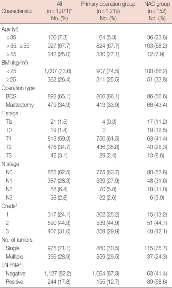
We investigated 1,786 breast cancer patients who under- went surgery during 1 year. There were 1,369 eligible patients with a median age of 49.5 years (range, 24–86 years). We ex- cluded 180 patients who underwent vacuum-assisted biopsy, excisional biopsy, or had previous breast surgery. Moreover, 232 patients were excluded for inadequate test results; for ex- ample, CNB sent from other hospitals after diagnosis and their specimens were inadequate to conduct IHC test. Five more patients were excluded because they received palliative chemotherapy. After diagnosis by CNB, 1,219 patients under- went primary surgery and 152 received NAC before surgery (Figure 1); there were two patients with bilateral breast cancer in each group. Patient characteristics and pathology results are shown in Table 1. Eight hundred ninety-two patients (65.1%)
underwent breast-conserving surgery and 479 (34.9%) under- went mastectomy. The rate of mastectomy was higher in the NAC group than in the primary surgery group.
Tumor pathology and IHC results
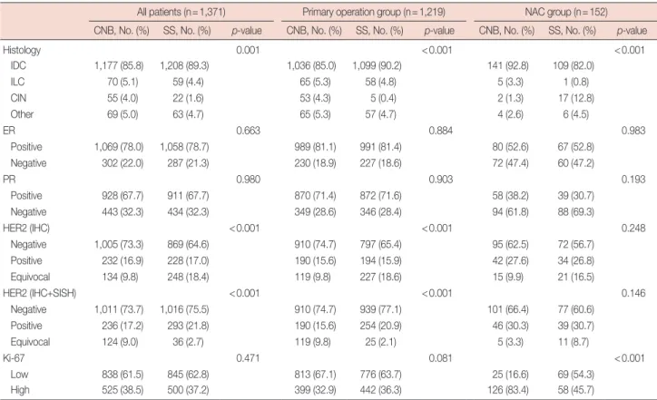
About 86% of patients were diagnosed with invasive ductal carcinoma using CNB. Expression of ER, PR, and Ki-67 was not significantly different between CNB and surgical speci- mens, except for HER2 in the primary surgery group. Patients receiving NAC showed a difference in the Ki-67 status. In contrast, ER, PR, and HER2 did not show significant differ- ences (Table 2).
Concordance of IHC results
ER and PR expression in CNB samples showed very good agreement with results for surgical samples; overall concor- dance rate was 96.7% for ER (κ=0.903) and 94.3% for PR (κ=0.870). HER2 and Ki-67 expression showed good agree- ment between CNB and surgical specimens; overall concor- dance rate was 84.8% for HER2 (κ=0.684) and 83.5% for Ki- 67 (κ=0.647).
We divided patients into primary surgery and NAC groups.
Concordance rates and κ-values for ER, PR, and HER2 showed similar tendency between the two groups. Meanwhile, in cases of primary surgery, Ki-67 expression also showed good agreement between CNB and surgical results with a con- cordance of 87.0% and κ-value of 0.712. The NAC group had poor agreement between CNB and surgical results with a con- cordance of 50.0% and κ-value of 0.056 (p=0.397) (Table 3).
Concordance among intrinsic biological subtypes
We classified surgical specimens and CNB into five intrinsic biological subtypes according to the 12th St. Gallen interna- tional breast cancer conference: luminal A, luminal B (HER2 negative and positive), HER2 positive, and triple negative.
Table 1. Patient characteristics and clinicopathological results Characteristic All
(n=1,371)*
No. (%)
Primary operation group (n=1,219)
No. (%)
NAC group (n=152)
No. (%) Age (yr)
≤35 100 (7.3) 64 (5.3) 36 (23.8)
>35, ≤55 927 (67.7) 824 (67.7) 103 (68.2)
>55 342 (25.0) 330 (27.1) 12 (7.9)
BMI (kg/m2)
<25 1,007 (73.6) 907 (74.5) 100 (66.2)
≥25 362 (26.4) 311 (25.5) 51 (33.8)
Operation type
BCS 892 (65.1) 806 (66.1) 86 (56.6)
Mastectomy 479 (34.9) 413 (33.9) 66 (43.4) T stage
Tis 21 (1.5) 4 (0.3) 17 (11.2)
T0 19 (1.4) 0 19 (12.5)
T1 813 (59.3) 750 (61.5) 63 (41.4)
T2 476 (34.7) 436 (35.8) 40 (26.3)
T3 42 (3.1) 29 (2.4) 13 (8.6)
N stage
N0 855 (62.5) 775 (63.7) 80 (52.6)
N1 387 (28.3) 339 (27.9) 48 (31.6)
N2 88 (6.4) 70 (5.8) 18 (11.8)
N3 38 (2.8) 32 (2.6) 6 (3.9)
Grade†
1 317 (24.1) 302 (25.2) 15 (13.2)
2 590 (44.9) 539 (44.9) 51 (44.7)
3 407 (31.0) 359 (29.9) 48 (42.1)
No. of tumors
Single 975 (71.1) 860 (70.5) 115 (75.7)
Multiple 396 (28.9) 359 (29.5) 37 (24.3) LN FNA‡
Negative 1,127 (82.2) 1,064 (87.3) 63 (41.4) Positive 244 (17.8) 155 (12.7) 89 (58.6) NAC=neoadjuvant chemotherapy; BMI=body mass index; BCS=breast- conserving surgery; LN FNA=lymph node fine needle aspiration.
*Some mismatches were caused by two patients who underwent surgery on both breasts. See Figure 1; †Grade and number of tumors evaluated by surgi- cal specimen; ‡Preoperative LN metastasis was determined by FNA.
1,786 Patients underwent operation for breast cancer in 2014
417 Excluded
180 Another procedure for diagnosis*
232 No IHC result from CNB 5 Palliative chemotherapy 1,369 Patients eligible for study†
1,219 Primary operation group‡ 152 NAC group‡
Figure 1. Flow chart of patients selection for analysis.
CNB=core needle biopsy; IHC=immunohistochemistry; NAC=neoadjuvant chemotherapy. *Underwent vacuum-assisted biopsy, excisional biopsy or had previous breast surgery; †Two patients diagnosed both breast cancer; ‡Each 1 patients who had both breast cancer underwent operation and NAC prior to surgery.
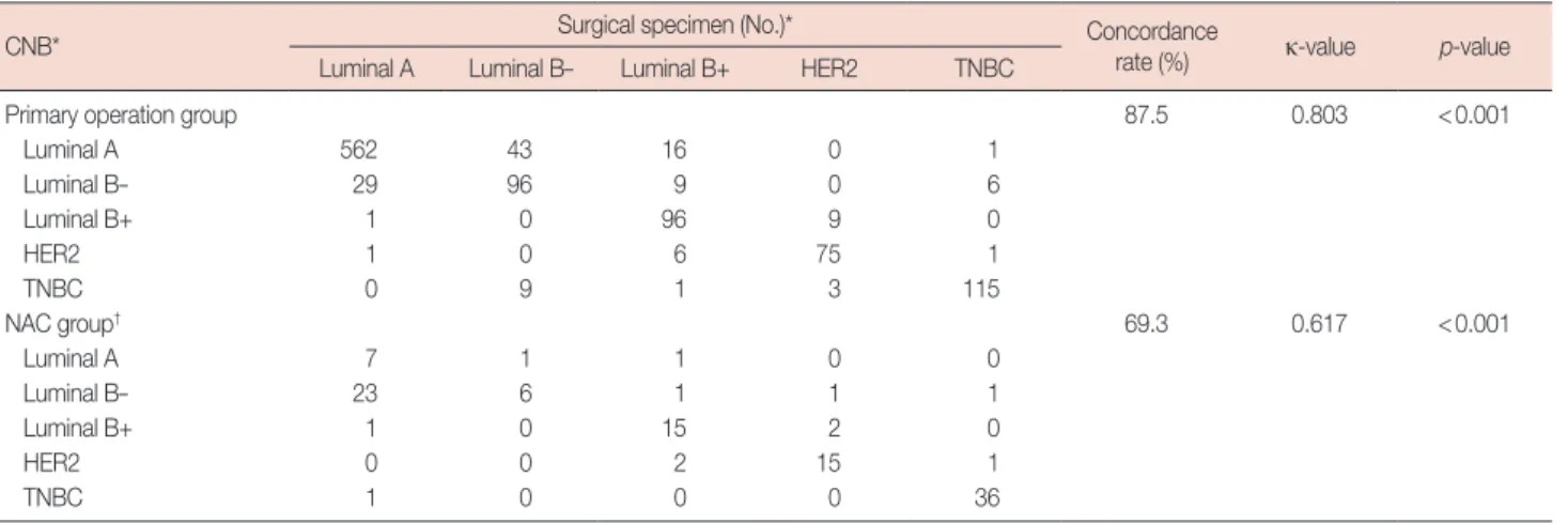
Concordance among the five subtypes was 85.8% and the
κ-value was 0.786 (p<0.001). Concordance rates of the sub- types were 87.5% for the primary surgery group and 69.3%
for the NAC group, and the κ-values between CNB and surgi- Table 2. Tumor pathology and IHC test results between the primary operation group and NAC group
All patients (n=1,371) Primary operation group (n=1,219) NAC group (n=152) CNB, No. (%) SS, No. (%) p-value CNB, No. (%) SS, No. (%) p-value CNB, No. (%) SS, No. (%) p-value
Histology 0.001 <0.001 <0.001
IDC 1,177 (85.8) 1,208 (89.3) 1,036 (85.0) 1,099 (90.2) 141 (92.8) 109 (82.0)
ILC 70 (5.1) 59 (4.4) 65 (5.3) 58 (4.8) 5 (3.3) 1 (0.8)
CIN 55 (4.0) 22 (1.6) 53 (4.3) 5 (0.4) 2 (1.3) 17 (12.8)
Other 69 (5.0) 63 (4.7) 65 (5.3) 57 (4.7) 4 (2.6) 6 (4.5)
ER 0.663 0.884 0.983
Positive 1,069 (78.0) 1,058 (78.7) 989 (81.1) 991 (81.4) 80 (52.6) 67 (52.8)
Negative 302 (22.0) 287 (21.3) 230 (18.9) 227 (18.6) 72 (47.4) 60 (47.2)
PR 0.980 0.903 0.193
Positive 928 (67.7) 911 (67.7) 870 (71.4) 872 (71.6) 58 (38.2) 39 (30.7)
Negative 443 (32.3) 434 (32.3) 349 (28.6) 346 (28.4) 94 (61.8) 88 (69.3)
HER2 (IHC) <0.001 <0.001 0.248
Negative 1,005 (73.3) 869 (64.6) 910 (74.7) 797 (65.4) 95 (62.5) 72 (56.7)
Positive 232 (16.9) 228 (17.0) 190 (15.6) 194 (15.9) 42 (27.6) 34 (26.8)
Equivocal 134 (9.8) 248 (18.4) 119 (9.8) 227 (18.6) 15 (9.9) 21 (16.5)
HER2 (IHC+SISH) <0.001 <0.001 0.146
Negative 1,011 (73.7) 1,016 (75.5) 910 (74.7) 939 (77.1) 101 (66.4) 77 (60.6)
Positive 236 (17.2) 293 (21.8) 190 (15.6) 254 (20.9) 46 (30.3) 39 (30.7)
Equivocal 124 (9.0) 36 (2.7) 119 (9.8) 25 (2.1) 5 (3.3) 11 (8.7)
Ki-67 0.471 0.081 <0.001
Low 838 (61.5) 845 (62.8) 813 (67.1) 776 (63.7) 25 (16.6) 69 (54.3)
High 525 (38.5) 500 (37.2) 399 (32.9) 442 (36.3) 126 (83.4) 58 (45.7)
IHC =immunohistochemistry; NAC =neoadjuvant chemotherapy; CNB =core needle biopsy; SS =surgical specimens; IDC =invasive ductal carcinoma;
ILC=invasive lobular carcinoma; CIN=carcinoma in situ; ER=estrogen receptor; PR=progesterone receptor; HER2=human epidermal growth factor receptor 2;
SISH=silver in situ hybridization.
Table 3. Agreement analysis of ER, PR, HER2, and Ki-67 between CNB and surgical specimens
CNB
All patients (n=1,371) Primary operation group (n=1,219) NAC group (n=152) No. of surgical
specimens Concordance
rate (%) κ-value p-value
No. of surgical
specimens Concordance
rate (%) κ-value p-value
No. of surgical
specimens Concordance
rate (%) κ-value p-value
Pos Neg Equ Pos Neg Equ Pos Neg Equ
ER 96.7 0.903 <0.001 97.1 0.906 <0.001 92.9 0.858 <0.001
Pos 1,035 21 - 972 16 - 63 5 -
Neg 23 266 - 19 211 - 4 55 -
PR 94.3 0.870 <0.001 95.0 0.877 <0.001 88.2 0.746 <0.001
Pos 878 43 - 840 29 - 38 14 -
Neg 33 391 - 32 317 - 1 74 -
HER2 (IHC) 84.8 0.684 <0.001 84.6 0.672 <0.001 86.6 0.762 <0.001
Pos 215 3 8 182 2 6 33 1 2
Neg 7 833 147 6 766 138 1 67 9
Equ 6 33 93 6 29 83 0 4 10
Ki-67 Low High 83.5 0.647 <0.001 Low High 87.0 0.712 <0.001 Low High 50.0 0.056 0.397
Low 727 109 712 100 15 9
High 112 389 58 341 54 48
ER=estrogen receptor; PR=progesterone receptor; HER2=human epidermal growth factor receptor 2; CNB=core needle biopsy; Pos=positive; Neg=negative;
Equ=equivocal; IHC=immunohistochemistry.
cal specimen results were 0.803 and 0.617, respectively (p<
0.001) (Table 4).
pCR rate according to the intrinsic biological subtypes in NAC patients
We included both no residual disease of any sort and resid- ual ductal carcinoma in situ (DCIS) without invasive disease and node metastasis in to define pCR. Among the 147 pa- tients in the NAC group, the total pCR rate was 22.4% (33/
147); the pCR rate of each intrinsic biological subtypes is shown in Table 5. Among the 33 patients who achieved pCR, 17 patients had no residual tumor and 16 patients had DCIS.
No residual tumor patients were excluded from the compari- son of the IHC results between CNB and surgical specimens Table 5. pCR rate according to the intrinsic biological subtypes from CNB in NAC patients
Subtype* No. of pCR patients NAC group† pCR rate (%)
Luminal A 0 10 0
Luminal B– 6 42 14.3
Luminal B+ 8 24 33.3
HER2 6 22 27.3
TNBC 13 49 26.5
Total 33 147 22.4
pCR =pathologic complete response; CNB =core needle biopsy; NAC = neoadjuvant chemotherapy; HER2=human epidermal growth factor receptor 2; TNBC=triple-negative breast cancer.
*Classified from CNB result. luminal A (ER+ and/or PR+, HER2-, Ki-67 low);
luminal B- (ER+ and/or PR+, HER2-, Ki-67 high); luminal B+ (ER+ and/or PR+, any Ki-67, HER2+); HER2 (ER-, PR-, HER2+); and TNBC (ER-, PR-, HER2-); †Five patients were equivocal HER2 result and cannot classified sub- type.
Table 6. Concordance rate between CNB and surgical specimens in previous studies
Author (year) No. Concordance (%)
ER PR HER2 Ki-67 NAC
Mann et al. (2005) [16] 100 86.0 83.0 80.0 NA No Arens et al. (2005) [17] 25 80.0 80.0 80.0 76.0 Yes Quddus et al. (2005) [18] 39 NA NA 61.5 NA Yes Burge et al. (2006) [19] 87 95.0 89.0 96.0 NA No Arnedos et al. (2009) [3] 336 98.2 84.5 98.8 NA No Tamaki et al. (2010) [6] 353 92.9 77.9 89.3 NA No Lorgis et al. (2011) [4] 175 84.0 78.3 98.3 NA No Ough et al. (2011) [7] 209 88.0 78.0 81.0 59.0 No Chen et al. (2013) [8] 298 93.6 85.9 96.3 79.5 No Dekker et al. (2013) [21] 122 99.1 NA 96.4 NA No Seferina et al. (2013) [22] 526 89.5 82.5 80.6 NA No CNB=core needle biopsy; ER=estrogen receptor; PR=progesterone recep- tor; HER2=human epidermal growth factor receptor 2; NAC=neoadjuvant chemotherapy; NA=not available.
Table 4. Agreement analysis between CNB and surgical specimens for intrinsic biological subtypes
CNB* Surgical specimen (No.)* Concordance
rate (%) κ-value p-value
Luminal A Luminal B– Luminal B+ HER2 TNBC
Primary operation group 87.5 0.803 <0.001
Luminal A 562 43 16 0 1
Luminal B– 29 96 9 0 6
Luminal B+ 1 0 96 9 0
HER2 1 0 6 75 1
TNBC 0 9 1 3 115
NAC group† 69.3 0.617 <0.001
Luminal A 7 1 1 0 0
Luminal B– 23 6 1 1 1
Luminal B+ 1 0 15 2 0
HER2 0 0 2 15 1
TNBC 1 0 0 0 36
CNB=core needle biopsy; NAC=neoadjuvant chemotherapy; HER2=human epidermal growth factor receptor 2; TNBC=triple-negative breast cancer.
*Luminal A (ER+ and/or PR+, HER2-, Ki-67 low); luminal B- (ER+ and/or PR+, HER2-, Ki-67 high); luminal B+ (ER+ and/or PR+, any Ki-67, HER2+); HER2 (ER-, PR-, HER2+); and TNBC (ER-, PR-, HER2-); †Total 114 patients after excluding 38 from 152 NAC group. Thirty-eight patients were group who were HER2 2+ and did not have silver in situ hybridization results, or pathologic complete response group who did not have immunohistochemistry results after NAC. Overall concor- dance rate 85.8%, κ-value of 0.786 and p-value <0.001.
because they had no available tissue sample and postoperative IHC assay could not be performed.
DISCUSSION
In this study, we investigated the concordance among IHC test results obtained using CNB and surgical specimens, as well as the agreement with subgroup classification. We found very good agreement for ER and PR with concordance rates of 96.7% (κ=0.903) and 94.3% (κ=0.870), respectively. HER2 and Ki-67 showed concordance rates of 84.8% (κ=0.684) and
83.5% (κ=0.647), respectively, which was interpreted as good agreement. Subgroup analysis showed 85.8% agreement and κ-value of 0.786, also indicating good agreement.
ER and PR status between CNB and surgical specimens showed very good agreement. These results were similar to those that were previously reported; the concordance rates between CNB and surgical specimens of these previous results are shown in Table 6 [3,4,6,7,16-22]. Hormone receptors are used as predictive factors for response to endocrine therapy and as prognostic factors [23]. We also found that overall agreement between CNB and surgical specimens is better for ER than PR. This result suggests that PR is more heteroge- neously distributed in tumors [24].
Approximately 15% to 20% of breast cancer patients have HER2 gene amplification [11]. This study showed a significant difference when comparing results with CNB and surgical specimens for HER2 (Table 2). As a result of the IHC test, concordance rate was 84.8% and the κ-value was 0.684.
Equivocal results were 9.8% and 18.4% for CNB and surgical specimens, respectively. We then performed additional SISH tests when HER2 2+ equivocal results were primarily obtained from surgical specimens, and defined HER2 positivity as SISH positive.
Ki-67 is currently the most representative marker for tumor proliferation [13]. The proliferating fraction of cells in tumors prior to treatment, as measured by the Ki-67 nuclear antigen and S-phase fraction (SPF), predict response to chemotherapy [25]. However, scoring procedures have varied, and standard- ization according to the specimen type is lacking [14]. In our institution, we divided the Ki-67 values at 10% intervals, and the cutoff value for high Ki-67 in this study was defined as 20%. In this study, no significant difference was observed in high and low Ki-67 values between CNB and surgical speci- mens, with a concordance rate of 83.5% and a κ-value of 0.647.
No significant difference was noted in the expression of hormone receptors in the NAC group. Concordance rate was 92.9% for ER (κ=0.858) and 88.2% for PR (κ=0.746). These results were slightly lower than those of the primary surgery group, but they still showed very good or good agreement as the κ-value was >0.6. HER2 results showed no significant dif- ference between CNB and surgical specimens. Concordance rate of the IHC results was 86.6% and the κ-value was 0.762.
However, the concordance rate for Ki-67 was 50.0% and the κ-value was 0.056 (p=0.397), indicating inconsistency be- tween the two groups. These findings suggest that NAC af- fected proliferative activity and the chemotherapeutic agents interfered with the signal transduction pathway associated with cell division, resulting in a diminished SPF [18].
The degree of agreement of each ER, PR, and HER2 was
generally good, except for results of Ki-67 in the NAC group (Table 3). The five intrinsic biological subtypes according to the 12th St. Gallen conference also showed high concordance rate in the NAC group (69.3%, κ=0.617, p<0.001) (Table 4).
Notably, 20% of patients (23/114) of the Luminal B (HER2 negative) group turned out to be luminal A after NAC due to a decrease in Ki-67 expression (Table 4). A previous study has found a significant decrease in Ki-67 expression after NAC with mitoxantrone, methotrexate ( ±mitomycin C), and tamoxifen [25], which has been confirmed in this study.
In the current study, the pCR rate was 0% in the luminal A, 14.3% in the luminal B/HER2 negative, 33.3% in the luminal B/HER2 positive, 27.3% in the HER2, and 26.5% in the triple- negative breast cancer group. These results were similar to those of a large German series and recent studies for the same pCR definition [26-28]. For this reason, it is important to de- termine the intrinsic biological subtypes of breast cancer with CNB before NAC for predicting clinical benefits and it is rec- ommended to some institutes that there are not conducting IHC of CNB specimens before NAC.
There are some limitations in this study. First, this was a single-center, retrospective study. Hence, the results could have selection bias and cannot be generalized. Second, many patients were initially diagnosed in other institutes and re- ferred to our institute for surgery. In this case, CNB was not reexamined, except in cases of vague diagnoses. CNB results were exposed to the possibility of several levels of technical problems that may arise during pathological examinations at hospitals. Third, several neoadjuvant regimens were used de- pending on the patient, which might cause bias when evaluat- ing the pCR rate among the intrinsic biological subtypes. Be- sides the limitations, to our knowledge, this study is one of the largest studies comparing concordance between CNB and surgical specimens with sufficient patients who were treated following the same guidelines in a short period. Second, we evaluated the factors of ER, PR, HER2, and Ki-67, which con- stitute the five intrinsic subtypes according to the 12th St.
Gallen international breast cancer conference (2011) [15].
Third, we compared the results of concordance between CNB and surgical specimens of the NAC group at the same time.
In conclusion, CNB showed high diagnostic accuracy when compared with surgical specimens, and good agreement for ER, PR, HER2, and Ki-67. Similarly, patients who underwent NAC also showed good agreement for receptor status, except for Ki-67. Our findings reaffirmed the recommendation of CNB as an initial procedure for breast cancer diagnosis, as well as the assessment of the receptor status and intrinsic biol- ogical subtypes to determine further treatment plans.
CONFLICT OF INTEREST
The authors declare that they have no competing interests.
REFERENCES
1. Bruening W, Fontanarosa J, Tipton K, Treadwell JR, Launders J, Schoelles K. Systematic review: comparative effectiveness of core-nee- dle and open surgical biopsy to diagnose breast lesions. Ann Intern Med 2010;152:238-46.
2. Pettine S, Place R, Babu S, Williard W, Kim D, Carter P. Stereotactic breast biopsy is accurate, minimally invasive, and cost effective. Am J Surg 1996;171:474-6.
3. Arnedos M, Nerurkar A, Osin P, A’Hern R, Smith IE, Dowsett M. Dis- cordance between core needle biopsy (CNB) and excisional biopsy (EB) for estrogen receptor (ER), progesterone receptor (PgR) and HER2 sta- tus in early breast cancer (EBC). Ann Oncol 2009;20:1948-52.
4. Lorgis V, Algros MP, Villanueva C, Chaigneau L, Thierry-Vuillemin A, Nguyen T, et al. Discordance in early breast cancer for tumour grade, estrogen receptor, progesteron receptors and human epidermal recep- tor-2 status between core needle biopsy and surgical excisional primary tumour. Breast 2011;20:284-7.
5. Al Sarakbi W, Salhab M, Thomas V, Mokbel K. Is preoperative core bi- opsy accurate in determining the hormone receptor status in women with invasive breast cancer? Int Semin Surg Oncol 2005;2:15.
6. Tamaki K, Sasano H, Ishida T, Miyashita M, Takeda M, Amari M, et al.
Comparison of core needle biopsy (CNB) and surgical specimens for accurate preoperative evaluation of ER, PgR and HER2 status of breast cancer patients. Cancer Sci 2010;101:2074-9.
7. Ough M, Velasco J, Hieken TJ. A comparative analysis of core needle biopsy and final excision for breast cancer: histology and marker ex- pression. Am J Surg 2011;201:692-4.
8. Chen X, Yuan Y, Gu Z, Shen K. Accuracy of estrogen receptor, proges- terone receptor, and HER2 status between core needle and open exci- sion biopsy in breast cancer: a meta-analysis. Breast Cancer Res Treat 2012;134:957-67.
9. Senkus E, Kyriakides S, Ohno S, Penault-Llorca F, Poortmans P, Rutgers E, et al. Primary breast cancer: ESMO Clinical Practice Guidelines for diagnosis, treatment and follow-up. Ann Oncol 2015;26 Suppl 5:v8-30.
10. Prowell TM, Pazdur R. Pathological complete response and accelerated drug approval in early breast cancer. N Engl J Med 2012;366:2438-41.
11. Wolff AC, Hammond ME, Hicks DG, Dowsett M, McShane LM, Allison KH, et al. Recommendations for human epidermal growth factor receptor 2 testing in breast cancer: American Society of Clinical Oncol- ogy/College of American Pathologists clinical practice guideline up- date. J Clin Oncol 2013;31:3997-4013.
12. Dowsett M, Bartlett J, Ellis IO, Salter J, Hills M, Mallon E, et al. Correla- tion between immunohistochemistry (HercepTest) and fluorescence in situ hybridization (FISH) for HER-2 in 426 breast carcinomas from 37 centres. J Pathol 2003;199:418-23.
13. Gerdes J, Lemke H, Baisch H, Wacker HH, Schwab U, Stein H. Cell cy- cle analysis of a cell proliferation-associated human nuclear antigen de- fined by the monoclonal antibody Ki-67. J Immunol 1984;133:1710-5.
14. Dowsett M, Nielsen TO, A’Hern R, Bartlett J, Coombes RC, Cuzick J, et al. Assessment of Ki67 in breast cancer: recommendations from the In- ternational Ki67 in Breast Cancer working group. J Natl Cancer Inst 2011;103:1656-64.
15. Goldhirsch A, Wood WC, Coates AS, Gelber RD, Thürlimann B, Senn HJ, et al. Strategies for subtypes: dealing with the diversity of breast can- cer: highlights of the St. Gallen International Expert Consensus on the Primary Therapy of Early Breast Cancer 2011. Ann Oncol 2011;22:
1736-47.
16. Mann GB, Fahey VD, Feleppa F, Buchanan MR. Reliance on hormone receptor assays of surgical specimens may compromise outcome in pa- tients with breast cancer. J Clin Oncol 2005;23:5148-54.
17. Arens N, Bleyl U, Hildenbrand R. HER2/neu, p53, Ki67, and hormone receptors do not change during neoadjuvant chemotherapy in breast cancer. Virchows Arch 2005;446:489-96.
18. Quddus RM, Sung JC, Zhang C, Pasqueriello T, Eklund M, Steinhoff MM. HER-2/neu expression in locally advanced breast carcinomas:
pre-and post-neoadjuvant chemotherapy. Breast Cancer 2005;12:294- 8.
19. Burge CN, Chang HR, Apple SK. Do the histologic features and results of breast cancer biomarker studies differ between core biopsy and sur- gical excision specimens? Breast 2006;15:167-72.
20. Chen X, Sun L, Mao Y, Zhu S, Wu J, Huang O, et al. Preoperative core needle biopsy is accurate in determining molecular subtypes in invasive breast cancer. BMC Cancer 2013;13:390.
21. Dekker TJ, Smit VT, Hooijer GK, Van de Vijver MJ, Mesker WE, Tollenaar RA, et al. Reliability of core needle biopsy for determining ER and HER2 status in breast cancer. Ann Oncol 2013;24:931-7.
22. Seferina SC, Nap M, van den Berkmortel F, Wals J, Voogd AC, Tjan- Heijnen VC. Reliability of receptor assessment on core needle biopsy in breast cancer patients. Tumour Biol 2013;34:987-94.
23. Allred DC, Harvey JM, Berardo M, Clark GM. Prognostic and predic- tive factors in breast cancer by immunohistochemical analysis. Mod Pathol 1998;11:155-68.
24. Zidan A, Christie Brown JS, Peston D, Shousha S. Oestrogen and pro- gesterone receptor assessment in core biopsy specimens of breast carci- noma. J Clin Pathol 1997;50:27-9.
25. Yerushalmi R, Woods R, Ravdin PM, Hayes MM, Gelmon KA. Ki67 in breast cancer: prognostic and predictive potential. Lancet Oncol 2010;
11:174-83.
26. von Minckwitz G, Untch M, Blohmer JU, Costa SD, Eidtmann H, Fasching PA, et al. Definition and impact of pathologic complete response on prognosis after neoadjuvant chemotherapy in various intrinsic breast cancer subtypes. J Clin Oncol 2012;30:1796-804.
27. Cortazar P, Zhang L, Untch M, Mehta K, Costantino JP, Wolmark N, et al. Pathological complete response and long-term clinical benefit in breast cancer: the CTNeoBC pooled analysis. Lancet 2014;384:164-72.
28. Bonnefoi H, Litière S, Piccart M, MacGrogan G, Fumoleau P, Brain E, et al. Pathological complete response after neoadjuvant chemotherapy is an independent predictive factor irrespective of simplified breast can- cer intrinsic subtypes: a landmark and two-step approach analyses from the EORTC 10994/BIG 1-00 phase III trial. Ann Oncol 2014;25:1128- 36.