Introduction
Aortic regurgitation (AR) resulted by primary disease of the aortic valve leaflets and/or the wall of aortic root such as bicus- pid aortic valve or rheumatic heart disease or secondary dis- ease due to dilatation of ascending aorta such as degenerative change with hypertension or aortic dissection, etc. In contrast to acute AR, chronic AR may maintain normal left ventricular (LV) function through the combination of chamber dilatation with hypertrophy.
The conventional echocardiographic parameters such as left ventricular ejection fraction (LVEF) have limitations in predict- ing early LV dysfunction. Recent clinical studies have dem- onstrated changes in myocardial function through regional myocardial deformation imaging, including speckle-tracking
ORIGINAL ARTICLE J Cardiovasc Ultrasound 2015;23(2):78-85
• Received: March 4, 2015 • Revised: May 16, 2015 • Accepted: May 19, 2015
• Address for Correspondence: Dong Heon Yang, Division of Cardiology, Department of Internal Medicine, Kyungpook National University Hospital, 130 Dongdeok-ro, Jung-gu, Daegu 700-212, Korea Tel: +82-53-200-5524, Fax: +82-53-424-6721, E-mail: ddhyang@knu.ac.kr
• This is an Open Access article distributed under the terms of the Creative Commons Attribution Non-Commercial License (http://creativecommons.org/licenses/by-nc/3.0) which permits unrestricted non-commercial use, distribution, and reproduction in any medium, provided the original work is properly cited.
echocardiography.1) We evaluated about prognostic value of myocardial longitudinal strain in patients with chronic AR.
Methods
This is single center non-randomized study and 314 patients with more than moderate chronic AR were enrolled retrospec- tively from January 2002 to December 2012. Following pa- tients excluded: other significant valvular heart disease such as aortic stenosis or mitral disease, acute AR with infective endo- carditis or aortic dissection, congenital heart disease, history of previous heart surgery and young age under 18 years old.
Therefore, most pathogenesis of AR was degenerative change.
The grade of AR defined as Doppler parameters including visual estimated density (dense regurgitation flow on continu-
Left Ventricular Strain as Predictor of Chronic Aortic Regurgitation
Sun Hee Park, MD, Young Ae Yang, MD, Kyu Yeon Kim, MD, Sang Mi Park, MD, Hong Nyun Kim, MD, Jae Hee Kim, MD, Se Yong Jang, MD, Myung Hwan Bae, MD, Jang Hoon Lee, MD, and Dong Heon Yang, MD
Division of Cardiology, Department of Internal Medicine, Kyungpook National University Hospital, Daegu, Korea
Background: It is not well known about the implication of left ventricular (LV) strain as a predictor for mortality in patients with chronic aortic regurgitation (AR). The purpose of this study was to investigate whether global longitudinal strain measured by two-dimensional speckle-tracking echocardiography could predict long-term outcome in patients with chronic AR.
Methods: This is a single center non-randomized retrospective observational study. The patients with chronic AR from January 2002 to December 2012 were retrospectively enrolled. Following patients were excluded; combined other significant valvular disease, previous heart surgery, aortic disease, congenital heart disease, acute AR and young age under 18 years old. Finally, 60 patients were analyzed and the LV global strain rate was measured on apical four chamber image (GS-4CH).
Results: During 64 months follow-up duration, 16 patients (26.7%) were deceased and 38 patients (63.3%) underwent aortic valve replacement (AVR). Deceased group was older (69 years old vs. 51 years old, p < 0.001) and had lower longitudinal strain (-12.05 ± 3.72% vs. -15.66 ± 4.35%, p = 0.005). Kaplan-Meier survival curve stratified by GS-4CH showed a trend of different event rate (log rank p = 0.001). On multivariate analysis by cox proportional hazard model adjusting for age, sex, body surface area, history of atrial fibrillation, blood urea nitrogen, LV dilatation, LV ejection fraction and AVR, decreased GS-4CH proved to be an independent predictor of mortality in patients with chronic AR (hazard ratio 1.313, 95% confidence interval 1.010–
1.706, p = 0.042).
Conclusion: GS-4CH may be a useful predictor of mortality in patient with chronic AR.
KEY WORDS: Chronic aortic regurgitation · Left ventricular strain · Mortality.
ous Doppler), diastolic flow reversal in descending thoracic aorta (mid- to holo-diastolic) and jet deceleration time (pres- sure halt time less than 500 ms) and/or quantitative parameter including width of vena contracta (more than 0.3 cm), jet/left ventricular outflow tract width ratio (more than 45%) and regur- gitation volume (more than 45 mL/beat).2-4) Patients didn’t satisfied one or more those criteria were excluded.
All echocardiographic studies were performed using Vivid 7 machines (GE Vingmed Ultrasound AS, Horten, Norway).
Measurement of LV dimension including LV end-diastolic di- ameter and LV end-systolic diameter was done with parasternal long axis image. LVEF, end-diastolic volume and end-systolic volume were measured using the biplane Simpson’s method.
Only apical four chamber images were used as strain rate data because of a number of patients’ image couldn’t be used due to poor image quality or too dilated LV cavity. Blinded offline analysis was performed using EchoPAC PC (GE Vingmed Ul- trasound AS, Horten, Norway).
For continuous variables, a Student t test was performed for
comparison between 2 groups. For categorical variables, the chi- square test or Fisher exact test were used for further analysis.
The cutoff value of the myocardial longitudinal strain for the prediction of all-cause mortality was determined in receiver- operating characteristic (ROC) curve analysis as maximum sum of sensitivity and specificity. Survival according to baseline LV longitudinal strain of apical 4 chamber images (GS-4CH) was determined with use of the Kaplan-Meier method. We per- formed a multivariate Cox regression analysis to examine the association of the GS-4CH value and mortality of moderate or severe AR with or without aortic valve replacement (AVR).
Statistical significance was assumed when the null hypothesis could be rejected at p < 0.05. Statistical analyses were conduct- ed with SPSS version 20 for Windows (SPSS Inc., Chicago, IL, USA).
Results
The data of total of 60 patients with chronic AR was ana- lyzed, finally. The severity of AR was assessed by mostly visual
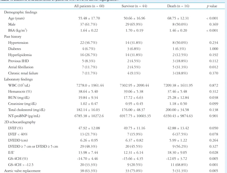
Table 1. Baseline characteristics of patients with chronic aortic regurgitation
All patients (n = 60) Survivor (n = 44) Death (n = 16) p value Demographic findings
Age (years) 55.48 ± 17.70 50.66 ± 16.96 68.75 ± 12.31 < 0.001
Male 37 (61.7%) 29 (65.9%) 08 (50.0%) 0.369
BSA (kg/m2) 1.64 ± 0.22 1.70 ± 0.19 1.46 ± 0.20 < 0.001
Past history
Hypertension 22 (36.7%) 14 (31.8%) 08 (50.0%) 0.234
Diabetes 04 (6.7%) 03 (6.8%) 01 (6.3%) 1.000
Hyperlipidemia 16 (26.7%) 14 (31.8%) 02 (12.5%) 0.192
Previous IHD 05 (8.3%) 02 (4.5%) 03 (18.8%) 0.112
Atrial fibrillation 07 (11.7%) 02 (4.5%) 05 (31.3%) 0.012
Chronic renal failure 07 (11.7%) 04 (9.1%) 03 (18.8%) 0.370
Laboratory findings
WBC (103/uL) 07278.0 ± 1961.44 7302.95 ± 2090.44 7209.38 ± 1611.95 0.872
Hematocrit (%) 38.64 ± 5.400 39.06 ± 5.380 37.46 ± 5.480 0.312
BUN (mg/dL) 19.84 ± 9.340 17.72 ± 6.630 25.28 ± 12.84 0.038
Creatinine (mg/dL) 1.02 ± 0.47 0.95 ± 0.45 1.18 ± 0.50 0.099
Total cholesterol (mg/dL) 182.14 ± 16.030 176.00 ± 48.370 200.00 ± 34.580 0.138
NT-proBNP (pg/mL) 6785.38 ± 10272.6 06917.75 ± 10603.35 6350.43 ± 9874.63 0.901
2D echocardiography
LVEF (%) 47.92 ± 12.08 49.75 ± 11.16 42.88 ± 13.42 0.050
LVEF < 40% 13 (21.7%) 07 (15.9%) 06 (37.5%) 0.078
LVEDD (cm) 6.26 ± 0.95 6.37 ± 0.82 5.99 ± 1.22 0.264
LVEDD ≥ 7 cm or LVESD ≥ 5 cm 29 (48.3%) 20 (45.5%) 09 (56.2%) 0.327
E/E’ 13.98 ± 7.440 12.31 ± 6.140 18.30 ± 9.050 0.028
GS-4CH (%) -14.70 ± 4.46-0 -15.66 ± 4.350- -12.05 ± 3.720- 0.005
GS-4CH > -12.5 20 (33.3%) 09 (20.5%) 11 (68.8%) 0.001
Aortic valve replacement 38 (63.3%) 33 (75.0%) 05 (31.3%) 0.005
BSA: body surface area, IHD: ischemic heart disease, WBC: white blood cell, BUN: blood urea nitrogen, NT-proBNP: N-terminal-pro B-type natriuretic pep- tide, LVEF: left ventricular ejection fraction, LVEDD: left ventricular end-diastolic diameter, LVESD: left ventricular end-systolic diameter, GS-4CH: global strain rate on apical four chamber image
estimated density, jet deceleration time and diastolic flow reversal, severe AR were 28 (47%), moderate to severe AR were 24 (40%) and moderate AR were 8 (13%) on the initial echocardiogram. During about 64 months follow-up period (median 63.87 ± 33.2 months; range 1 day–10.2 years), 38 patients (63%) underwent AVR and 16 patients (27%) were deceased. The most causes of death were cardiovascular death [sudden cardiac death (n = 3), heart failure (n = 3)] and acute cerebral accident (n = 2) in mainly who received conservative treatment (n = 7 vs. 1). The mean age was 55 years (range 24–
82) and 61.7% was male. The mechanism of AR was mostly degenerative change, 9 patients (15%) had combined dilated ascending aorta (diameter more than 5.7 cm) and 1 patient had right coronary cusp tear probably related Behcets’ disease. Acute AR patients who related aortic dissection or infective endocar- ditis were excluded when screening.
The deceased group was significantly older (69 ± 12 years old vs. 51 ± 17 years old, p < 0.001), more thin [body surface area (BSA) 1.46 ± 0.20 kg/m2 vs. 1.70 ± 0.19 kg/m2, p <
0.001], higher blood urea nitrogen (BUN) level (25.28 ± 12.84 mg/dL vs. 17.72 ± 6.63 mg/dL, p = 0.038) and fewer had AVR [5 patients (31.3%) vs. 33 patients (75.0%), p = 0.005]. The history of atrial fibrillation was more frequent in deceased group [5 patients (31.3%) vs. 2 patients (4.5%), p = 0.012]. However, incidence of other past history including hy- pertension, diabetes, hyperlipidemia, ischemic heart disease (IHD) and chronic renal failure was not significantly different between two groups. In aspect of two-dimensional echocardio- graphic parameters, deceased group had more decreased LV function (LVEF 42.9 ± 13.4% vs. 49.8 ± 11.2%, p = 0.050), increased E/E’ ratio (18.3 ± 9.1 vs. 12.3 ± 6.1, p = 0.028), and decreased GS-4CH value (-12.1 ± 3.7% vs. -15.7 ± 4.4%, p = 0.005) (Table 1).
The patients who received AVR (AVR group) were younger than patients who received conservative treatment (conserva- tive group) (51.8 ± 14.7 years old vs. 61.9 ± 20.8 years old, p = 0.053). In AVR group, the patients had bigger body surface area (1.70 ± 0.19 kg/m2 vs. 1.53 ± 0.22 kg/m2, p = 0.002),
Table 2. Baseline characteristics according to treatment
Conservative (n = 22) AVR (n = 38) p value
Demographic findings
Age (years) 61.86 ± 20.77 51.79 ± 14.71 0.053
Male 12 (54.5%) 25 (65.8%) 0.421
BSA (kg/m2) 1.53 ± 0.22 1.70 ± 0.19 0.002
Past history
Hypertension 08 (36.4%) 14 (36.8%) 1.000
Diabetes 01 (4.5%) 03 (7.9%) 1.000
Hyperlipidemia 06 (27.3%) 10 (26.3%) 1.000
Previous IHD 02 (9.1%) 03 (7.9%) 1.000
Atrial fibrillation 05 (22.7%) 02 (5.3%) 0.088
Chronic renal failure 03 (13.6%) 04 (10.5%) 0.700
Laboratory findings
WBC (103/uL) 7209.09 ± 1540.47 7317.89 ± 2198.95 0.838
Hematocrit (%) 38.18 ± 4.720 38.90 ± 5.810 0.622
BUN (mg/dL) 23.06 ± 10.58 17.96 ± 8.110 0.046
Creatinine (mg/dL) 1.16 ± 0.41 0.99 ± 0.51 0.628
Total cholesterol (mg/dL) 195.18 ± 33.830 177.66 ± 49.200 0.281
NT-proBNP (pg/mL) 09140.20 ± 10572.79 05607.97 ± 10185.20 0.384
2D echocardiography
LVEF (%) 45.73 ± 12.68 49.18 ± 11.71 0.289
LVEF < 40% 07 (31.8%) 06 (15.8%) 0.130
LVEDD (cm) 6.09 ± 0.88 6.38 ± 0.98 0.204
LVEDD ≥ 7 cm or LVESD ≥ 5 cm 11 (50.0%) 18 (47.4%) 0.528
E/E’ 15.28 ± 7.630 12.67 ± 7.220 0.298
GS-4CH (%) -14.84 ± 4.120- -14.61 ± 4.700- 0.849
GS-4CH > -12.5 08 (36.4%) 12 (31.6%) 0.780
All cause death 11 (50.0%) 05 (13.2%) 0.005
AVR: aortic valve replacement, BSA: body surface area, IHD: ischemic heart disease, WBC: white blood cell, BUN: blood urea nitrogen, NT-proBNP: N-ter- minal-pro B-type natriuretic peptide, LVEF: left ventricular ejection fraction, LVEDD: left ventricular end-diastolic diameter, LVESD: left ventricular end-sys- tolic diameter, GS-4CH: global strain rate on apical four chamber image
lower BUN level (17.96 ± 8.11 mg/dL vs. 23.06 ± 10.58 mg/dL, p = 0.046) and lower mortality [5 patients (13.2%) vs.
11 patients (50.0%), p = 0.005] than conservative group. The mean follow-up duration was longer in AVR group than con- servative treatment group (40 ± 29 months vs. 71 ± 30 months, p < 0.001). Otherwise, there was no significant difference be- tween the two groups (Table 2).
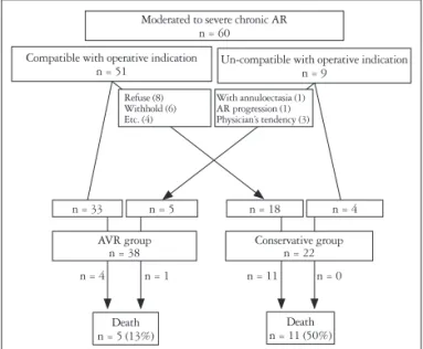
Decision of surgery often depended on demographic factor such as age, tendency of physician or will of the patient or the family rather than classical operative indication. The 5 (13.2%) of 38 patients who received AVR were not correspond with indication, while 18 (81.8%) of 22 patients who received con-
servative management although appropriate with surgical indi- cation. The most common causes of deferred surgery were pa- tient’s refusal (n = 8, 44%) and withholding of physicians due to old age or poor condition (n = 6, 33%) (Fig. 1).
The median value of longitudinal GS-4CH was -14.70 ± 4.46% (range -23.9–-6.3%, frame rate 34.63 ± 14.31/sec, heart rate 74 ± 16 bpm) (Fig. 2). In ROC curve for mortality of patients with chronic AR, classical markers such as LVEF or LV dimension didn’t show significant predicting power [area under the curve (AUC) 0.360 vs. 0.425]. On the contrary, glob- al longitudinal strain representing as GS-4CH has impact to mortality of chronic AR and the most powerful predicting lev- el was -12.5% (sensitivity 0.688, specificity 0.795, AUC 0.744) (Fig. 3). In AVR group, GS-4CH was more predictive than conservative group (AUC 0.894 vs. 0.682).
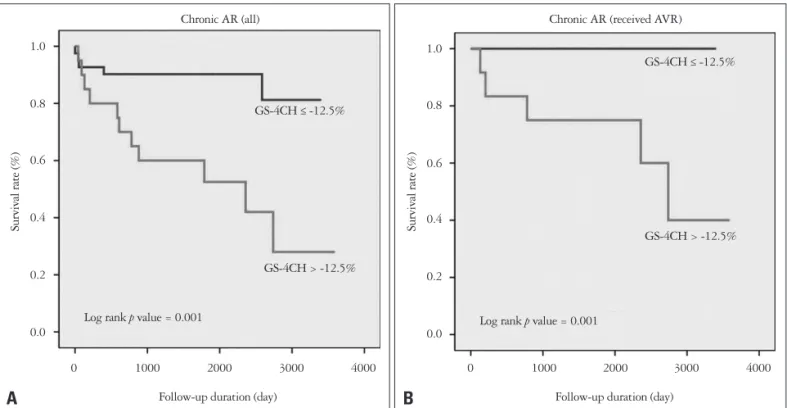
In cumulative survival curves using Kaplan-Meier method, patients with decreased GS-4CH value (> -12.5%) showed sig- nificantly increased mortality (log rank p = 0.001) (Fig. 4). In decreased GS-4CH group, the patients were older (64 ± 13 years old vs. 51 ± 18 years old, p = 0.002) and thin (BSA 1.56 ± 0.22 kg/m2 vs. 1.68 ± 0.21 kg/m2, p = 0.039). They had more frequently previous IHD (4 patients, 20% vs. 1 patient, 2.5%, p = 0.038) and lower LVEF (38.60 ± 12.37 vs. 52.58 ± 8.92, p < 0.001). However, other manifestations such as sex, comor- bidity and laboratory finding didn’t show significant difference between low and high GS-4CH group (Table 3).
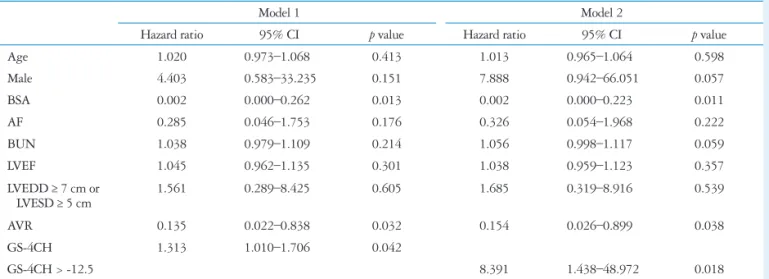
On multivariate analysis by Cox proportional hazard model adjusting for age, sex, BSA, past history of atrial fibrillation, level of BUN, LVEF, LV dilatation and AVR, decreased global longitudinal strain rate on apical four chamber image proved
Moderated to severe chronic AR n = 60
Death
n = 5 (13%) Death
n = 11 (50%) n = 0 n = 1
n = 4 n = 11
Un-compatible with operative indication n = 9
Compatible with operative indication n = 51
n = 33 AVR group
n = 38 Conservative group
n = 22
n = 5 n = 18 n = 4
With annuloectasia (1) AR progression (1) Physician’s tendency (3) Refuse (8)
Withhold (6) Etc. (4)
Fig. 1. Flow diagram of patients in study. AR: aortic regurgitation, AVR:
aortic valve replacement.
12
10
8
6
4
2
0
-25.0 -20.0 -15.0 -10.0 -5.0 Global longitudinal strain (%)
Number of patients
Fig. 2. Histogram of the distribution of baseline global longitudinal strain rate on apical 4 chamber image (-14.70 ± 4.46%, range -23.9–
-6.3%, frame rate 34.63 ± 14.31/sec, heart rate).
1.0
0.8
0.6
0.4
0.2
0.0
0.0 0.2 0.4 0.6 0.8 1.0 1-specificity
GS-4CH LVEF LVEDD
Cut-off value of GS-4CH: -12.5%
AUC 0.744 (95% CI 0.605–0.884) Sensitivity 0.688
Specificity 0.795
Sensitivity
Fig. 3. Receiver operating characteristic curve showing the best cutoffs for prediction of all-cause mortality in patients with chronic aortic regurgitation. GS-4CH: global strain rate on apical four chamber image, LVEF: left ventricular ejection fraction, LVEDD: left ventricular end- diastolic diameter, AUC: area under the curve, CI: confidence interval.
to be an independent predictor of all-cause mortality in patient with chronic AR (hazard ratio 1.313, 95% confidence interval 1.010–1.706, p = 0.042) (Table 4).
Discussion
In chronic AR, adaptation to increased preload and afterload results in increase in end-diastolic volume with eccentric and concentric hypertrophy. LV hypertrophy inhibits the increase of wall stress and it enable to maintain normal LVEFs and as- ymptomatic state for considerable period. According to AR per- sist, the wall thickening fails to cope the hemodynamic over- load and systolic function insidiously decreased. In this stage, symptom such as dyspnea or exertional chest pain may repre- sent, and some patients remained asymptomatic until severe LV dysfunction has developed. Ten-year survival rate was 69 ± 9% and cardiac event free survival rate was 37 ± 8% in asymp- tomatic patients with even severe AR in study of Detaint et al.5)
In natural history of AR, progression to symptoms and/or LV dysfunction is less than 6% per year among asymptomatic patients with normal LV systolic function. Whereas, greater than 25% per year of asymptomatic patients with LV dysfunc- tion progress to cardiac symptoms and the mortality rate of symptomatic patients is greater than 10% per year.2)6)
LV systolic dysfunction is initially reversible and it is possible to fully recovery of LV size and function with AVR. The classic indications for valve replacement are development of symp- tom such as dyspnea more than NYHA class 3, LV dysfunction (LVEF < 50%) or dilated LV dimension (LV end diastolic di- mension > 75 mm or end-systolic dimension > 55 mm).2) How- ever, there is no single parameter to detect this period and the
timing of AVR in patient with chronic AR is still debated. And once myocardial dysfunction developed, it could cause high risk for post-operative heart failure and death.
The factors predictive of reduced postoperative survival and recovery of LV function in patients with AR are severity of pre- operative symptoms or reduced exercise tolerance, severity of depression of LVEF and duration of preoperative LV systolic dysfunction.2) Tornos et al.7) studied about 101 patients with asymptomatic chronic AR and normal LVEF for up to 10 years.
During the follow-up period, 14 patients needed surgery (8 patients because of symptoms and 6 patients because of LV impairment), baseline end-systolic diameter > 50 mm and LVEF < 60% were independent predictors for cardiac symp- toms or LV dysfunction.7)
Guideline recommendations for AVR have focused on LVEF and LV end systolic dimension as measures of LV pump perfor- mance, and LV end-diastolic dimension as a measure of the se- verity of the volume overload.2)
In volume overloading state such as chronic AR, it tends to overestimate LV systolic function. The conventional echocar- diographic parameters such as LV dimensions and the LVEF have limitation in predicting early LV dysfunction, thus re- cently many studies investigated about quantitative measure- ment of LV function used speckle-tracking echocardiography.
Myocardial strain rate imaging derived from tissue Doppler echocardiography estimates spatial gradients of myocardial ve- locities and allow accurate quantification of regional myocar- dial function, therefore it facilitate early detection of subclini- cal myocardial disease.8-11) Another study about myocardial deformation in 59 asymptomatic patients with isolated wide
1.0
0.8
0.6
0.4
0.2
0.0
0 1000 2000 3000 4000 Follow-up duration (day)
Chronic AR (received AVR)
Log rank p value = 0.001
GS-4CH > -12.5%
Survival rate (%)
B
GS-4CH ≤ -12.5%
1.0
0.8
0.6
0.4
0.2
0.0
0 1000 2000 3000 4000 Follow-up duration (day)
Chronic AR (all)
Log rank p value = 0.001
GS-4CH > -12.5%
Survival rate (%)
A
GS-4CH ≤ -12.5%
Fig. 4. Kaplan-Meier curve for all-cause mortality stratified by GS-4CH in all patients with chronic AR (A) and patients received AVR (B). GS-4CH:
global strain rate on apical four chamber image, AR: aortic regurgitation, AVR: aortic valve replacement.
Table 3. Baseline characteristics according to global strain
Preserved GS-4CH
≤ -12.5% (n = 40) Decreased GS-4CH
> -12.5% (n = 20) p value Demographic findings
Age (years) 51.05 ± 18.16 64.35 ± 13.10 0.002
Male 26 (65.0%) 11 (55.0%) 0.575
BSA (kg/m2) 1.68 ± 0.21 1.56 ± 0.22 0.039
Past history
Hypertension 13 (32.5%) 09 (45.0%) 0.401
Diabetes 02 (5.0%) 02 (10.0%) 0.595
Hyperlipidemia 11 (27.5%) 05 (25.0%) 1.000
Previous IHD 01 (2.5%) 04 (20.0%) 0.038
Atrial fibrillation 04 (10.0%) 03 (15.0%) 0.676
Chronic renal failure 03 (7.5%) 04 (20.0%) 0.208
Laboratory findings
WBC (103/uL) 07390.0 ± 9747.30 07054.0 ± 1512.03 0.536
Hematocrit (%) 39.11 ± 5.320 37.69 ± 5.580 0.340
BUN (mg/dL) 19.47 ± 9.520 20.58 ± 9.200 0.675
Creatinine (mg/dL) 1.01 ± 0.39 1.03 ± 0.61 0.835
Total cholesterol (mg/dL) 183.67 ± 43.490 178.62 ± 53.160 0.745
NT-proBNP (pg/mL) 6518.35 ± 9747.30 7519.71 ± 12300.8 0.818
2D echocardiography
LVEF (%) 52.58 ± 8.920 38.60 ± 12.37 < 0.001
LVEF < 40% 04 (10.0%) 09 (45.0%) 0.003
LVEDD (cm) 6.15 ± 0.85 6.50 ± 1.12 0.187
LVEDD ≥ 7 cm or LVESD ≥ 5 cm 15 (37.5%) 14 (70.0%) 0.028
E/E’ 12.44 ± 7.200 17.04 ± 7.240 0.080
Aortic valve replacement 26 (65.0%) 12 (60.0%) 0.780
All cause death 05 (12.5%) 11 (55.0%) 0.001
GS-4CH: global strain rate on apical four chamber image, BSA: body surface area, IHD: ischemic heart disease, WBC: white blood cell, BUN: blood urea nitro- gen, NT-proBNP: N-terminal-pro B-type natriuretic peptide, LVEF: left ventricular ejection fraction, LVEDD: left ventricular end-diastolic diameter, LVESD:
left ventricular end-systolic diameter
Table 4. Multivariate analysis on Cox proportional hazard model for all-cause mortality in patients with chronic aortic regurgitation
Model 1 Model 2
Hazard ratio 95% CI p value Hazard ratio 95% CI p value
Age 1.020 0.973–1.068 0.413 1.013 0.965–1.064 0.598
Male 4.403 0.583–33.235 0.151 7.888 0.942–66.051 0.057
BSA 0.002 0.000–0.262 0.013 0.002 0.000–0.223 0.011
AF 0.285 0.046–1.753 0.176 0.326 0.054–1.968 0.222
BUN 1.038 0.979–1.109 0.214 1.056 0.998–1.117 0.059
LVEF 1.045 0.962–1.135 0.301 1.038 0.959–1.123 0.357
LVEDD ≥ 7 cm or
LVESD ≥ 5 cm 1.561 0.289–8.425 0.605 1.685 0.319–8.916 0.539
AVR 0.135 0.022–0.838 0.032 0.154 0.026–0.899 0.038
GS-4CH 1.313 1.010–1.706 0.042
GS-4CH > -12.5 8.391 1.438–48.972 0.018
CI: confidence interval, BSA: body surface area, AF: atrial fibrillation, BUN: blood urea nitrogen, LVEF: left ventricular ejection fraction, LVEDD: left ventric- ular end-diastolic diameter, LVESD: left ventricular end-systolic diameter, AVR: aortic valve replacement, GS-4CH: global strain rate on apical four chamber image
range of severity of AR showed that strain rate imaging is a sen- sitive tool in detecting changes of contractility.12)
Di Salvo et al.13) studied about asymptomatic 26 patients aged ≤ 16 years with isolated moderate to severe AR. The pa- tients were divided to stable and progressive AR (defined as development of symptoms, increased LV volume more than 15%, or decreased LVEF more than 10% during follow-up) group. In patients with progressive AR group, LV average lon- gitudinal strain was significantly reduced (-17.8 ± 3.9% vs.
-22.7 ± 2.7%, p = 0.001) although baseline LVEF was similar.
Among echocardiographic parameters including LV end dia- stolic dimension and LV volume, E/E’ ratio and jet area/LV out- flow tract area ratio, LV average longitudinal strain was only sig- nificant risk factor for progressive AR (cut-off value > -19.5%, sensitivity 77.8%, specificity 94.1%, AUC 0.889).13)
Olsen et al.14) performed longitudinal study in 64 patients with moderate to severe AR and demonstrated that reduced myocardial systolic strain was associated with disease progres- sion during conservative management and impaired outcome after surgery. The present study showed no significant differ- ent echocardiographic findings including LVEF, LV dimension and GS-4CH between conservative and AVR group. In AVR group (n = 38), 5 patients (13%) were deceased and they had significant lower GS-4CH level (-9.02 ± 1.64% vs. -15.46 ± 4.42%, p = 0.003; AUC 0.897) although LVEF and LV end- diastolic dimension weren’t (43.40 ± 16.79% vs. 50.06 ± 10.83%, p = 0.241; AUC 0.397, and 6.26 ± 1.74 cm vs. 6.40
± 0.85 cm, p = 0.865; AUC 0.539). In conservative group, de- ceased patients were 11 (50%), GS-4CH didn’t show signifi- cant difference between survivors and deceased patients in common with LVEF and dimension.
In these days, speckle-tracking deformation imaging has be- come routine echocardiographic examination. Echocardio- graphic analysis of LV longitudinal strain rate with speckle- tracking echocardiography is useful to early detect LV dysfunction and determine the right timing of operative management in patients with chronic, isolated AR. Measurement of GS-4CH is easier and faster than global longitudinal strain because it is possible with apical 4 chamber image alone. And the present study showed GS-4CH has sufficient predicting value for mor- tality in patients with chronic aortic regurgitation. GS-4CH may be a simple and useful surrogate marker in predicting the fate of chronic AR.
Limitations
This number of enrolled patients was small because it is based on single center data and isolated chronic AR without other valvular heart disease is not common. The follow-up du- ration of patients was very various (1 day–123 months, mean 60 ± 33 months) and moreover, there are lack of information about mechanism of AR, concomitant coronary artery disease and medical treatment. Few patients had done cardiac cathe- terization and/or coronary angiography. We assumed etiology
of most patients is degenerative change as echocardiographic findings. Because of some patients’ LV chambers were so dilat- ed and frame rates were lower than optimal range, data collec- tion for strain measurement was limited. Further evaluation with global circumferential and radial strain may strengthen pre- dicting power to prognosis of patients with chronic AR.
Conclusions
In this study, global strain represent as GS-4CH had inde- pendently significant predictive value for all-cause mortality after adjusting for other affecting variables while conventional parameter such as LVEF or LV dimension did not. GS-4CH seems to be useful parameters to support decision making and predict prognosis in patients with chronic AR.
References
1. Iida N, Seo Y, Ishizu T, Nakajima H, Atsumi A, Yamamoto M, Machino-Ohtsuka T, Kawamura R, Enomoto M, Kawakami Y, Aonuma K. Transmural compensation of myocardial deformation to pre- serve left ventricular ejection performance in chronic aortic regurgitation. J Am Soc Echocardiogr 2012;25:620-8.
2. Bonow RO, Carabello BA, Chatterjee K, de Leon AC Jr, Faxon DP, Freed MD, Gaasch WH, Lytle BW, Nishimura RA, O’Gara PT, O’Rourke RA, Otto CM, Shah PM, Shanewise JS; American Col- lege of Cardiology/American Heart Association Task Force on Prac- tice Guidelines. 2008 focused update incorporated into the ACC/AHA 2006 guidelines for the management of patients with valvular heart disease:
a report of the American College of Cardiology/American Heart Association Task Force on Practice Guidelines (Writing Committee to revise the 1998 guidelines for the management of patients with valvular heart disease). En- dorsed by the Society of Cardiovascular Anesthesiologists, Society for Car- diovascular Angiography and Interventions, and Society of Thoracic Sur- geons. J Am Coll Cardiol 2008;52:e1-142.
3. Ekery DL, Davidoff R. Aortic regurgitation: quantitative methods by echocardiography. Echocardiography 2000;17:293-302.
4. The Korean Society of Echocardiography. The Text Book of Clinical Echocardiography. 3rd ed. Seoul: Daehan Medical Books;2013. p. 329-33.
5. Detaint D, Messika-Zeitoun D, Maalouf J, Tribouilloy C, Mahoney DW, Tajik AJ, Enriquez-Sarano M. Quantitative echocardiographic de- terminants of clinical outcome in asymptomatic patients with aortic regurgita- tion: a prospective study. JACC Cardiovasc Imaging 2008;1:1-11 6. American College of Cardiology/American Heart Association Task
Force on Practice Guidelines; Society of Cardiovascular Anesthesiol- ogists; Society for Cardiovascular Angiography and Interventions;
Society of Thoracic Surgeons, Bonow RO, Carabello BA, Kanu C, de Leon AC Jr, Faxon DP, Freed MD, Gaasch WH, Lytle BW, Nishimura RA, O’Gara PT, O’Rourke RA, Otto CM, Shah PM, Shanewise JS, Smith SC Jr, Jacobs AK, Adams CD, Anderson JL, Antman EM, Faxon DP, Fuster V, Halperin JL, Hiratzka LF, Hunt SA, Lytle BW, Nishimura R, Page RL, Riegel B. ACC/AHA 2006 guidelines for the management of patients with valvular heart disease: a re- port of the American College of Cardiology/American Heart Association Task Force on Practice Guidelines (writing committee to revise the 1998 Guidelines for the Management of Patients With Valvular Heart Disease):
developed in collaboration with the Society of Cardiovascular Anesthesiolo- gists: endorsed by the Society for Cardiovascular Angiography and Interven- tions and the Society of Thoracic Surgeons. Circulation 2006;114:e84- 231.
7. Tornos MP, Olona M, Permanyer-Miralda G, Herrejon MP, Cam-
precios M, Evangelista A, Garcia del Castillo H, Candell J, Soler-Sol- er J. Clinical outcome of severe asymptomatic chronic aortic regurgitation: a long-term prospective follow-up study. Am Heart J 1995;130:333-9.
8. Onishi T, Kawai H, Tatsumi K, Kataoka T, Sugiyama D, Tanaka H, Okita Y, Hirata K. Preoperative systolic strain rate predicts postoperative left ventricular dysfunction in patients with chronic aortic regurgitation. Circ Cardiovasc Imaging 2010;3:134-41.
9. Chaliki HP, Mohty D, Avierinos JF, Scott CG, Schaff HV, Tajik AJ, Enriquez-Sarano M. Outcomes after aortic valve replacement in patients with severe aortic regurgitation and markedly reduced left ventricular func- tion. Circulation 2002;106:2687-93.
10. Edvardsen T, Gerber BL, Garot J, Bluemke DA, Lima JA, Smiseth OA. Quantitative assessment of intrinsic regional myocardial deformation by Doppler strain rate echocardiography in humans: validation against three- dimensional tagged magnetic resonance imaging. Circulation 2002;106:
50-6.
11. Yu CM, Sanderson JE, Marwick TH, Oh JK. Tissue Doppler imaging a new prognosticator for cardiovascular diseases. J Am Coll Cardiol 2007;
49:1903-14.
12. Marciniak A, Sutherland GR, Marciniak M, Claus P, Bijnens B, Ja- hangiri M. Myocardial deformation abnormalities in patients with aortic regurgitation: a strain rate imaging study. Eur J Echocardiogr 2009;10:
112-9.
13. Di Salvo G, Rea A, Mormile A, Limongelli G, D’Andrea A, Pergola V, Pacileo G, Caso P, Calabrò R, Russo MG. Usefulness of bidimension- al strain imaging for predicting outcome in asymptomatic patients aged ≤ 16 years with isolated moderate to severe aortic regurgitation. Am J Cardiol 2012;110:1051-5.
14. Olsen NT, Sogaard P, Larsson HB, Goetze JP, Jons C, Mogelvang R, Nielsen OW, Fritz-Hansen T. Speckle-tracking echocardiography for pre- dicting outcome in chronic aortic regurgitation during conservative manage- ment and after surgery. JACC Cardiovasc Imaging 2011;4:223-30.