80
80 THE EWHA MEDICAL JOURNAL THE EWHA MEDICAL JOURNAL
More Effective Way to Perform Complete Percutaneous Vertebroplasty for Patients in Kummell’s Disease: A Case Report
Seung Hee Yoo , Ji Seon Chae 1 , Minjin Lee , Bo Kyung Kang , Hahck Soo Park 2 , Won-Joong Kim
Department of Anesthesiology and Pain Medicine, Ewha Womans University Mokdong Hospital,
1Ewha Womans University Seoul Hospital, Ewha Womans University School of Medicine,
2Parkhahcksoo Pain Clinic, Seoul, Korea
Case Report
Ewha Med J 2021;44(3):80-83
https://doi.org/10.12771/emj.2021.44.3.80 eISSN 2234-2591
An 84-year-old woman visited our pain clinic with complaints of low back pain and severe radiating pain in the right lower extremity during walking. The patient dem- onstrated subacute compression fracture of L3 with vacuum change in lumbar spine plain radiographs and MRI which suggest Kummell’s disease. Despite our conservative treatments, she had little back pain relief. Therefore, we planned a percutaneous verte- broplasty. Manual compression could help perform percutaneous vertebroplasty more effectively by expanding the vertebral body. In addition, the spontaneous recovery of vacuum cleft width using negative pressure could help perform the technique more effectively. We successfully performed percutaneous vertebroplasty using these combi- nation therapies for our patient. (Ewha Med J 2021;44(3):80-83)
Received March 23, 2021 Revised April 16, 2021 Accepted April 29, 2021 Corresponding author Won-Joong Kim
Department of Anesthesiology and Pain Medicine, Ewha Womans University Mokdong Hospital, Ewha Womans University School of Medicine, 1071 Anyangcheon-ro, Yangcheon- gu, Seoul 07985, Korea
Tel: 82-2-2650-2689, Fax: 82-2-2655-2924 E-mail: [email protected]
*Seung Hee Yoo and Ji Seon Chae contributed equally to this study.
Key Words
Fractures, compression; Osteonecrosis;
Osteoporosis; Vertebroplasty
This is an Open Access article distributed under the terms of the Creative Commons Attribution Non-Commercial License (http://creativecommons.org/licenses/by-nc/4.0) which permits unrestricted non-commercial use, distribution, and reproduction in any medium, provided the original work is properly cited.
Introduction
Kummell’s disease is an avascular necrosis disorder of the vertebral body after osteoporotic vertebral compression frac- ture. This disease involves delayed healing of a fractured verte- bral body which can be seen in the intravertebral vacuum cleft with fluid or air inside it and needs more aggressive treatment [1,2]. The term was originally coined by Hermann Kummell for a condition wherein a minor asymptomatic spinal trauma leads to symptomatic, progressive painful back pain and func- tional limitations [2]. This condition is common in the elderly,
who are susceptible to vertebral fractures owing to osteoporosis or osteonecrosis which are initially asymptomatic but progress and become symptomatic with change of vertebral body image [2].
Most vertebral fractures do not need aggressive treatment
and can be treated by bed rest using braces and pain medica-
tions. However, some patients diagnosed with Kummell’s dis-
ease complain of severe back pain, deformity, and inability to
walk, all of these lowering their quality of life [3,4]. Percutane-
ous vertebroplasty (PVP) and percutaneous kyphoplasty (PKP)
are effective minimally invasive treatments. Bone cement inject-
81
THE EWHA MEDICAL JOURNAL Percutaneous Vertebroplasty for Patients in Kummell’s Disease
ed during these treatments appropriately agglutinates fractures and resolves the pseudoarthrosis-caused instability by filling the vacuum cleft; this is thus expected to provide remarkable pain relief [3-5]. Among these, PVP is an option including shorter operating time, decreased cost that still provides comparable pain relief and vertebral height restoration as PKP for treating Kummell’s disease [6,7].
Several studies have reported on ways to perform PVP more effectively: spontaneous re-expansion of the vertebral body was reported when inserting the needle into the fractured ver- tebral body in Kummell’s disease [5,8]. In addition, manual compression after inserting cement into the vertebral body may help maintain the extended spine position and increase hyper- lordosis [9-12].
Since no studies have implemented PVP in combination with the methods presented in previous studies, we performed PVP in our patient who had an osteoporotic compression fracture with osteonecrosis using the abovementioned combination therapy.
Case
An 84-year-old female (height 155 cm, weight 59 kg) visited our pain clinic with complaints of low back pain and severe radiating pain in the right lower extremity during walking (nu- meric rating scale [NRS], 7–8). Our patient had osteoporosis, hypertension, and stroke history and had taken anticoagulant
medication.
Physical examination revealed no specific sign except for the tenderness of the spinous process at L3 level. Lumbar spine plain radiographs readings were compression fracture at L3 and L4, degenerative spondylosis, decrease in height of L3 vertebral body and vacuum change, all of which suggest Kum- mell’s disease (Fig. 1A, B). MRI showed subacute compression fracture of L3 with mild central stenosis and old compression fracture of L4 and L5 (Fig. 1C). From these findings, we con- cluded that her condition corresponded to Kummell’s disease with instability of L3.
Despite our conservative treatments including right L3/4 transforaminal epidural steroid injections and bilateral facet joint blocks with medications such as NSAIDs, afloqualone, and gabapentin, she had little back pain relief (NRS 6–7).
Therefore, we planned a PVP considering her age, life expec- tancy, and general conditions.
On the day of the procedure, she was given a prophylactic antibiotic (1 g cefazoline) and a durogesic patch was attached for analgesia. She was laid in prone position on the operating table (Spinal Table HM 604; Handok Medical, Seoul, Ko- rea). We monitored her blood pressure, heart rate, and oxygen saturation, and the area was prepared and draped in an aseptic method with betadine. After disinfecting the skin, skin infiltra- tion with 1% lidocaine was performed, and a vertebroplasty needle (HMN-NVP1103; Hyun Medics, Bucheon, Korea) was inserted targeting the right L3 vertebra pedicle. We re- moved the inlet cannula carefully and intentionally waited for about 5 minutes expecting the spontaneous reexpansion of the
A B C
Fig. 1. Antero-posterior (A) and lateral (B) images of L spine plain ra- diography. They show severe vertebral collapse with vertebral vacuum cleft (arrow) at L3. T1-weighted lumbar MR image (C) shows low sig- nal area in collapsed L3 vertebra. Informed consent for publication of the clinical images was obtained from the patient.
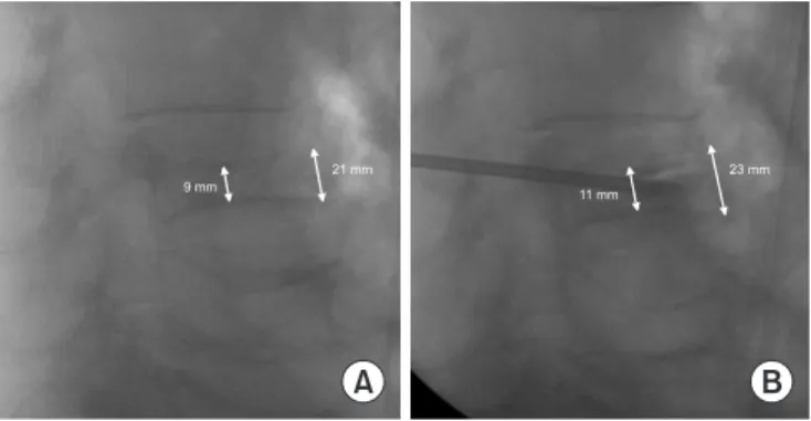
Fig. 2. The lateral image of C-arm fluoroscopy. There is the phenom- enon of spontaneous recovery of the collapsed vertebral body. The height of the vertebral column increases from 9 to 11 mm centrally and from 21 to 23 mm anteriorly (A, B). Informed consent for publica- tion of the clinical images was obtained from the patient.
A B
9 mm
21 mm
11 mm
23 mm