Prevalence of Congenital Heart Defects Associated with Down Syndrome in Korea
Congenital heart defect (CHD) is common in infants with Down syndrome (DS), which is the principle cause of mortality. However, there is no data available for the frequency and types of CHD in infants with DS in Korea. We investigated the frequency of CHD in infants with DS in Korea. After the survey on birth defects was conducted throughout the country, the prevalence of CHD in DS in 2005-2006 was calculated. This study was conducted based on the medical insurance claims database of the National Health Insurance Corporation.
The number of total births in Korea was 888,263 in 2005-2006; of them, 25,975 cases of birth defects were identified. The prevalence of DS was 4.4 per 10,000 total births, accounting for 1.5% of all birth defects. Of the 394 infants with DS, 224 (56.9%) had a CHD. Atrial septal defect was the most common defect accounting for 30.5% of DS followed by ventricular septal defect (19.3%), patent duct arteriosus (17.5%), and atrioventricular septal defect (9.4%). Our study will be helpful to demonstrate the current status of DS and to identify the distribution of CHD in infants with DS in Korea.
Keywords: Down Syndrome; Heart Defects, Congenital; Prevalence, Korea Min-A Kim,1 You Sun Lee,1 Nan Hee Yee,2
Jeong Soo Choi,2 Jung Yun Choi,3 and Kyung Seo1
1Division of Maternal-Fetal Medicine, Department of Obstetrics and Gynecology, Gangnam Severance Hospital, Institute of Women’s Medical Life Science, Yonsei University College of Medicine, Seoul;
2Health Policy Research Division, Korea Institute for Health and Social Affairs, Seoul; 3Department of Pediatrics, Seoul National University Bundang Hospital, Seongnam, Korea
Received: 6 August 2014 Accepted: 12 August 2014 Address for Correspondence:
Kyung Seo, MD
Department of Obstetrics and Gynecology, Gangnam Severance Hospital, Yonsei University College of Medicine, 211 Eonju-ro, Gangnam-gu, Seoul 135-720, Korea
Tel: +82.2-2019-3433, Fax: +82.2-3462-8209 E-mail: [email protected]
http://dx.doi.org/10.3346/jkms.2014.29.11.1544 • J Korean Med Sci 2014; 29: 1544-1549
INTRODUCTION
Down syndrome (DS) is a well-known genetic disorder that places affected individuals at increased risk for several medical morbidities, especially congenital heart defect (CHD). The prev- alence of CHD among infants born with DS is reported to range from 40% to 60% compared with 0.8% in the general population (1). The prevalence of DS, the most frequent chromosomal ab- normality, has remained stable over time with increasing aver- age maternal age despite higher prenatal detection rates. How- ever, there are few reports concerning the prevalence of CHD in infants with DS in Korea.
The frequency of CHD in infants with DS varies among geo- graphical locations. In the United States of America and Europe, an atrioventricular septal defect (AVSD) is reportedly the most common CHD associated with DS (2-8). However, in Asian com- munities, a ventricular septal defect (VSD) is the most common defect (9, 10), whereas in Latin America, an atrial septal defect (ASD) is reportedly the most common defect (11). For Korea, however, there are no data available for the frequency and types of CHD in infants with DS in Korea.
The aim of this study was to identify the frequency and distri- bution of CHD in infants with DS in Korea and compare it with
the prevalence in infants with CHD who have non-chromosom- al abnormalities. To our knowledge, this study was planned to establish epidemiologic data about the distribution of CHD in infants with DS. We believe this study will provide baseline data for future studies on the epidemiology and management of in- fants with DS in Korea.
MATERIALS AND METHODS
This study analyzed data from the Congenital Anomaly Survey and the Infant and Maternal Mortality Survey, which were both performed by the Korea Institute for Health and Social Affairs (12, 13). These surveys were conducted using data from all Ko- rean medical institutes from January 1, 2005 to December 31, 2006, and based on the medical insurance claims database of the National Health Insurance Corporation.
Research categories and subjects were selected in advance through pilot surveys, and a web-based research system was regulated to improve data acquisition and screening. During the data collection period, progress was monitored in real time, and corrective actions were implemented to respond to issues raised by the participating institutes. The data collected through these processes were reviewed and re-checked for accuracy, Obstetrics & Gynecology
and differences or overlaps in birth defect diagnoses among the medical institutes were adjusted for uniform reporting.
Since the possible period of diagnosis for birth defects is wide, we only included data from the first postnatal year. This approach is in agreement with other international bodies, such as Inter- national Clearinghouse for Birth Defects Surveillance and Re- search (ICBDSR) and European Surveillance of Congenital Ano- malies (EUROCAT), which generally include only data from the first postnatal year in their analysis. The Korean Medical Record Association integrated the database of birth defects with ongo- ing surveillance at each medical institute.
Birth defects were classified by Q code, according to the 10th revision of the International Classification of Diseases. Defects were analyzed according to the methods of EUROCAT, ICBDSR, and the National Birth Defects Prevention Network (NBDPN), which are global leaders in this field. In this study, only study subjects who were confirmed by chromosomal analysis were included and cases with rule-out diagnoses of birth defects and mismatching between disease diagnoses and medical records were excluded. Regarding fetal cardiac anomalies, which are closely accompanied by other heart defects, we focused only on the major anomalies, and divided the results into 3 groups with the assistance of the fetal heart specialist.
In this study, we calculated the total birth prevalence rate of each anomaly per 10,000 births, with the number of total births as the denominator, and the number of each anomaly as the numerator. We collected data regarding birth defects, in the first postnatal year, from the medical insurance claims database (sorted by main disease code and sub-code), and attempted to collect all information prior to analysis. Among the research categories, only easily accessible data regarding the risk factors of birth defects were utilized. Collected variables were maternal age, paternal age, parental occupation, parental education lev- el, gestational age, birth weight, and pregnancy with or without multiple gestations.
Statistical analysis
We reviewed maternal and paternal age, parental job, education- al background, gestational age at delivery, birthweight, and fetal sex as variables for analysis and performed the Poisson distri- bution to calculate 95% confidence intervals of DS prevalence.
Ethics statement
The study protocol was approved by the institutional review board of Gangnam Severance Hospital (IRB No. 3-2011-0286).
Informed consent was waived by the board.
RESULTS
The number of total births in Korea was 888,263 in 2005-2006;
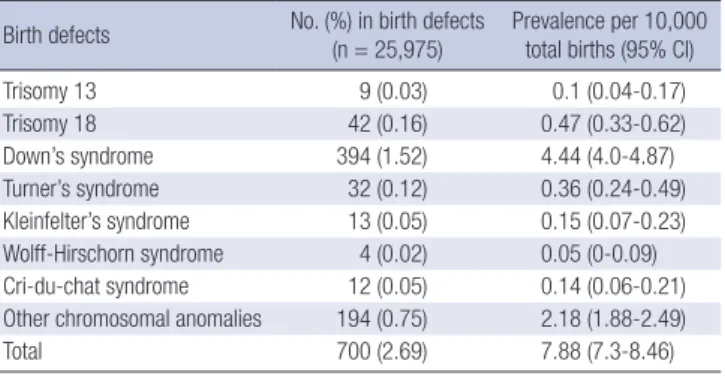
of them, 25,975 had birth defects with a prevalence of 292.4 per 10,000 total births. The prevalence of chromosomal anomalies was 7.9 per 10,000 total births, accounting for 2.7% of all birth defects in 2005-2006. DS was the most common chromosomal anomaly and its prevalence was 4.4 per 10,000 total births, ac- counting for 1.5% of all birth defects (Table 1).
Baseline demographic characteristics of infants with DS, chro- mosomal abnormalities, and non-chromosomal abnormalities are presented in Table 2. There were significant differences in maternal age, paternal age, gestational age at delivery, and birth weights among infants with DS and those with non-chromoso- mal abnormalities.
Of the 394 infants with DS, 224 (56.9%) had a CHD (Table 3).
There were significant differences in gestational age at delivery and birth weights between DS without CHD and those with CHD.
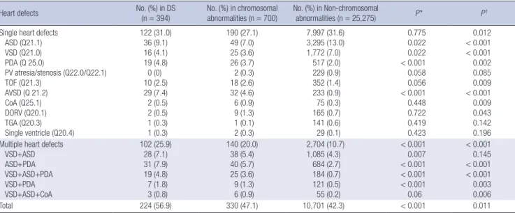
The data for CHD in infants with DS are summarized in Table 4. Atrial septal defect (ASD) was the most common defect re- ported as 30.5%, ventricular septal defect (VSD) 19.3%, patent ductus arteriosus (PDA) 17.5%, atrioventricular septal defect (AVSD) (9.4%), and tetralogy of Fallot (TOF) 2.5%. There were
Table 1. Prevalence of major chromosomal anomalies in Korea, 2005-2006 Birth defects No. (%) in birth defects
(n = 25,975) Prevalence per 10,000 total births (95% CI)
Trisomy 13 9 (0.03) 0.1 (0.04-0.17)
Trisomy 18 42 (0.16) 0.47 (0.33-0.62)
Down’s syndrome 394 (1.52) 4.44 (4.0-4.87)
Turner’s syndrome 32 (0.12) 0.36 (0.24-0.49)
Kleinfelter’s syndrome 13 (0.05) 0.15 (0.07-0.23) Wolff-Hirschorn syndrome 4 (0.02) 0.05 (0-0.09)
Cri-du-chat syndrome 12 (0.05) 0.14 (0.06-0.21)
Other chromosomal anomalies 194 (0.75) 2.18 (1.88-2.49)
Total 700 (2.69) 7.88 (7.3-8.46)
CI, confidence interval.
Table 2. Demographic characteristics of Down syndrome, chromosomal abnormalities, and non-chromosomal abnormalities in Korea
Parameters DS
(n = 394)
Chromosomal abnormalities (n = 700)
Non- chromosomal abnormalities
(n = 25,275) P* P†
Maternal age (yr) 33.0 ± 5.4 32.2 ± 5.2 30.5 ± 4.0 < 0.001 < 0.001
Paternal age (yr) 35.6 ± 5.5 34.7 ± 5.1 33.2 ± 4.3 < 0.001 < 0.001
Gestational age (weeks) 35.1 ± 6.0 34.7 ± 6.2 37.9 ± 3.5 < 0.001 < 0.001
Birth weight (grams) 2,585 ± 925 2,417 ± 1,002 3,040 ± 683 < 0.001 < 0.001
Fetal sex ratio (M/F) 1.3 1.1 1.3 0.812 0.118
*Comparison between groups with DS and non-chromosomal abnormalities; †Comparison between groups with chromosomal abnormalities and non-chromosomal abnormali- ties. M, male; F, female; DS, Down syndrome.
Table 3. Demographic characteristics of Down syndrome without congenital heart defect and those with congenital heart defect
Parameters
Total patients with Down syndrome without CHD P
(n = 170) with CHD
(n = 224)
Maternal age (yr) 32.9 ± 5.8 33.0 ± 5.2 0.912
Paternal age (yr) 35.9 ± 4.9 35.4 ± 5.7 0.511
Gestational age (weeks) 31.6 ± 7.5 37.7 ± 2.1 < 0.001 Birthweight (grams) 2,128 ± 1,167 2,897 ± 525 < 0.001
Fetal sex ratio (M/F) 1.6 1.1 0.116
M, male; F, female; CHD, congenital heart defect.
Table 4. Distribution of major congenital heart defects in Down syndrome
Heart defects No. (%) in DS
(n = 394) No. (%) in chromosomal
abnormalities (n = 700) No. (%) in Non-chromosomal
abnormalities (n = 25,275) P* P†
ASD (Q21.1) 120 (30.5) 163 (23.3) 5719 (22.6) < 0.001 0.681
VSD (Q21.0) 76 (19.3) 109 (15.6) 3304 (13.1) < 0.001 0.054
PDA (Q 25.0) 69 (17.5) 81(11.6) 1194 (4.7) < 0.001 < 0.001
PV atresia/stenosis (Q22.0/Q22.1) 4 (1.0) 6 (0.9) 426 (1.7) 0.304 0.091
TOF (Q21.3) 10 (2.5) 20 (2.9) 361 (1.4) 0.067 0.002
AVSD (Q 21.2) 37 (9.4) 43 (6.1) 271 (1.1) < 0.001 < 0.001
CoA (Q25.1) 8 (2.0) 18 (2.6) 204 (0.8) 0.008 < 0.001
DORV (Q20.1) 2 (0.5) 9 (1.3) 168 (0.7) 0.703 0.049
TGA (Q20.3) 1 (0.3) 2 (0.3) 143 (0.6) 0.411 0.327
Single ventricle (Q20.4) 1 (0.3) 2 (0.3) 29 (0.1) 0.423 0.196
*Comparison between groups with DS and non-chromosomal abnormalities; †Comparison between groups with chromosomal abnormalities and non-chromosomal abnormali- ties. DS, Down syndrome; ASD, atrial septal defect; VSD, ventricular septal defect; PDA, patent ductus arteriosus; PV, pulmonary valve; TOF, tetralogy of Fallot; AVSD, atrioven- tricular septal defect; CoA, coarctation of aorta; DORV, double outlet right ventricle; TGA, transposition of great arteries.
Table 5. Frequency of congenital heart defects in patients with Down syndrome, chromosomal abnormalities, and non-chromosomal abnormalities in Korea
Heart defects No. (%) in DS
(n = 394)
No. (%) in chromosomal abnormalities (n = 700)
No. (%) in Non-chromosomal
abnormalities (n = 25,275) P* P†
Single heart defects ASD (Q21.1) VSD (Q21.0) PDA (Q 25.0)
PV atresia/stenosis (Q22.0/Q22.1) TOF (Q21.3)
AVSD (Q 21.2) CoA (Q25.1) DORV (Q20.1) TGA (Q20.3) Single ventricle (Q20.4)
122 (31.0) 36 (9.1) 16 (4.1) 19 (4.8) 0 (0) 10 (2.5) 29 (7.4) 2 (0.5) 2 (0.5) 1 (0.3) 1 (0.3)
190 (27.1) 49 (7.0) 25 (3.6) 26 (3.7) 2 (0.3) 18 (2.6) 32 (4.6) 6 (0.9) 9 (1.3) 1 (0.1) 2 (0.3)
7,997 (31.6) 3,295 (13.0) 1,772 (7.0)
517 (2.0) 229 (0.9) 352 (1.4) 233 (0.9) 75 (0.3) 165 (0.7) 141 (0.6) 29 (0.1)
0.775 0.022 0.022
< 0.001 0.058 0.056
< 0.001 0.448 0.722 0.419 0.423
0.012
< 0.001
< 0.001 0.002 0.085 0.009
< 0.001 0.009 0.043 0.142 0.196 Multiple heart defects
VSD+ASD ASD+PDA VSD+ASD+PDA VSD+PDA VSD+ASD+CoA
102 (25.9) 28 (7.1) 31 (7.9) 19 (4.8) 7 (1.8) 3 (0.8)
140 (20.0) 38 (5.4) 40 (5.7) 25 (3.6) 9 (1.3) 6 (0.9)
2,704 (10.7) 1,085 (4.3)
684 (2.7) 184 (0.7) 121 (0.5) 55 (0.2)
< 0.001 0.007
< 0.001
< 0.001
< 0.001 0.06
< 0.001 0.145
< 0.001
< 0.001 0.003 0.006
Total 224 (56.9) 330 (47.1) 10,701 (42.3) < 0.001 0.011
*Comparison between groups with DS and non-chromosomal abnormalities; †Comparison between groups with chromosomal abnormalities and non-chromosomal abnormali- ties. DS, Down syndrome; ASD, atrial septal defect; VSD, ventricular septal defect; PDA, patent ductus arteriosus; PV, pulmonary valve; TOF, tetralogy of Fallot; AVSD, atrioven- tricular septal defect; CoA, coarctation of aorta; DORV, double outlet right ventricle; TGA, transposition of great arteries.
significant differences in ASD, VSD, PDA, AVSD, and coarcta- tion of the aorta (CoA) between infants with DS and those with non-chromosomal abnormalities and in PDA, TOF, AVSD, and CoA between infants with chromosomal abnormalities and those with non-chromosomal abnormalities.
Of the 394 infants with DS, 122 (31.0%) had single heart de- fects and 102 (25.9%) had multiple heart defects. There was no significant difference in single heart defects between infants with DS and those with non-chromosomal abnormalities, wher- eas there was a significant difference in multiple heart defects between infants with DS and those with non-chromosomal ab- normalities. CHD was significantly more frequent in infants with DS than in infants with non-chromosomal abnormalities (Table 5).
With regard to CHD, the frequencies of total CHD, single heart defects, and multiple heart defects were higher in infants with DS than in infants with other chromosomal abnormalities. How- ever, extracardiac anomalies were seen more frequently in in- fants with Trisomy 13 or Turner syndrome than in those with DS (Table 6).
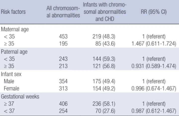
Table 7 shows the relative risk of all infants with chromosom- al abnormalities and CHD according to maternal age, paternal age, infant sex, and gestational age at delivery. The proportion of infants with DS who had a CHD did not differ by paternal age, infant sex, or gestational age. However, infants with DS born to mothers who were ≥ 35 yr of age were more likely to have a CHD than infants with DS born to mothers who were < 35 yr of age (relative risk, 1.467; 95% confidence interval [CI], 0.6-1.7).
DISCUSSION
This is the first nationwide study of CHDs in infants with DS in Korea. The total number of births in Korea was 888,263 in 2005- 2006, with 25,975 birth defects in 25,335 live births and 640 still- births. The prevalence of DS was 4.4 per 10,000 total births, ac- counting for 1.5% for all birth defects. We found that CHDs oc- curred in 56.3% of infants with DS, which is similar with the prev- alence presented in the previous literature (44%-58%). In our study, ASD was the most common CHD, occurring in 30.5% of all DS, followed by VSD (19.3%), PDA (17.5%), and AVSD (9.4%).
DS is the most common chromosomal abnormality in infants who survived birth, and its association with CHD is well estab- lished. Despite the increasing prenatal detection rate and ter-
mination of pregnancies due to DS diagnosis, there has been no change in the prevalence of DS for the last decade (14-17).
The prevalence of DS in our study was considerably lower than in previous studies conducted in Korea. The prevalence of DS was 14 and 26.7 per 10,000 total births in Shin et al. (18) and Jun et al. (19), respectively. However, since two previous studies were limited to only a few institutions and not nationwide, it may not reflect the overall prevalence of DS in Korea. Also, prenatal di- agnostic tests may have increased the rate of terminations of pregnancy after prenatal diagnosis of DS in Korea. That may have contributed low frequency of the present study.
AVSD, VSD, and ASD were the most common types of CHD, and previous studies have reported that the distribution of CHDs in infants with DS may vary according to ethnicity. Most studies regarding the distribution of CHD in DS were conducted in Cau- casian populations, where AVSD was reported to be the most common CHD. The few Asian studies have reported varied re- sults. For example, VSD has been reported in the range of 43.6%- 52.9% of all CHDs in the Asian population, while AVSD has been reported in the range of 11.8%-22.0% (20-22). In contrast, Cau- casian studies have reported that AVSD occurs in 35.7%-60.1%
of DS cases, while VSD occurs in 15.6-35.0% of cases (23, 24).
Our results were unique compared to those from prior Asian studies. ASD (30.5%) was the most common CHD in infants with DS, followed in frequency by VSD, PDA, and AVSD. We also found that AVSD accounted for only 9.4% of all CHDs, which is in stark contrast to the prevalence of AVSD in Caucasian studies.
Similar to findings in Mexican cases of DS, we found that ASD was the most common CHD, while AVSD was the least common, as reported in previous Asian studies. AVSD occurred in only 9% of all CHDs in Mexican infants with DS, whereas ASD, VSD, and PDA were more common. Furthermore, the total number of VSD cases (19.3%) in our results was much lower than in pre- vious Asian studies (6, 20, 25).
In our study, the AVSD was the fourth most prevalent CHD, accounting for 9.4% of CHDs in infants with DS. However, as a single heart defect, AVSD was the second most prevalent defect, occurring in 7.4% of all single heart defects, second only to ASD.
Table 6. Chromosome anomalies detected in infants with single heart defect, multiple heart defect, and extracardiac anomalies Birth defects No. (%) in birth defects
(n = 25,975) Congenital heart defects
(n = 11,031) Single heart defects
(n = 8,187) Multiple heart defects
(n = 2,844) Extracardiac anomalies (n = 15,986)
Trisomy 13 9 (0.03) 3 (33.3) 2 (22.2) 1 (11.1) 5 (55.6)
Trisomy 18 42 (0.17) 11 (26.2) 7 (16.7) 4 (9.5) 10 (23.8)
Down’s syndrome 394 (1.52) 224 (56.8) 122 (31.0) 102 (25.9) 64 (16.2)
Turner’s syndrome 32 (0.12) 6 (18.8) 4 (12.5) 3 (9.4) 13 (40.6)
Kleinfelter’s syndrome 13 (0.05) 3 (23.1) 2 (15.4) 1 (7.7) 5 (38.5)
Wolff-Hirschorn syndrome 4 (0.02) 1 (25.0) 1 (25.0) 0 (0) 3 (75)
Cri-du-chat syndrome 12 (0.05) 5 (41.7) 3 (25.0) 2 (16.7) 4 (33.3)
Other chromosomal anomalies 194 (0.74) 77 (39.7 ) 49 (25.3) 28 (14.4) 90 (46.4)
Total 700 (2.7) 330 (47.1) 190 (27.1) 140 (20.0) 195 (27.9)
Data are presented as No. (%) in each chromosomal anomaly unless otherwise specified.
Table 7. Adjusted risk factors of infants with CHD and chromosomal abnormalities Risk factors All chromosom-
al abnormalities
Infants with chromo- somal abnormalities
and CHD RR (95% CI)
Maternal age < 35 ≥ 35
453 195
219 (48.3) 85 (43.6)
1 (referent) 1.467 (0.611-1.724) Paternal age
< 35 ≥ 35
243 213
144 (59.3) 121 (56.8)
1 (referent) 0.931 (0.589-1.474) Infant sex
Male Female
354 313
175 (49.4) 154 (49.2)
1 (referent) 0.996 (0.674-1.467) Gestational weeks
≥ 37 < 37
406 254
236 (58.1) 70 (27.6)
1 (referent) 0.987 (0.612-1.467) CHD, congenital heart defect; RR, relative risk; CI, confidence interval.
Regarding the prevalence of CHD in infants with DS born to wo- men < 35 yr and those ≥ 35 yr, we found that mothers ≥ 35 yr were more likely to give birth to a DS child with a CHD.
It has been reported that the prevalence of CHD varies, de- pending on the presence of DS and other non-chromosomal abnormalities. For instance, the Baltimore-Washington Infant Study found AVSD in only 2.8% of non-DS cases, compared to 60.1% of DS cases. As indicated by various studies, several CHDs observed in non-chromosomal abnormalities are rare, although they are common in DS cases.
Advanced maternal age is a well-known risk factor for DS. In Korea, there is an ongoing trend towards delayed childbearing, and older mothers are known to have an increased risk of preg- nancies with chromosomal anomalies, including DS. An increase in the prevalence of DS will result in a significant increase in the cost of early diagnosis and effective management of CHD, as the current trend is to treat these infants in the same manner as non-syndromic individuals. In our study, maternal age, paternal age, and fetal sex ratio were not significantly different between DS infants with or without CHD.
Differences in the prevalence of ASD, VSD, PDA, AVSD, and CoA among patients with DS or non-chromosomal abnormali- ties were found to statistically significant. However, differences in the prevalence of PDA, TOF, AVSD, CoA, and DORV among patients with DS or non-chromosomal abnormalities were not statistically significant. Unfortunately, we cannot accurately perform more detailed statistical analysis, as the majority of the data had insufficient detail regarding medication taken during pregnancy, or maternal employment and living environment, which are all important environmental factors that could affect the prevalence of DS. Therefore, future research is needed for more accurate and objective analysis.
The reported frequencies of CHD may vary with the variabil- ity of classification criteria, the conduct of postmortem exami- nation, examiner experience, social and economic factors and so on. With advanced ultrasound technology and increasing experience and interest in prenatal screening, it may increase the detection rate of CHD. However, since postmortem exami- nations of stillborn infants are conducted less commonly in Ko- rea than in other countries, this may result in the underestima- tion of the frequency of CHD.
Our results indicate a need for timely identification and in- creased awareness of the development of CHD in DS, which might help decrease neonatal morbidity and increase overall health and wellness. It is necessary to assess and collect period- ic data, so that trends can be monitored for early recognition and optimal management of CHD in infants with DS. Our study contributes to the understanding of the effects of DS, and pro- vides the basis for identifying the distribution of CHD in infants with DS in Korea.
ACKNOWLEDGMENT
All of the authors made equal and significant contributions to acquisition of data, analysis and interpretation of data, writing the manuscript and final decision to submit for publication.
DISCLOSURE
All authors declare that they have no potential conflicts of interest.
ORCID
Min-A Kim http://orcid.org/0000-0002-6199-2010 You Sun Lee http://orcid.org/0000-0003-0336-4478 Nan Hee Lee http://orcid.org/0000-0002-2026-3321 Jeong Soo Choi http://orcid.org/0000-0002-7827-1930 Kyung Seo http://orcid.org/0000-0001-6657-3889 REFERENCES
1. Khoury MJ, Erickson JD. Improved ascertainment of cardiovascular mal- formations in infants with Down’s syndrome, Atlanta, 1968 through 1989.
Implications for the interpretation of increasing rates of cardiovascular malformations in surveillance systems. Am J Epidemiol 1992; 136: 1457- 64.
2. Hoffman JI, Kaplan S. The incidence of congenital heart disease. J Am Coll Cardiol 2002; 39: 1890-900.
3. Greenwood RD, Nadas AS. The clinical course of cardiac disease in Down’s syndrome. Pediatrics 1976; 58: 893-7.
4. Grech V. Epidemiology, and diagnostic and surgical trends in atrioven- tricular septal defect in Malta. Eur J Epidemiol 1999; 15: 403-5.
5. de Rubens Figueroa J, del Pozzo Magaña B, Pablos Hach JL, Calderón Jiménez C, Castrejón Urbina R. Heart malformations in children with Down syndrome. Rev Esp Cardiol 2003; 56: 894-9.
6. Jacobs EG, Leung MP, Karlberg J. Distribution of symptomatic congeni- tal heart disease in Hong Kong. Pediatr Cardiol 2000; 21: 148-57.
7. Busacca P, Pozzolini A, Minutiello L. Association between parachute mitral valve and Down’s syndrome. Report of a case. G Ital Cardiol 1998;
28: 1144-8.
8. Hoe TS, Chan KC, Boo NY. Cardiovascular malformations in Malaysian neonates with Down’s syndrome. Singapore Med J 1990; 31: 474-6.
9. Hamerton JL, Briggs SM, Giannelli F, Carter CO. Chromosome studies in detection of parents with high risk of second child with Down’s syn- drome. Lancet 1961; 2: 788-91.
10. Castilla EE, Rittler M, Dutra MG, Lopez-Camelo JS, Campaña H, Paz JE, Orioli IM. Survival of children with Down syndrome in South America.
ECLAMC-Downsurv Group. Latin American Collaborative Study of Con- genital Malformations. Am J Med Genet 1998; 79: 108-11.
11. Frid C, Drott P, Lundell B, Rasmussen F, Annerén G. Mortality in Down’s syndrome in relation to congenital malformations. J Intellect Disabil Res 1999; 43: 234-41.
12. Choi JS, Seo K, Han YJ, Lee SW, Bu YK, Lee SW, Shin CW, Yee NH. Con- genital anomaly survey and statistics. Seoul: Ministry of Heath & Welfare,
2009, p306.
13. Han YJ, Choi JS, Seo K, Shin SM, Oh HC, Lee SO, Lee SU, Hong JS, Lee ES, Kim ON. Infant and maternal mortality survey in 2005-2006. Seoul:
Korea Institute for Health and Social Affairs, Korean Medical Record As- sociation, Ministry for Health, Welfare and Family Affairs, 2008.
14. Parker SE, Mai CT, Canfield MA, Rickard R, Wang Y, Meyer RE, Ander- son P, Mason CA, Collins JS, Kirby RS, et al. Updated National Birth Prev- alence estimates for selected birth defects in the United States, 2004-2006.
Birth Defects Res A Clin Mol Teratol 2010; 88: 1008-16.
15. Morris JK, Alberman E. Trends in Down’s syndrome live births and an- tenatal diagnoses in England and Wales from 1989 to 2008: analysis of data from the National Down Syndrome Cytogenetic Register. BMJ 2009;
339: b3794.
16. Bittles AH, Glasson EJ. Clinical, social, and ethical implications of chang- ing life expectancy in Down syndrome. Dev Med Child Neurol 2004; 46:
282-6.
17. Matthews TJ, Hamilton BE. Delayed childbearing: more women are hav- ing their first child later in life. NCHS Data Brief 2009: 1-8.
18. Kwon JY, Park IY, Park YG, Lee Y, Lee G, Shin JC. Korean-specific param- eter models for calculating the risk of Down syndrome in the second tri- mester of pregnancy. J Korean Med Sci 2011; 26: 1619-24.
19. Park JW, Jun JK, Koo JN, Seo DK, Moon JB, Suh YH, Kim SI, OH KJ, Hong
JS, Kim BJ, et al. Prevalence of congenital anomalies in Korea: multi-cen- ter study. Korean J Ultra Obstet Gynecol 2011; 13: 148-156.
20. Lo NS, Leung PM, Lau KC, Yeung CY. Congenital cardiovascular mal- formations in Chinese children with Down’s syndrome. Chin Med J (Engl) 1989; 102: 382-6.
21. Hijii T, Fukushige J, Igarashi H, Takahashi N, Ueda K. Life expectancy and social adaptation in individuals with Down syndrome with and without surgery for congenital heart disease. Clin Pediatr (Phila) 1997;
36: 327-32.
22. Tandon R, Moller JH, Edwards JE. Tetralogy of Fallot associated with persistent common atrioventricular canal (endocardial cushion defect).
Br Heart J 1974; 36: 197-206.
23. Freeman SB, Taft LF, Dooley KJ, Allran K, Sherman SL, Hassold TJ, Kho- ury MJ, Saker DM. Population-based study of congenital heart defects in Down syndrome. Am J Med Genet 1998; 80: 213-7.
24. Park SC, Mathews RA, Zuberbuhler JR, Rowe RD, Neches WH, Lenox CC. Down syndrome with congenital heart malformation. Am J Dis Child 1977; 131: 29-33.
25. Matsuo N, Oshima M, Masuyoshi N, Shimizu K, Okada R. Major and minor anomalies in Japanese children with Down’s syndrome. Jpn Heart J 1972; 13: 307-16.