http://dx.doi.org/10.4174/astr.2014.86.5.232 Annals of Surgical Treatment and Research
Laparoscopic treatment of hepatic cysts located in the posterosuperior segments of the liver
Doo-Ho Lee, Jai Young Cho, Ho-Seong Han, Yoo-Seok Yoon, Dae Wook Hwang, Kyuwhan Jung, Young Ki Kim, Hong Kyung Shin, Woohyung Lee
Department of Surgery, Seoul National University Bundang Hospital, Seoul National University College of Medicine, Seongnam, Korea
INTRODUCTION
Since the first report of laparoscopic treatment of hepatic cysts in 1991 [1], this approach has gained wide acceptance and is frequently performed. Laparoscopy should therefore be considered for the treatment of hepatic cysts because of its low morbidity and cosmetic advantages in young patients [2].
However, the development of laparoscopic liver resection has been hindered by technical difficulties related to maintaining hemostasis of the transection plane, controlling hemorrhage, and visualizing and working with the deeper regions of the
liver [3,4].
Regarding cyst location, cysts located in the peripheral portion of the anterolateral segments of the liver (AL; segments II, III, V, VI, and the inferior part of IV) are considered suitable for laparoscopy [5-7]. However, lesions located in the posterosuperior segments of the liver (PS; segments I, VII, VIII, and the superior part of IV) are considered poor candidates for laparoscopic resection because of limited visualization and the difficulty of controlling bleeding [8-13]. Although laparoscopic left lateral sectionectomy is a routine approach, laparoscopic major liver resection is still preserved for experienced surgeons Purpose: Laparoscopy is considered the treatment of choice for hepatic cysts, especially those located in anterolateral segments (AL; segments II, III, IVb, V, and VI) because of the ease of laparoscopic access. Here, we evaluated the feasibility and safety of laparoscopic treatment of hepatic cysts in posterosuperior segments (PS; segments I, IVa, VII, and VIII).
Methods: We retrospectively analyzed clinical data for 34 patients who underwent laparoscopic treatment of hepatic cysts between September 2004 and December 2012. Patients were divided into two groups depending on whether the main largest cyst was located in AL (n = 20) or PS (n = 14). Laparoscopic cyst unroofing was performed in 29 patients with symptomatic simple cysts. Laparoscopic resection was performed in 5 patients with suspected cystic neoplasms.
Results: There were no deaths or major complications. The mean operation time was 110 minutes and the mean hospital stay was 4.4 days. The mean cyst size was not significantly different (P = 0.511) but the frequency of multiple cysts was significantly greater in group PS (P = 0.003). The predominant type of resection was unroofing in both groups (P = 0.251).
The mean blood loss (P = 0.747), mean hospital stay (P = 0.812), mean operation time (P = 0.669), morbidity rate (P = 0.488), and relapse rate (P = 0.448) were not significantly different. Relapse occurred in one patient who underwent reunroofing 17 months later. The median follow-up is 62 months.
Conclusion: Laparoscopy is a safe procedure for hepatic cysts located in posterosuperior segments.
[Ann Surg Treat Res 2014;86(5):232-236]
Key Words: Laparoscopy, Hepatic cyst, Posterosuperior
Received November 29, 2013, Reviewed December 8, 2013, Accepted December 20, 2013
Corresponding Author: Jai Young Cho
Department of Surgery, Seoul National University Bundang Hospital, 82 Gumi-ro 173beon-gil, Bundang-gu, Seongnam 463-707, Korea
Tel: +82-31-787-7098, Fax: +82-31-787-4078 E-mail: [email protected]
Copyright ⓒ 2014, the Korean Surgical Society
cc Annals of Surgical Treatment and Research is an Open Access Journal. All articles are distributed under the terms of the Creative Commons Attribution Non- Commercial License (http://creativecommons.org/licenses/by-nc/3.0/) which permits unrestricted non-commercial use, distribution, and reproduction in any medium, provided the original work is properly cited.
[14]. Therefore, laparoscopy was often avoided when possible malignant hepatic cysts were detected in the posterosuperior segments of the liver because major liver resection is sometimes required for such lesions [15].
However, improved laparoscopic techniques, better surgical field visualization with flexible laparoscopes, and routine use of laparoscopic cavitron ultrasonic surgical aspirators in transecting deeper portions of the liver parenchyma have helped address the major limitations of laparoscopy [15].
Although many reports have described laparoscopic treat- ment of hepatic cysts, few have analyzed the feasibility and outcomes of laparoscopic treatment according to cyst location.
Given these facts, we performed a retrospective study to evaluate the feasibility and operative outcomes of laparoscopic resection for hepatic cysts located in the PS segments of the liver compared with those for cysts located in the AL segments.
METHODS
Indications for operation
In our institution, laparoscopy is routinely considered as the treatment of choice for hepatic cysts. Treatment of hepatic cyst is indicated for symptomatic cysts (severe pain, nausea, vomiting, or jaundice), rapidly growing cysts, or if a malignancy is suspected. Laparoscopic unroofing was performed for benign cysts (n = 29), while laparoscopic liver resection was performed for suspected malignant cystic neoplasms, including biliary cystadenoma/carcinoma or intraductal papillary neoplasms (n = 5).
Operative technique
Briefly, laparoscopic unroofing was performed under general anesthesia with the patient placed in the supine or lithotomy position. For cysts located in the posterosuperior segments, the
surgeon usually stood between the lower limbs of the patient.
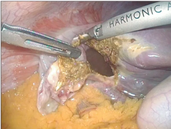
For cysts located in the left segments of the liver, laparoscopy was performed with the patient in the supine position, with a 30o Trendelenburg adjustment. The surgeon stood on the right side of the patient. Pneumoperitoneum was established through an 11 mm umbilical port and was maintained at <12 mmHg to reduce the risk of air embolism. A flexible laparoscope was routinely used. The dome of the cyst was opened using a harmonic scalpel (Ethicon, Cincinnati, OH, USA) after aspirating the fluid in the cyst using a laparoscopic aspirator (Fig. 1). The cystic cavity was examined to identify possible indentations that could indicate neoplastic changes; in such cases, liver resection was necessary. The wall of the cyst was excised, including the 3– to 4-mm cystic rim of the hepatic parenchyma (Fig. 2). The cyst and liver parenchyma was sent for pathology. A hemostatic clip or tie suture was applied if the surgeon detected the presence of bile along the cystic edge, indicating injury to the septal bile ducts [16]. Hemostasis of the cyst edge was achieved using electrocautery and an argon beam coagulator. If feasible, the greater omentum was packed into larger cavities to decrease dead space and prevent fluid collection. Examination of frozen tissue sections revealed no evidence of cystadenoma or carcinoma in any case.
Laparoscopic hepatectomy was performed if the cysts had malignant features such as mural nodules, an intracystic septum, or a honeycomb appearance. We performed laparo- scopic left lateral sectionectomy in three patients, right hemihepatectomy in one patient, and left hemihepatectomy in one patient. The operative techniques for laparoscopic liver resection used at our institution have been described elsewhere [15,17,18]. Laparoscopic anatomical liver resection was preferred to clear all of the affected segments.
Fig. 1. Aspiration of cystic fluid without spillage before
laparoscopic unroofing. Fig. 2. Resection of the cystic rim of the liver parenchyma using ultrasonic shears.
Study population and patient categorization
Between September 2004 and December 2012, laparoscopy was performed to treat 34 patients with hepatic cysts at our hospital. Laparoscopic unroofing was performed in 29 pa- tients, laparoscopic left lateral sectionectomy in 3 patients, laparoscopic left hemihepatectomy in 1 patient, and laparo- scopic right hemihepatectomy in 1 patient. The patients were classified into two groups according to whether the main cyst(s) were located in the anterolateral segments (group AL, n = 20) or in the posterosuperior segments (group PS, n = 14).
Statistics
Continuous, normally distributed variables are presented as the mean ± standard deviation, while discontinuous vari ables are expressed as the median (range). Continuous para meters were compared between the anterolateral and postero superior groups using independent-samples t-test, and catego rical parameters were compared using the chi-square test.
All analyses were performed using PASW ver. 18.0 (SPSS Inc., Chicago, IL, USA. Differences were considered significant at values of P < 0.05.
RESULTS
Preoperative demographic data
The preoperative characteristics of patients are summarized in Table 1. The mean age of patients was 64.5 years. There were more females than males, and single large cysts were more frequently found than multiple cysts. There were no differences in the mean cyst size between the two groups (P = 0.511).
However, the frequency of multiple cysts was significantly greater in group PS than in the group AL (P = 0.003).
Procedures performed
Table 1 lists the procedures performed in these patients.
Unroofing was performed in 16 and 13 patients in group AL and group PS, respectively. Laparoscopic hepatectomy was performed in 5 patients, which included laparoscopic left lateral sectionectomy in 3, right hemihepatectomy in 1, and left hemihepatectomy in 1. Four laparoscopic hepatectomies were performed in group AL and one laparoscopic hepatectomy was performed in group PS (P = 0.251). The predominant type of resection was unroofing in both groups.
Intraoperative and postoperative outcomes
There were no deaths or major complications. The intraopera-
Table 1. Characteristics of patients who underwent laparoscopic treatment of hepatic cysts (n = 34)
Characteristic Total (n = 34) Group AL (n = 20) Group PS (n = 14) P-value
Demographic factors
Age (yr), mean ± SD 64.5 ± 12.4 65.5 ± 10.3 63 ± 15.3 0.366
Sex, male/female 8/26 6/14 2/12 0.288
Cyst characteristics
Size (cm), mean ± SD 9.6 ± 4.8 9.75 ± 5.8 9.4 ± 3.6 0.511
Single/multiple 22/12 17/3 5/9 0.003
Type of operation 0.251
Unroofing 29 16 13
Left lateral sectionectomy 3 3 0
Right hemihepatectomy 1 0 1
Left hemihepatectomy 1 1 0
AL, anterolateral; PS, posterosuperior; SD, standard deviation.
Table 2. Intraoperative and postoperative outcomes (n = 34)
Variable Total (n = 34) Group AL (n = 20) Group PS (n = 14) P-value
Conversion 0 0 0
Operation time (min) 110.7 (25–525) 131.7 (25–525) 80.7 (35–245) 0.669
Blood loss (mL) 120.5 (5–900) 161.2 (5–900) 62.5 (5–600) 0.747
Postoperative hospital stay (day) 5 (2–11) 5 (2–11) 4 (1–7) 0.812
Morbidity 1 1 0 0.488
Relapse 1 0 1 0.448
AL, anterolateral; PS, posterosuperior.
tive and postoperative results are listed in Table 2. Relapse occurred in one patient and reunroofing of the recurred hepatic cysts was performed 17 months after the initial unroofing. The median follow-up is 62 months. The mean overall operation time was 110 minutes and estimated blood loss was 120.5 mL.
Pathologic results showed that all of the resected cysts were benign simple cysts. Mean operation time (P = 0.669) and mean blood loss (P = 0.747) were not significantly different between the two groups. The mean postoperative hospital stay was 4.4 ± 2.4 days. One patient in group AL who developed hospital-acquired pneumonia at postoperative day 5 was treated with intravenous antibiotics and discharged 7 days later. Mean hospital stay (P = 0.812), the morbidity rate (P = 0.488), and the relapse rate (P = 0.448) were not significantly different between the two groups.
DISCUSSION
Simple hepatic cysts are usually asymptomatic and are not associated with abnormal hepatic function. However, they may become symptomatic as they expand [19]. Complications such as rupture, infection, or intracystic hemorrhage may occur.
Simple aspiration results in recurrence in all patients, and has been abandoned in clinical practice. Aspiration combined with sclerotherapy shows limited short-term success and can result in abscess formation. The goal of surgical treatment of giant solitary cysts is to decompress the cyst and avoid recurrence.
Current management involves cyst unroofing [20], which is commonly performed laparoscopically [21,22].
Despite increasing laparoscopic and hepatic surgery experien- ce, laparoscopic resection of hepatic cysts is limited to patients with cysts localized to the anterolateral segments of the liver [23]. Limitations of laparoscopic resection compared with open resection of hepatic cysts include poor visualization and difficulty in controlling bleeding, especially for cysts located in the posterosuperior segments of the liver [23]. The most common reasons for conversion to laparotomy include the presence of cysts deep within the hepatic parenchyma, cysts located in segments VI, VII, and VIII, uncontrolled bleeding, or major biliary injury [10,24,25] Lesions located in the posterior or superior parts of the liver (segments I, VII, VIII, and the superior part of IV) have long been considered poor candidates for laparoscopic resection because of limited visualization and difficulty in controlling bleeding [8,9].
The findings of the present study showed that the outcomes of laparoscopic resection of hepatic cysts located in the
posterosuperior segments were comparable with those of cysts in the anterolateral segments. In particular, we found no significant differences between the two patient groups in terms of median operation time, median blood loss, median hospital stay, morbidity rate, or relapse rate. Moreover, there were no conversions during laparoscopic treatment, regardless of the location of the cysts. Morbidity occurred in one patient in the anterolateral group: this patient developed hospital-acquired pneumonia at postoperative day 5. Meanwhile, one patient in the posterosuperior group experienced relapse
Solitary cysts located in segments VII and VIII of the liver can be safely treated by laparoscopic unroofing. Cyst recurrence may be prevented by complete excision of the cystic roof together with the adjacent hepatic parenchyma [16]. Laparoscopic resection of tumors located in the posterosuperior segments of the liver is more difficult than that in the anterolateral segments, but the posterosuperior location does not compro- mise accessibility or surgical outcome [2]. Laparoscopic surgery can be safely performed in patients with giant solitary cysts, regardless of their location [13].
We performed laparoscopic liver resection for suspected neoplasms, although the pathologic results confirmed that all of the cysts were benign. Although it is easy to attribute symptoms to the presence of a large hepatic cyst, the possibility of a coexisting malignancy must be excluded. The main problem is that it is difficult to detect neoplasms before and during surgery, despite improved imaging techniques [26]. It is also difficult to distinguish between congenital and neoplastic liver cysts, leading to inappropriate treatment that may increase the risk of tumor recurrence and reoperation [27]. Indeed, in patients with complicated liver cysts, with hemorrhage or superin fection for example, radiologic differentiation of benign and neoplastic cysts may be especially difficult [15,17]. Thus, during laparoscopic treatment of such lesions, surgeons must be especially vigilant for the appearance of unusual cystic fluid and must carefully inspect the internal portion of the cystic wall; if abnormalities are found, liver resection should be performed instead of unroofing [17,28].
In conclusion, our results indicate that laparoscopic treatment of hepatic cysts located in the posterosuperior segments of the liver is safe and feasible.
CONFLICTS OF INTEREST
No potential conflict of interest relevant to this article was reported.
1. Paterson-Brown S, Garden OJ. Laser- assisted laparoscopic excision of liver cyst. Br J Surg 1991;78:1047.
2. Koffron A, Geller D, Gamblin TC, Abecassis M. Laparoscopic liver surgery:
Shifting the management of liver tumors.
Hepatology 2006;44:1694-700.
3. Buell JF, Thomas MJ, Doty TC, Gersin KS, Merchen TD, Gupta M, et al. An initial experience and evolution of laparoscopic hepatic resectional surgery. Surgery 2004;136:804-11.
4. Morino M, Morra I, Rosso E, Miglietta C, Garrone C. Laparoscopic vs open hepatic resection: a comparative study. Surg Endosc 2003;17:1914-8.
5. Cherqui D, Husson E, Hammoud R, Malassagne B, Stephan F, Bensaid S, et al.
Laparoscopic liver resections: a feasi bility study in 30 patients. Ann Surg 2000;232:
753-62.
6. Gigot JF, Glineur D, Santiago Azagra J, Goergen M, Ceuterick M, Morino M, et al.
Laparoscopic liver resection for malignant liver tumors: preliminary results of a multi center European study. Ann Surg 2002;236:90-7.
7. Kaneko H, Takagi S, Shiba T. Laparoscopic partial hepatectomy and left lateral segmentectomy: technique and results of a clinical series. Surgery 1996;120:468-75.
8. Dulucq JL, Wintringer P, Stabilini C, Berticelli J, Mahajna A. Laparoscopic liver resections: a single center experience.
Surg Endosc 2005;19:886-91.
9. Laurent A, Cherqui D, Lesurtel M, Brunetti F, Tayar C, Fagniez PL. Laparoscopic liver resection for subcapsular hepatocellular carcinoma complicating chronic liver disease. Arch Surg 2003;138:763-9.
10. Morino M, De Giuli M, Festa V, Garrone C. Laparoscopic management of sympto- matic nonparasitic cysts of the liver. Indi-
cations and results. Ann Surg 1994;219:
157-64.
11. Gigot JF, Metairie S, Etienne J, Horsmans Y, van Beers BE, Sempoux C, et al. The surgical management of congenital liver cysts. Surg Endosc 2001;15:357-63.
12. Katkhouda N, Hurwitz M, Gugenheim J, Mavor E, Mason RJ, Waldrep DJ, et al.
Laparoscopic management of benign solid and cystic lesions of the liver. Ann Surg 1999;229:460-6.
13. Petri A, Hohn J, Makula E, Kokai EL, Savanya GK, Boros M, et al. Experience with different methods of treatment of nonparasitic liver cysts. Langenbecks Arch Surg 2002;387:229-33.
14. Buell JF, Cherqui D, Geller DA, O'Rourke N, Iannitti D, Dagher I, et al. The interna- tional position on laparoscopic liver sur- gery: The Louisville Statement, 2008. Ann Surg 2009;250:825-30.
15. Cho JY, Han HS, Yoon YS, Shin SH. Feasi- bility of laparoscopic liver resection for tumors located in the posterosuperior seg ments of the liver, with a special referen ce to overcoming current limita- tions on tumor location. Surgery 2008;
144:32-8.
16. Weber T, Sendt W, Scheele J. Laparoscopic unroofing of nonparasitic liver cysts within segments VII and VIII: technical considerations. J Laparoendosc Adv Surg Tech A 2004;14:37-42.
17. Han HS, Cho JY, Yoon YS. Techniques for performing laparoscopic liver resection in various hepatic locations. J Hepatobiliary Pancreat Surg 2009;16:427-32.
18. Cho JY, Han HS, Yoon YS, Shin SH.
Outcomes of laparoscopic liver resection for lesions located in the right side of the liver. Arch Surg 2009;144:25-9.
19. Sanchez H, Gagner M, Rossi RL, Jenkins RL, Lewis WD, Munson JL, et al. Surgical
management of nonparasitic cystic liver disease. Am J Surg 1991;161:113-8.
20. Litwin DE, Taylor BR, Langer B, Greig P.
Nonparasitic cysts of the liver. The case for conservative surgical management.
Ann Surg 1987;205:45-8.
21. Krahenbuhl L, Baer HU, Renzulli P, Z’grag- gen K, Frei E, Buchler MW. Laparo scopic management of nonparasitic symp tom- producing solitary hepatic cysts. J Am Coll Surg 1996;183:493-8.
22. Fabiani P, Katkhouda N, Iovine L, Mouiel J.
Laparoscopic fenestration of biliary cysts.
Surg Laparosc Endosc 1991;1:162-5.
23. Kaneko H, Takagi S, Otsuka Y, Tsuchiya M, Tamura A, Katagiri T, et al. Laparoscopic liver resection of hepatocellular carcino- ma. Am J Surg 2005;189:190-4.
24. Yoon YS, Han HS, Cho JY, Kim JH, Kwon Y.
Laparoscopic liver resection for centrally located tumors close to the hilum, major hepatic veins, or inferior vena cava. Sur- gery 2013;153:502-9.
25. Park JS, Han HS, Hwang DW, Yoon YS, Cho JY, Koh YS, et al. Current status of laparoscopic liver resection in Korea. J Korean Med Sci 2012;27:767-71.
26. Yoon YS, Han HS, Cho JY, Ahn KS. Total laparoscopic liver resection for hepato- cellular carcinoma located in all segments of the liver. Surg Endosc 2010;24:1630-7.
27. Kim MH, Lee J, Yoo DS, Lee YG, Byeon SE, Hong EK, et al. Protective effect of stress- induced liver damage by saponin fraction from Codonopsis lanceolata. Arch Pharm Res 2009;32:1441-6.
28. Choi JH, Ahn BM, Yi J, Lee JH, Lee JH, Nam SW, et al. MRP2 haplotypes confer differential susceptibility to toxic liver injury. Pharmacogenet Genomics 2007;17:
403-15.