Letter to the Editor
138 Ann Dermatol
Received November 24, 2014, Revised April 2, 2015, Accepted for publication April 8, 2015
Corresponding author: Dong Hoon Shin, Department of Dermatology, Yeungnam University College of Medicine, 170 Hyeonchung-ro, Nam-gu, Daegu 42415, Korea. Tel: 82-53-620-3160, Fax: 82-53-622-2216, E-mail: dhshin@med.yu.ac.kr
This is an Open Access article distributed under the terms of the Creative Commons Attribution Non-Commercial License (http://
creativecommons.org/licenses/by-nc/4.0) which permits unrestricted non-commercial use, distribution, and reproduction in any medium, pro- vided the original work is properly cited.
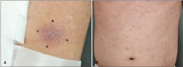
Fig. 1. (A) A 2×2-cm-sized hyperpigmented nodule on the left arm. (B) Multiple miliary grain to rice-grain-sized erythematous to brownish macules and papules on the trunk.
http://dx.doi.org/10.5021/ad.2016.28.1.138
Secondary Granulomatous Cutaneous Involvement in Peripheral T-cell Lymphoma
Byeong Su Kim, Yeon Woong Kim, Jin Hwa Choi, Dong Hoon Shin, Jong Soo Choi
Department of Dermatology, Yeungnam University College of Medicine, Daegu, Korea
Dear Editor:
A 40-year-old Korean man was referred to our department with a 1-month history of brownish macules and papules on the trunk, and a 10-day history of 2 walnut-sized brownish nodules on the left arm (Fig. 1). The patient had been experiencing persistent coughing for 1 month, and computed tomography of the chest revealed multiple re- active mediastinal lymph nodes. A skin biopsy examina- tion of nodules on the left arm revealed small-to-me- dium-sized pleomorphic lymphocytes in the upper der- mis, which were positive for cluster of differentiation CD3 and CD4, and negative for CD8, epidermotropism, and granuloma formation (Fig. 2). Some histiocytic cells were positive for CD68. A biopsy of the abdomen revealed epi-
dermotropism and lymphocytes containing hyper- chromatic and indented nuclei, but without granuloma formation. Molecular biological investigation of the skin biopsy samples showed monoclonal T-cell receptor-γ gene rearrangement. Bone marrow biopsy findings were nor- mal, but positron emission tomography scanning showed multiple lymphadenopathies and testicular involvement.
Biopsy of the testes showed T-cell lineage malignant lym- phoma without granulomatous features. Based on the clin- ical, histological, and molecular genetic features, periph- eral T-cell lymphoma, not otherwise specified, World Health Organization classification with secondary gran- ulomatous cutaneous involvement, was diagnosed.
Microbiological (bacterial, fungal, mycobacterial) studies
Letter to the Editor
Vol. 28, No. 1, 2016 139 Fig. 2. (A) Dermal, perivascular nodular inflammatory cell infiltra- tion. (B) Epidermotropism of lym- phocytes, with hyperchromatic and indented nucleus. (C) Many his- tiocytes and atypical lymphocytes with hyperchromatic nuclei form sarcoid-like granuloma (H&E; A:
×40, B: ×200, C: ×200).
could not detect any specific infectious agent. Sarcoidosis or pulmonary tuberculosis was not evident in the com- plete hematological, biochemical, and radiological exa- minations. The patient was treated with narrow band ultra- violet B phototherapy (twice a week, suberythemal dose) and systemic chemotherapy (cyclophosphamide, doxor- ubicin, vincristine, prednisolone). Fourteen days after the initial treatment, the macules on the abdomen subsided and the sizes of the two nodules on the left arm were reduced.
Granuloma formation is rarely observed in secondary cu- taneous lesions from T-cell lymphomas whose original le- sions showed no granulomatous features. Granulomatous lymphomas (both primary and secondary) are reported to represent 1.6% of all lymphomas1-3. Sarcoid-like gran- ulomas are most common, but tuberculoid, granuloma an- nulare-like, granulomatous rosacea, or granulomatous panniculitis types have also been reported2. In addition, various granulomatous patterns may coexist in the same patient1.
The exact pathogenesis of secondary granulomatous cuta- neous involvement in peripheral T-cell lymphoma remains unclear. It has been proposed that epithelioid cell reaction indicates secretion of chemotactic or migration-inhibiting lymphokines by the neoplastic T-cell, and these cytokines may in turn be responsible for attracting monocytes, there- by resulting in granuloma formation1. However, the prog- nostic value of this epithelioid response remains con-
troversial. In contrast, some reports have suggested that granulomatous inflammation represents a host response against the tumor and may represent a good prognostic factor for the clinical behavior of cutaneous lymphoma4. However, because of the limited number of cases reported to date, determining a direct relationship between the presence of a granulomatous inflammatory infiltrate and the prognosis of secondary cutaneous lymphomas is difficult.
REFERENCES
1. Gallardo F, García-Muret MP, Servitje O, Estrach T, Bielsa I, Salar A, et al. Cutaneous lymphomas showing prominent granulomatous component: clinicopathological features in a series of 16 cases. J Eur Acad Dermatol Venereol 2009;
23:639-647.
2. Pujol RM, Gallardo F, Servitje O, Martí RM, Bordes R, García-Muret MP, et al. Peripheral T-cell lymphoma with secondary epithelioid granulomatous cutaneous involve- ment: a clinicopathologic study of four cases. J Dermatol 2005;32:541-548.
3. Patsouris E, Noël H, Lennert K. Histological and immu- nohistological findings in lymphoepithelioid cell lymphoma (Lennert's lymphoma). Am J Surg Pathol 1988;12:341-350.
4. Flaxman BA, Koumans JA, Ackerman AB. Granulomatous mycosis fungoides. A 14-year follow-up of a case. Am J Dermatopathol 1983;5:145-151.