INTRODUCTION
Recent large randomized controlled studies have
demonstrated the usefulness of multi-parametric mag- netic resonance imaging (mpMRI) for prostate cancer (PCa) detection [1,2]. Target biopsy based on mpMRI
Received: May 19, 2019 Revised: Jun 24, 2019 Accepted: Jun 26, 2019 Published online Jul 30, 2019 Correspondence to: Sung-Woo Park https://orcid.org/0000-0002-9895-3461
Department of Urology, Pusan National University Yangsan Hospital, 20 Geumo-ro, Mulgeum-eup, Yangsan 50612, Korea.
Tel: +82-55-360-2672, Fax: +82-55-360-2164, E-mail: [email protected] Copyright © 2020 Korean Society for Sexual Medicine and Andrology
Usefulness of Bi-Parametric Magnetic Resonance Imaging with b=1,800 s/mm 2 Diffusion-Weighted Imaging for Diagnosing Clinically Significant
Prostate Cancer
Seung Soo Lee1 , Dong Hoon Lee1, Won Hoon Song1 , Jong Kil Nam1 , Ji Yeon Han1 , Hyun Jung Lee2 , Tae Un Kim3 , Sung-Woo Park1,4
Departments of 1Urology, 2Pathology, and 3Radiology, 4Research Institute for Convergence of Biomedical Science and Technology, Pusan National University Yangsan Hospital, Yangsan, Korea
Purpose:
Purpose: This study was conducted to compare the accuracy of bi-parametric magnetic resonance imaging (bpMRI) with high b-value (b=1,000 s/mm2, b1000) diffusion-weighted imaging (DWI) to that of bpMRI with ultra-high b-value (b=1,800 s/mm2, b1800) DWI to detect clinically significant prostate cancer (csPCa).
Materials and Methods:
Materials and Methods: A total of 408 patients with suspected PCa were evaluated by bpMRI prior to biopsy. One reader ret- rospectively reviewed all images for confirmation of Prostate Imaging–Reporting and Data System (PI-RADS) score. Cognitive magnetic resonance/ultrasound fusion target biopsy was done for all visible lesions (PI-RADS 3–5). Systematic biopsy was done for all cases. The csPCa detection rates were compared according to the bpMRI protocol (with/without b1800 DWI) or PI-RADS score. The accuracy of PI-RADS score was estimated using receiver operating characteristics curve. The signal inten- sity (SI) ratio (visible lesion/surrounding background) was evaluated.
Results:
Results: Among 164 men confirmed having PCa, 102 had csPCa (Gleason score≥7). Proportions of PI-RADS score 1–2/3/4/5 without b1800 DWI (n=133) and with b1800 DWI (n=275) were 19.5%/57.9%/15.8%/6.8% and 21.1%/48.7%/22.2%/8.0%, respectively. csPCa detection rates with/without b1800 DWI were 27.6%/19.5% (p=0.048), respectively. Areas under the curve of PI-RADS grading with/without b1800 DWI for csPCa detection were 0.885 and 0.705, respectively. The SI ratio in b1800 DWI was higher than that in b1000 DWI (p<0.001).
Conclusions:
Conclusions: Adding b1800 DWI to bpMRI protocol improved the diagnostic accuracy and detection rate of csPCa. The higher SI ratio (lesion/background) in b1800 DWI enabled clearer identification of lesions.
Keywords:
Keywords: Diagnostic imaging; Image-guided biopsy; Magnetic resonance imaging; Prostatic neoplasms
This is an Open Access article distributed under the terms of the Creative Commons Attribution Non-Commercial License (http://creativecommons.org/licenses/by-nc/4.0) which permits unrestricted non-commercial use, distribution, and reproduction in any medium, provided the original work is properly cited.
pISSN: 2287-4208 / eISSN: 2287-4690 World J Mens Health 2020 Jul 38(3): 370-376 https://doi.org/10.5534/wjmh.190079
has a higher detection rate for clinically significant PCa (csPCa) than conventional random systematic bi- opsy used as a standard tool so far [1]. In the past few years, the use of mpMRI has made tremendous chang- es in the diagnosis of PCa.
Prostate Imaging–Reporting and Data System (PI- RADS) version 2 is the most widely used evaluation criteria for magnetic resonance imaging (MRI) find- ings of PCa [3]. Following PI-RADS version 2 guideline, T2-weighted imaging (T2WI), apparent diffusion coef- ficient (ADC) maps, high b-value diffusion-weighted imaging (DWI), and dynamic contrast-enhanced (DCE) imaging are used. T2WI is a key image for transitional zone lesion, while ADC map using various b-value DWI is the most valuable technique for characterizing pe- ripheral zone lesion. Compared to PI-RADS version 1 guideline, the need for DCE is minimized in PI-RADS version 2. DCE images are useful only for peripheral zone lesion with PI-RADS 3 to distinguish PI-RADS 4 from 3. Biopsy is even necessary in both scores. Howev- er, burdens of mpMRI including additional cost, acqui- sition time, and side effects of contrast agent are still present. For these reasons, a simple and standardized MRI protocol is needed to replace mpMRI including DCE. Recently, bi-parametric MRI (bpMRI) including T2WI and DWI has a similar diagnostic accuracy and does not use contrast agent [4,5].
High b-value DWI has been emphasized in PI-RADS version 2 guideline. The signal intensity (SI) of the normal prostate gland is often not suppressed in DWI, even when b-value=1,000 s/mm2 (b1000) are used [6].
Higher b-value DWI would be more advantageous in detecting relatively small diffusion changes such as those between the normal prostate parenchyma and foci of PCa, thus yielding greater tissue diffusivity and less T2 shine-through effect [7]. Unfortunately, there is still no consensus regarding the optimal b-value as a high b-value DWI. Several small-scale studies have determined the accuracy of PCa detection using vari- ous b-value DWI [8-12]. They found that b-values in the range of 1,500 to 2,500 s/mm2 were optimal for PCa detection [12]. Although higher b-value DWI clearly shows the lesion from the background, it has lower sensitivity for detecting PCa.
With recent development of MRI technology, most institutions have been able to use ultra-high b-value (b≥1,800 s/mm2) DWI. Therefore, creating a simple and widely available standard MRI protocol using ultra-
high b-value DWI is possible. The objective of this study was to determine the benefit of using ultra-high b-value (b=1,800 s/mm2, b1800) DWI and to compare this bpMRI protocol with high b-value (b=1,000 s/mm2, b1000) DWI to detect csPCa.
MATERIALS AND METHODS
1. Ethics statement
This retrospective, single-center study was approved by Pusan National University Yangsan Hospital In- stitutional Review Board (IRB) (No. 05-2016-007). Data analysis was waived review and consent by the IRB Board, as all data was being analyzed retrospectively, after de-identification.
2. Subject and study methods
From January 2014 to July 2018, 408 men who un- derwent prostate biopsy and bpMRI before biopsy at Pusan National University Yangsan Hospital were enrolled. Two urologists (SWP and DHL) who had 8 and 5 years of experience in prostate MRI, respectively, performed systematic biopsy in all men and cognitive target biopsy for visible lesion (PI-RADS 3–5). A geni- tourinary pathologist reviewed and described all biopsy cores. For each positive biopsy core of PCa, Gleason score and cancer core length were reported. A csPCa was defined as the presence of a single biopsy core in- dicating disease of Gleason score 3+4 (Gleason sum of 7) or greater [2].
In our institute, all patients underwent 3.0 T MRI (InteraAchieva 3.0 T; Phillips Medical System, Best, Netherlands) on an instrument equipped with a phased-array coil (six channels). For bpMRI, axial T2WI for anatomical evaluation and DWI scans were acquired within 4 and 8 minutes, respectively. Using three b-values (50, 400, and 1,000), diffusion restriction was quantified as ADC map. The b1800 DWIs were obtained by additional sequence. We divided the MRI protocol and compared the results of the radiologic and pathologic results in each protocol. MRI protocol A (n=133) included T2WI, ADC, and b1000 DWI, while MRI protocol B (n=275) included T2WI, ADC, b1000, and b1800 DWI. MRI protocol A was performed until December 2015. Subsequent patients were evaluated using MRI protocol B since January 2016. Without any clinical information, one reader (SWP) retrospectively reviewed all images for confirmation of PI-RADS score.
In men who were evaluated by MRI protocol B, SIs of visible lesions (PI-RADS 3–5) and adjacent back- ground were evaluated manually by tracing a region of interest within an identified lesion on b1000 and b1800 DWIs corresponding to the location of the index lesion on the ADC map [12]. The SI ratio of the lesion/
background was then calculated for each patient.
All patients were prepared with local gel anesthet- ics using a BK ultrasound scanner (BK Medical, Pea- body, MA, USA), an endfire transducer, a needle guide, and an 18G 25 cm biopsy needle. After the operator reviewed the MRI, any suspicious lesions on MRI (PI- RADS 3–5 lesion) were visually matched and registered on the corresponding axial transrectal ultrasonography image based on zonal anatomy. All target biopsy cores were marked and kept in separate bottles according to the location of the suspicious lesion on MRI. After tar- get biopsy, systematic biopsy cores were collected from 12 prostatic regions and marked separately. Patients without suspicious lesions (PI-RADS 1–2) on MRI un- derwent systematic biopsy only.
Demographic, pathologic, and radiologic results of patients were compared between MRI protocol A and MRI protocol B groups using chi-square test and t-test for categorical and continuous variables, respectively.
PCa detection rates and proportions of csPCa according to MRI protocol and PI-RADS score were compared.
The odds ratio of b1000 and b1800 SI ratio (lesion/
background) for csPCa detection in PI-RADS 3–5 lesion with MRI protocol B were evaluated. For binary logis- tic regression test, the cutoff of the SI ratio was 1.3 and 1.5 for b1000 and b1800 DWI, respectively. The distri- bution of SIs are shown using boxplots. All comparing analyses were performed using PASW ver. 18.0 (IBM Corp., Armonk, NY, USA). Statistical significance was considered at p≤0.05. Using MedCalc ver. 18.6 (MedCalc Software, Ostend, Belgium), the receiver operating characteristic curve for csPCa detection was analyzed.
For each MRI protocol, specificity, sensitivity, positive predictive value, and negative predictive value were also evaluated.
RESULTS
The median age, prostate-specific antigen, and pros- tate volume of the overall cohort were 64 years old (interquartile range [IQR]: 58–71 years old), 6.8 ng/mL (IQR: 5.0–9.5 ng/mL), and 36.2 mL (IQR: 27–50 mL), respectively. Table 1 shows no significant demographic characteristics difference in both groups. Among 164 men confirmed with PCa, 102 had csPCa. Although the detection rate of overall PCa in each group was not significantly different (38.3% vs. 41.1%, p=0.667), the csPCa detection rate using MRI protocol B (27.6%) was
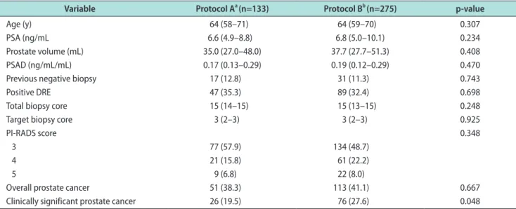
Table 1. Demographic, radiological, and pathological characteristics according to magnetic resonance protocol
Variable Protocol Aa (n=133) Protocol Bb (n=275) p-value
Age (y) 64 (58–71) 64 (59–70) 0.307
PSA (ng/mL 6.6 (4.9–8.8) 6.8 (5.0–10.1) 0.234
Prostate volume (mL) 35.0 (27.0–48.0) 37.7 (27.7–51.3) 0.408
PSAD (ng/mL/mL) 0.17 (0.13–0.29) 0.19 (0.12–0.29) 0.470
Previous negative biopsy 17 (12.8) 31 (11.3) 0.743
Positive DRE 47 (35.3) 89 (32.4) 0.698
Total biopsy core 15 (14–15) 15 (13–15) 0.248
Target biopsy core 3 (2–3) 3 (2–3) 0.925
PI-RADS score 0.348
3 77 (57.9) 134 (48.7)
4 21 (15.8) 61 (22.2)
5 9 (6.8) 22 (8.0)
Overall prostate cancer 51 (38.3) 113 (41.1) 0.667
Clinically significant prostate cancer 26 (19.5) 76 (27.6) 0.048
Values are presented as median (interquartile range) or number (%).
PSA: prostate specific antigen, PSAD: prostate specific antigen density, DRE: digital rectal examination, PI-RADS: Prostate Imaging–Reporting and Data System.
aMagnetic resonance imaging (MRI) protocol A included T2-weighted imaging (T2WI), apparent diffusion coefficient (ADC), and b1000 diffusion- weighted imaging (DWI), bMRI protocol B included T2WI, ADC, b1000, and b1800 DWI.
higher (p=0.048) than that using MRI protocol A (19.5%).
Proportions of PI-RADS 1–2/3/4/5 in MRI protocol A and B cohorts were 19.5%/57.9%/15.8%/6.8% and 21.1%/48.7%/22.2%/8.0%, respectively (Table 1). Fig. 1 shows csPCa detection rates using MRI protocol B in men with PI-RADS 4/5 lesions were 63.9%/100%, it is significantly higher than those of MRI protocol A (38.1%/66.7%) (p=0.004/0.001, respectively). However, csPCa detection rates in men with PI-RADS 3 lesion is not different in both group.
Areas under the curve of PI-RADS score for scPCa detection were 0.705 and 0.885 in MRI protocol A and
B cohorts, respectively. In MRI protocol A/B cohort, sensitivity, specificity, positive predictive value, and negative predictive values were 0.538/0.803, 0.850/0.889, 0.467/0.735, and 0.883/0.922, respectively, for PI-RADS 1–3 vs. 4–5 lesions. In MRI protocol A/B cohort, those were 0.885/0.987, 0.215/0.286, 0.215/0.346, and 0.885/0.983, respectively, for PI-RADS 1–2 vs. 3–5 lesions. The cut- off of PI-RADS 1–3 vs. 4–5 lesions using MRI protocol B was the most accurate among variable conditions.
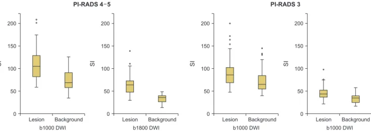
Fig. 2 shows the lesion distribution and background SI in PI-RADS 3–5 lesions. Except for PI-RADS 4–5 lesions in b1800 DWI, visible lesion and background
Fig. 1. Detection rate of overall and clinically significant cancer according to Prostate Imaging–Reporting and Data System (PI-RADS) score in each magnetic resonance imaging protocol. aMagnetic resonance imaging (MRI) protocol A included T2-weighted imaging (T2WI), apparent diffusion coefficient (ADC), and b1000 diffusion-weighted imaging (DWI), bMRI protocol B included T2WI, ADC, b1000, and b1800 DWI.
Fig. 2. Signal intensity (SI) of Prostate Imaging–Reporting and Data System (PI-RADS) 4–5 lesion was higher than SI of background in b-value 1,800 s/mm2 diffusion-weighted imaging (DWI). Severe overlap of values with SI was observed under other conditions.
SI values severely overlapped with each DWI. The mean SI ratios (lesion/background) were 1.36 and 1.60 in b1000 and b1800 DWI, respectively (p<0.001). The odds ratio of the SI ratio for predicting csPCa in b1000 and b1800 DWI were 3.2 (95% confidence interval [CI], 1.7–6.0; p<0.001) and 5.1 (95% CI, 3.2–9.0; p<0.001), re- spectively.
DISCUSSION
The most important result of our study was that bpMRI including b1800 DWI increased the csPCa de- tection rate from 19.5% to 27.8%. Higher detection rate of csPCa in PI-RADS 4–5 lesions is the most important cause of this increase. Comparing SI ratio (lesion/back- ground) of b1000 DWI, higher those of b1800 in PI- RADS 4–5 make it easier to identify lesions.
Standardization of MRI protocol is crucial for broad application of MRI. PI-RADS version 2 guideline is based on T2W, DWI, and DCE [3]. DWIs are used to calculate ADC maps with mono-exponential fitting of DWIs acquired at various b-values (≤1,000 s/mm2). High b-value (≥1,400 s/mm2) DWI should be obtained by direct acquisition or computed from the source DWI.
However, ultra-high b-value (≥1,800 s/mm2) DWI is not mandatory. Several studies have suggested the benefit of b1800 or higher b-value DWI for csPCa detection [8- 12]. In the past, it was infeasible to measure ultra-high b-value DWIs with an acceptable image quality. Long echo times were required, causing significant loss of signal-to-noise ratio. To reduce these effects, various b- values DWI with an acceptable signal-to-noise ratio were generally obtained and virtual high b-values DWI were mathematically extrapolated [13].
A quality-assured MRI protocol and experienced reader are essential for constant csPCa detection rate according to PI-RADS score. In the PROMIS study, csPCa detection rates with PI-RADS 3, 4, and 5 lesions were 21%, 58%, and 81%, respectively [1]. Similarly, those in the PRICISION study were 12%, 60%, and 83%, respectively [2]. Results of this study in the overall co- hort were 12%, 56%, and 90%, respectively, consistent with results of two past large studies. However, results in MRI protocol A cohort were relatively disappoint- ing, with detection rates of 12%, 38%, and 67%, respec- tively. Those with MRI protocol B were 10%, 64%, and 100%, respectively. The csPCa detection rates in MRI protocol B were higher than those in MRI protocol A
for PI-RADS 4 and 5 lesions. Compared with b1000 DWI, b1800 DWI could discriminate lesion from adja- cent background for PI-RADS 4–5 lesions more clearly.
However, lesion and background SI values in both MRI protocol cohort severely overlapped in PI-RADS 3 and, even in PI-RADS 4–5 lesions in b1000 DWI (Fig. 2).
In csPCa patients, the SI ratio (lesion/background) of b1800 DWI had higher odds ratio than that of b1000 DWI (5.1 vs. 3.2). Therefore, more accurate localization of csPCa in PI-RADS 4–5 lesions could lead to higher csPCa detection rates.
In addition to accuracy, cost-effectiveness is more important in the application of MRI to biopsy-naïve patients. To date, most guidelines recommend that MRI should be used when the first biopsy is negative [14].
However, in recent PROMIS and Pahwa et al’s study [15], the use of MRI in the first session biopsy improves the accuracy of csPCa detection, and the cost-effec- tiveness is also advantageous, considering prostate spe- cific antigen follow-up, re-biopsy, and additional costs because of delayed diagnosis [16].
We did not compare the cost-effectiveness of mpMRI and bpMRI. The cost of mpMRI in our institution was approximately 600 US dollars, and about 45 minutes of imaging time was required to obtain all necessary images. Our bpMRI protocol consisted of only two se- quences: axial T2WI for anatomical evaluation and DWI. These sequences were acquired within 4 minutes and 8 minutes, respectively. The cost for bpMRI pro- cedure was reduced to 300 US dollars. As we described in the previous studies, the overall detection rates of csPCa were not different between the mpMRI and bpMRI [17]. In addition, several studies used simple bpMRI for cost-effectiveness [15,17-19]. bpMRI avoids additional cost, acquisition time, and side effects caused by contrast media. Cheikh et al [4] reported that DCE did not have any significant differences compared with T2WI. In their study, DCE was significantly less spe- cific (83.5% vs. 89.7%, p<0.002) than T2WI. Moreover, while DCE was more sensitive (52.4% vs. 32.1%), the dif- ference was not significant (p=0.09). Delongchamps et al [5] also reported that DCE did not increase the accu- racy, compared with DWI, for PCa detection in either the prostate peripheral or transitional zone. Therefore, DCE only plays a role as the minor sequence when pe- ripheral zone cancer is equivocally suspected.
Several inevitable limitations exist in this retrospec- tive study. First, accuracies of MRI protocols A and B
were analyzed with different populations. However, baseline characteristics including age, prostate-specific antigen, or prostate volume were not significantly different between the two MRI protocols. Second, PI- RADS version 2 guideline was updated during the study period. Therefore, we added b1800 DWI since 2016. Third, the learning curve could be a limitation because MRI protocols A and B were performed in different periods. However, we already used mpMRI for over 100 cases for target biopsy before this study period. Forth, we did not analyzed according to zonal anatomy, peripheral and transitional zone lesion. The optimal sequence is different for detection of csPCa in peripheral and transitional zone [3].
CONCLUSIONS
Adding b1800 DWI to bpMRI protocol improved the diagnostic accuracy and detection rate of csPCa. For PI-RADS 4 and 5 lesions, the csPCa detection rates us- ing bpMRI with b1800 DWI were higher than those in bpMRI with b1000 DWI. A higher SI ratio of lesion/
background in b1800 DWI makes it possible to distin- guish suspected lesions more clearly. Among men with PI-RADS score 4–5 lesions, over 70% were diagnosed with csPCa after prostate biopsy in the present study.
ACKNOWLEDGEMENTS
This study was supported by Research Institute for Convergence of Biomedical Science and Technology grant (30-2017-017), Pusan National University Yang- san Hospital.
Conflict of Interest
The authors have nothing to disclose.
Author Contribution
Conceptualization: SSL, DHL, JYH, TUK, SWP. Data cura- tion: all authors. Formal analysis: SSL, TUK, SWP. Funding acquisition: SWP. Investigation: SWP. Methodology: SSL, SWP.
Project administration: SSL, SWP. Resources: HJL, TUK, SWP.
Software: TUK, SWP. Supervision: TUK, SWP. Validation: TUK, SWP. Visualization: TUK, SWP. Writing–original draft: SSL, SWP. Writing–review & editing: SSL, HJL, TUK, SWP.
Data Sharing Statement
The data analyzed for this study have been deposited in HARVARD Dataverse and are available at https://doi.
org/10.7910/DVN/R35JV9.
REFERENCES
1. Ahmed HU, El-Shater Bosaily A, Brown LC, Gabe R, Kaplan R, Parmar MK, et al.; PROMIS study group. Diagnostic ac- curacy of multi-parametric MRI and TRUS biopsy in prostate cancer (PROMIS): a paired validating confirmatory study.
Lancet 2017;389:815-22.
2. Kasivisvanathan V, Rannikko AS, Borghi M, Panebianco V, Mynderse LA, Vaarala MH, et al.; PRECISION Study Group Collaborators. MRI-targeted or standard biopsy for prostate- cancer diagnosis. N Engl J Med 2018;378:1767-77.
3. Weinreb JC, Barentsz JO, Choyke PL, Cornud F, Haider MA, Macura KJ, et al. PI-RADS prostate imaging - reporting and data system: 2015, version 2. Eur Urol 2016;69:16-40.
4. Cheikh AB, Girouin N, Colombel M, Maréchal JM, Gelet A, Bissery A, et al. Evaluation of T2-weighted and dynamic contrast-enhanced MRI in localizing prostate cancer before repeat biopsy. Eur Radiol 2009;19:770-8.
5. Delongchamps NB, Rouanne M, Flam T, Beuvon F, Libera- tore M, Zerbib M, et al. Multiparametric magnetic resonance imaging for the detection and localization of prostate cancer:
combination of T2-weighted, dynamic contrast-enhanced and diffusion-weighted imaging. BJU Int 2011;107:1411-8.
6. Katahira K, Takahara T, Kwee TC, Oda S, Suzuki Y, Morishita S, et al. Ultra-high-b-value diffusion-weighted MR imaging for the detection of prostate cancer: evaluation in 201 cases with histopathological correlation. Eur Radiol 2011;21:188- 96.
7. DeLano MC, Cooper TG, Siebert JE, Potchen MJ, Kuppusamy K. High-b-value diffusion-weighted MR imaging of adult brain: image contrast and apparent diffusion coefficient map features. AJNR Am J Neuroradiol 2000;21:1830-6.
8. Ohgiya Y, Suyama J, Seino N, Hashizume T, Kawahara M, Sai S, et al. Diagnostic accuracy of ultra-high-b-value 3.0-T dif- fusion-weighted MR imaging for detection of prostate cancer.
Clin Imaging 2012;36:526-31.
9. Hausmann D, Aksöz N, von Hardenberg J, Martini T, Westhoff N, Buettner S, et al. Prostate cancer detection among readers with different degree of experience using ultra-high b-value diffusion-weighted imaging: Is a non- contrast protocol sufficient to detect significant cancer? Eur Radiol 2018;28:869-76.
10. Barral M, Cornud F, Neuzillet Y, Lonchampt E, Lassalle L, Delonchamp NB, et al. Characteristics of undetected prostate cancer on diffusion-weighted MR Imaging at 3-Tesla with a b-value of 2000s/mm(2): imaging-pathologic correlation. Di- agn Interv Imaging 2015;96:923-9.
11. Agarwal HK, Mertan FV, Sankineni S, Bernardo M, Senegas J, Keupp J, et al. Optimal high b-value for diffusion weighted MRI in diagnosing high risk prostate cancers in the periph- eral zone. J Magn Reson Imaging 2017;45:125-31.
12. Rosenkrantz AB, Parikh N, Kierans AS, Kong MX, Babb JS, Taneja SS, et al. Prostate cancer detection using computed very high b-value diffusion-weighted imaging: How high should we go? Acad Radiol 2016;23:704-11.
13. Bittencourt LK, Attenberger UI, Lima D, Strecker R, de Oliveira A, Schoenberg SO, et al. Feasibility study of comput- ed vs measured high b-value (1400 s/mm2) diffusion-weight- ed MR images of the prostate. World J Radiol 2014;6:374-80.
14. Mottet N, Bellmunt J, Bolla M, Briers E, Cumberbatch MG, De Santis M, et al. EAU-ESTRO-SIOG guidelines on prostate cancer. Part 1: screening, diagnosis, and local treatment with curative intent. Eur Urol 2017;71:618-29.
15. Pahwa S, Schiltz NK, Ponsky LE, Lu Z, Griswold MA, Gulani
V. Cost-effectiveness of MR Imaging-guided strategies for detection of prostate cancer in biopsy-naive men. Radiology 2017;285:157-66.
16. Faria R, Soares MO, Spackman E, Ahmed HU, Brown LC, Kaplan R, et al. Optimising the Diagnosis of prostate cancer in the era of multiparametric magnetic resonance imaging: a cost-effectiveness analysis based on the Prostate MR Imaging Study (PROMIS). Eur Urol 2018;73:23-30.
17. Lee DH, Nam JK, Lee SS, Han JY, Lee JW, Chung MK, et al.
Comparison of multiparametric and biparametric MRI in first round cognitive targeted prostate biopsy in patients with PSA levels under 10 ng/mL. Yonsei Med J 2017;58:994-9.
18. Junker D, Steinkohl F, Fritz V, Bektic J, Tokas T, Aigner F, et al. Comparison of multiparametric and biparametric MRI of the prostate: Are gadolinium-based contrast agents needed for routine examinations? World J Urol 2019;37:691-9.
19. Di Campli E, Delli Pizzi A, Seccia B, Cianci R, d’Annibale M, Colasante A, et al. Diagnostic accuracy of biparametric vs multiparametric MRI in clinically significant prostate cancer:
comparison between readers with different experience. Eur J Radiol 2018;101:17-23.