Introduction
Chronic angle-closure glaucoma (CACG), in which closure of the anterior chamber angle leads to increased intraocular pressure (IOP) and glaucomatous optic neu- ropathy, is a leading cause of blindness throughout the world.1-4 A thick and anteriorly positioned crystalline lens plays a pivotal role in the pathogenesis of CACG.5 There-
fore, lens extraction itself has a favorable outcome for the control of IOP and vision improvement in eyes with CACG and cataracts.6,7 Recent studies have shown that results of phacoemulsification alone were comparable to those of combined phacoemulsification and trabeculectomy in terms of IOP control in eyes with CACG and cataracts.8,9
In cases of advanced glaucoma with a visually significant cataract, combined phacoemulsification and trabeculectomy is usually advocated because uncontrolled IOP after cataract extraction alone carries a risk of postoperative visual field loss, which may involve fixation with central vision loss.10 However, trabeculectomy in end-stage glaucoma carries a risk of sudden vision loss (“wipe-out phenomenon”) as well as vision-threatening complications like prolonged hypo- tony, choroidal detachment, and malignant glaucoma.11-13 In addition, combined surgery requires additional skill and
Safety and Efficacy of Phacoemulsification and Intraocular Lens Implantation in Eyes with End-Stage Chronic Angle-Closure Glaucoma
Seok Hwan Kim, MD1,2, Young Keun Han, MD1,2
1Department of Ophthalmology, Seoul National University College of Medicine, Seoul, Korea
2Department of Ophthalmology, Seoul National University Boramae Hospital, Seoul, Korea
Purpose: To evaluate surgical outcomes of phacoemulsification and intraocular lens implantation in eyes with end-stage chronic angle-closure glaucoma (CACG).
Methods: Medical records of patients with end-stage CACG (mean deviation, <-19 dB) and clinically significant cataracts who underwent phacoemulsification and intraocular lens implantation were retrospectively reviewed. During at least 12 months of follow-up, changes in visual acuity, intraocular pressure, visual field parameters, and number of antiglaucoma agents used were assessed. Intraoperative and postoperative complications were documented. Multiple regression analysis was performed to identify independent predictors of postoperative visual outcome.
Results: Forty-two eyes of 38 subjects were enrolled. The mean follow-up period was 19.7 months (12–33 months). Corrected visual acuity improved from 0.63 ± 0.53 logarithm of the minimum angle of resolution (logMAR) preoperatively to 0.21 ± 0.33 logMAR postoperatively (p < 0.001).
Intraocular pressure decreased from 15.50 ± 3.23 mmHg to 11.05 ± 1.76 mmHg (p < 0.001). No change was found in visual field parameters or number of antiglaucoma agents used. During surgery, 4 eyes had zonular dialysis and 1 eye had posterior capsule rupture. After surgery, 2 eye required additional trabeculectomy due to uncontrolled intraocular pressure after phacoemulsification. Preoperative visual acuity and mean sensitivities of 4 central points of the visual field were independent predictors of postoperative visual outcome.
Conclusions: Phacoemulsification and intraocular lens implantation was performed safely and effectively in end-stage CACG eyes with cataracts.
Key words: Phacoemulsification; Cataract; Chronic angle-closure glaucoma; End-stage glaucoma
Received: 2015. 9. 20. Revised: 2015. 10. 28.
Accepted: 2015. 11. 11.
Corresponding Author: Seok Hwan Kim , MD
Department of Ophthalmology, Seoul National University Boramae Hospital, 20 Boramae-ro 5-gil, Dongjak-gu, Seoul 07061, Korea Tel: +82-2-870-2415, Fax: +82-2-831-2826
E-mail: [email protected]
* None of the authors have any financial/conflicting interests to disclose.
a longer surgical duration, and may have prolonged visual recovery. Therefore, in eyes with end-stage CACG and cat- aracts, phacoemulsification alone might be an alternative to conventional combined surgery because phacoemulsifica- tion can be expected to reduce IOP in eyes with CACG.
In this study, we evaluated the safety and efficacy of phacoemulsification and intraocular lens implantation in terms of visual outcome and IOP in eyes with end-stage CACG and cataracts.
Patients and Methods
Medical records of patients who had end-stage CACG and clinically significant cataracts, and underwent phacoemul- sification between August 2008 and July 2012 at Boramae Medical Center were retrospectively reviewed. The patients with at least 12 months of postoperative follow-up were in- cluded. This study was approved by the institutional review board of Boramae Medical Center.
CACG was defined by the presence of all of the following factors: glaucomatous optic neuropathy with compatible visual field defects, 2 or more quadrants of angle-closure obscuring the pigmented part of the trabecular meshwork under gonioscopy in the presence of patent laser iridotomy, and IOP > 21 mmHg without use of antiglaucoma medi- cations.8 End-stage glaucoma was defined on the basis of visual field results if the mean deviation (MD) on Hum- phrey automated perimetry 24-2 (Carl Zeiss Meditec, Inc., Dublin, CA) was -19 dB or worse.14 Eyes with a clinically significant cataract and visual acuity of 30/50 or worse were included. Eyes with a history of intraocular surgery, except laser iridotomy and laser peripheral iridoplasty, were ex- cluded. Eyes with secondary angle-closure glaucoma were excluded. We also excluded all eyes with macular disease or other intraocular eye diseases affecting the visual field (e.g., age-related macular degeneration, diabetic retinopa- thy, retinal vein occlusion, and optic neuropathy other than glaucoma). Only patients with end-stage CACG who had an IOP of ≤25 mmHg with use of 3 or fewer topical drugs were included in this study because patients with end-stage CACG and cataract who had an IOP of >25 mmHg despite
topical medications underwent combined cataract surgery and trabeculectomy.
A preoperative medical history was obtained for deter- mining presence of previous intraocular surgery, coexisting systemic disease and use of systemic medications. A base- line ophthalmic examination was performed within 3 days before surgery; this examination included a visual acuity test, manifest refraction and correction, IOP measurements by Goldmann applanation tonometry, slit-lamp exami- nation, gonioscopy, and fundus examination. Humphrey Swedish interactive thresholding algorithm (SITA) 24-2 perimetry and optical biometer (IOLMaster, Carl Zeiss Meditec, Inc., Dublin, CA) were performed. The number of preoperative antiglaucoma medications used by the patient was documented.
Patients were examined postoperatively at 1 day, 1 week, 1 month, 3 months, and 6 months and thereafter, at an inter- val of 3 to 6 months; the examination included assessment of best-corrected visual acuity, IOP, intraocular lens status, and the number of antiglaucoma agents that was required to achieve an optimal IOP level. Additional visits were sched- uled as clinically warranted. The incidence of intraoperative and postoperative complications such as posterior capsular rupture, zonular dialysis, and IOP spike (IOP > 21 mmHg) were documented at each visit. Assessment of visual fields was repeated at an interval of 6 months after surgery.
Main outcome measures included the change in logarithm of the minimum angle of resolution (logMAR) and IOP. The MD and mean sensitivity of the 4 central visual field points in the latest assessment of the visual field after surgery were compared with preoperative values. Factors related to the final visual acuity outcome were also identified.
Surgical technique
The surgical procedure was standard for all subjects, and all procedures were performed by 1 experienced surgeon (SH Kim). Phacoemulsification was performed using topical or sub-Tenon anesthesia of 2% lidocaine. A 3.2-mm clear corneal incision was placed temporally. After instillation of 1.0% sodium hyaluronate into the anterior chamber, a con- tinuous curvilinear capsulorhexis was created with a bent
27-gauge needle. Hydrodissection and phacoemulsification of the nucleus was performed using the stop-and-chop tech- nique, and the residual cortex was subsequently removed. A foldable acrylic intraocular lens (Bausch & Lomb; Akreos Adapt, New Jersey, USA) was placed, and the viscoelas- tic material was aggressively evacuated from the anterior chamber to avoid an immediate postoperative increase in the IOP. The intraocular lens was implanted in the capsular bag in all but 1 patient, in whom the intraocular lens was placed with scleral fixation because of severe zonular dial- ysis and posterior capsule rupture. In an eye with zonular dialysis less than 90°, the intraocular lens was placed in the capsular bag after a capsular tension ring was inserted to stabilize zonular tension. To prevent a postoperative IOP spike, 1 drop of 0.5% timolol maleate was applied immedi- ately after cataract surgery. Use of antiglaucoma agents was continued for the patients on the day of surgery.
Statistical analysis
A paired t-test was performed to compare preoperative and postoperative values of visual acuity, IOP, number of
antiglaucoma agents used, MD, and the mean sensitivity of the 4 central visual field points. Multiple regression analysis was performed to identify independent predictors of postoperative visual outcome. The correlation between postoperative visual acuity and preoperative variables was analyzed using the Pearson correlation. Variables with a p value less than 0.05 were included in a backward stepwise multiple linear regression after assessment of collinearity.
Statistical analysis was performed using SPSS version 18.0 (SPSS Inc., Chicago, IL, USA). A p value of less than 0.05 was accepted as statistically significant.
Results
Initially, 47 eyes of 43 patients who were diagnosed with end-stage CACG and clinically significant cataracts, and underwent phacoemulsification were enrolled. Among se- lected 47 eyes, 5 eyes of 5 patients were excluded because of follow-up loss. Finally, the study involved 42 eyes of 38 patients. Table 1 shows the patients’ demographic charac- teristics. The mean age of patients at the time of surgery
Table 1. Demographic features of 42 eyes of 38 patients with end-stage chronic angle-closure glaucoma and cataract who underwent phacoemulsification and intraocular lens implantation
Parameters Values
Age (years, mean ± SD) Gender (male:female) Right eye : Left eye
Follow-up (months, mean ± SD) Extent of synechial angle closure
Eyes with history of acute angle closure attack
74.37 ± 5.81 (range: 65 to 86 years) 14:24
19:23
19.68 ± 7.89 (range: 12 to 33 months) 291.4 ± 71.0 (range: 180 to 360)
12/42 SD = standard deviation.
Table 2. Preoperative and postoperative clinical status of 42 eyes of 38 patients with end-stage chronic angle-closure glaucoma and cataract who underwent phacoemulsification and intraocular lens implantation
Baseline
(mean ± SD) Final visit
(mean ± SD) p-value*
IOP (mmHg) 15.50 ± 3.23 11.05 ± 1.76 <0.001
Visual acuity (logMAR) 0.63 ± 0.53 0.21 ± 0.33 <0.001
Mean deviation (dB) -26.08 ± 3.23 -25.87 ± 3.34 0.516
Mean sensitivity of 4 central visual field points (dB) 11.92 ± 7.72 12.15 ± 6.75 0.622
Number of medications 1.95 ± 1.09 1.95 ± 0.72 0.999
*Paired t-test.
was 74.37 ± 5.81 years (range: 65 to 86 years). The mean follow-up period was 19.68 ± 7.89 (range: 12 to 33 months).
No eyes had mature or white cataracts.
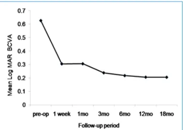
Table 2 shows preoperative and postoperative clinical sta- tus of 42 eyes. We observed a significant reduction in IOP from 15.50 ± 3.23 mmHg (range: 11 to 23 mmHg) to 11.05 ± 1.76 mmHg (range: 8 to 16 mmHg). (Fig. 1) The mean log- MAR visual acuity was also significantly improved from 0.63 ± 0.53 to 0.21 ± 0.33 after phacoemulsification. (Fig. 2) Significant changes were not observed in MD, the mean sensitivity of the 4 central visual field points, or the number of antiglaucoma agents used.
Table 3 lists intraoperative and postoperative compli-
cations. During surgery, zonular dialysis was found in 4 eyes. Among 4 eyes, three eyes had capsular tension ring implantation because zonular dialysis of less than 90° was identified during cataract surgery. The other eye had ante- rior vitrectomy and scleral fixation of the intraocular lens due to severe zonular dialysis and posterior capsule rup- ture. For this eye, intravenous mannitol was administered immediately after surgery to prevent a postoperative IOP spike. After surgery, 2 eyes showed severe anterior cham- ber inflammation with fibrin exudate, which was controlled by frequent application of topical 1% prednisolone acetate.
Two eyes of 2 patients showed IOP elevation despite use of antiglaucoma agents at 1 month and at 2 months respec- tively after cataract surgery, and the patients underwent additional trabeculectomy. No immediate IOP spike was observed 1 day after cataract surgery in all patients.
In multiple linear regression analysis, preoperative log- MAR visual acuity and preoperative mean sensitivity of the 4 central visual field points were identified as independent predictors of postoperative logMAR visual acuity after adjusting for preoperative IOP and MD (R2 = 0.649, p <
0.001).
Discussion
This retrospective case series suggests that phacoemulsifi- cation and intraocular lens implantation is a safe and effec- tive option for the treatment of eyes with end-stage CACG and cataracts. We also showed that preoperative vision and the mean sensitivity of the 4 central points of the visual field were independent predictors of postoperative visual Figure 1. Intraocular pressure changes after phacoemulsifica-
tion and intraocular lens implantation in 42 eyes with end-stage chronic angle-closure glaucoma and cataract. Mo, month.
Figure 2. Log MAR BCVA (best corrected visual acuity) changes after phacoemulsification and intraocular lens implantation in 42 eyes with end-stage chronic angle-closure glaucoma and cata- ract. mo, month.
Table 3. Intraoperative and postoperative complications of phaco- emulsification and intraocular lens implantation in 42 eyes with end-stage chronic angle-closure glaucoma
Complications Eyes Rate
Intraoperative Posterior capsule tear Zonular dialysis Iris synechiolysis Postoperative Uncontrolled IOP Anterior uveitis
1 4 7 2 2
2.4%
9.5%
16.7%
4.8%
4.8%
outcome.
Combined cataract surgery and trabeculectomy is usually advocated for eyes with advanced glaucoma and clinically significant cataracts.10 The advantages of combined surgery includes the patients’ convenience of undergoing 2 surger- ies on 1 day, the apparent IOP-lowering effect and reduced requirement of antiglaucoma medications due to trabec- ulectomy, and prevention of the postoperative IOP spike as- sociated with cataract surgery. However, combined surgery has several disadvantages. Combined surgery has more complications than cataract surgery alone, such as post- operative hypotony, hyphema, choroidal detachment, and malignant glaucoma.11-13 Although most of these complica- tions are transient and treatable, they hamper early visual rehabilitation and might induce permanent vision loss. Tra- beculectomy in end-stage glaucoma also carries the risk of sudden, unexplained vision loss (“wipe-out phenomenon”), although it is very rare.13,15 In addition, combined surgery requires additional skill for trabeculectomy, longer surgical duration, more follow-up, and additional surgical interven- tion in the postoperative period to maintain bleb function and to manage complications.8 Recent studies showed that the results of phacoemulsification alone were comparable to those of combined phacoemulsification and trabeculectomy for the control of IOP in eyes with CACG and cataracts.8,9 In this study, we showed that for eyes with end-stage CACG, phacoemulsification alone provides favorable visual out- come and control of IOP. Because this study was a case series, further studies comparing the outcome between phacoemulsification alone and combined surgery should be performed for eyes with end-stage CACG. Furthermore, since we included eyes with relatively medically controlled CACG and cataracts in this study, additional studies are warranted for eyes with medically uncontrolled end-stage CACG and cataracts.
Previous reports have showed that in eyes with CACG, cataract surgery reduced the number of antiglaucoma med- ications used as well as IOP;8,9,16 in contrast, in the present study, there was no difference in the number of antiglau- coma agents used preoperatively and postoperatively. This can be partly explained by the end-stage visual field status
of the study subjects. Given the possibility of an IOP spike after discontinuation of antiglaucoma agents, which might involve fixation with central vision loss, the author hesitated to discontinue antiglaucoma agents despite good IOP con- trol.
Only 2 eye among 42 eyes required additional trabec- ulectomy to control IOP after cataract surgery alone in this study. IOPs of both eyes were well controlled after trabec- ulectomy without any complications. Our results imply that additional trabeculectomy can be performed successfully in cases with uncontrolled IOP after performing phacoemulsi- fication alone.
An early postoperative IOP spike can be minimized with topical or systemic antiglaucoma medications.17,18 A recent randomized clinical trial reported that immediate postop- erative treatment with 0.5% timolol maleate reduced the occurrence of IOP spikes.19 In this study, we applied 0.5%
timolol maleate immediately after phacoemulsification to prevent an early postoperative IOP spike. In addition, we did not discontinue antiglaucoma agents even on the day of surgery, and we judiciously removed viscoelastic agents af- ter intraocular lens implantation. Prophylactic intravenous mannitol was administered in the case of an eye with pos- terior capsule rupture and scleral fixation of the intraocular lens; this eye had a high chance of an IOP spike. Among 42 eyes in this study, none showed IOP elevation at 1 day or 1 week after surgery.
In eyes with end-stage glaucoma and a coexisting cataract, visual outcome is often difficult to predict after cataract surgery because glaucoma also contributes to the vision de- crease. Herein, we performed multiple regression analysis and found that preoperative vision and the mean sensitivity of 4 central points of the visual field were independent pre- dictors of postoperative visual outcome.
Our study does not suggest that phacoemulsification alone should be a substitute for combined surgery for the treat- ment of eyes with end-stage CACG and cataracts. We found that the IOP-lowering effect of phacoemulsification was modest (mean, 4.5 mmHg) and that the number of antiglau- coma agents used was not different postoperatively. There- fore, the risks and benefits of combined surgery should be
weighed carefully against those of cataract surgery alone for the treatment of eyes with end-stage CACG. In addition, patients should be informed preoperatively that an addition- al trabeculectomy may be required.
In conclusion, this study showed that phacoemulsification and intraocular lens implantation can be performed safely and effectively in eyes with medically controlled end-stage CACG. Further studies comparing the outcome between phacoemulsification alone and combined surgery in end- stage CACG should be warranted.
References
1. Foster PJ. The epidemiology of primary angle closure and associated glaucomatous optic neuropathy. Semin Oph- thalmol 2002;17:50-8.
2. Foster PJ, Baasanhu J, Alsbirk PH, et al. Glaucoma in Mongolia. A population-based survey in Hövsgöl province, northern Mongolia. Arch Ophthalmol 1996;114:1235-41.
3. Johnson GJ, Foster PJ. Can we prevent angle-closure glau- coma? Eye. 2005;19:1119-24.
4. Dandona L, Dandona R, Mandal P, et al. Angle-closure glaucoma in an urban population in southern India. The Andhra Pradesh eye disease study. Ophthalmology.
2000;107:1710-6.
5. Tarongoy P, Ho CL, Walton DS. Angle-closure glaucoma:
the role of the lens in the pathogenesis, prevention, and treatment. Surv Ophthalmol 2009;54:211-25.
6. Hayashi K, Hayashi H, Nakao F, Hayashi F. Effect of cata- ract surgery on intraocular pressure control in glaucoma patients. J Cataract Refract Surg 2001;27:1779-86.
7. Yang CH, Hung PT. Intraocular lens position and anterior chamber angle changes after cataract extraction in eyes with primary angle-closure glaucoma. J Cataract Refract Surg 1997;23:1109-13.
8. Tham CC, Kwong YY, Leung DY, et al. Phacoemulsifica- tion versus combined phacotrabeculectomy in medically controlled chronic angle closure glaucoma with cataract.
Ophthalmology 2008;115:2167-73.
9. Tham CC, Kwong YY, Leung DY, et al. Phacoemulsifica- tion versus combined phacotrabeculectomy in medically uncontrolled chronic angle closure glaucoma with cata- racts. Ophthalmology 2009;116:725-31.
10. Khatana AK, Cohen JS, Osher RH. Combined cataract implant and filtering surgery. In:Steinert RF, ed, Cataract Surgery. Maryland Heights, Elsevier, Inc.; 2010; 259-81 11. Bellucci R, Perfetti S, Babighian S, et al. Filtration and
complications after trabeculectomy and after phaco-trab- eculectomy. Acta Ophthalmol Scand Suppl 1997;224:44- 5.
12. Law SK, Nguyen AM, Coleman AL, Caprioli J. Severe loss of central vision in patients with advanced glau- coma undergoing trabeculectomy. Arch Ophthalmol 2007;125:1044-50.
13. Costa VP, Smith M, Spaeth GL, et al. Loss of visual acuity after trabeculectomy. Ophthalmology 1993;100:599-612.
14. Much JW, Liu C, Piltz-Seymour JR. Long-term survival of central visual field in end-stage glaucoma. Ophthalmology 2008;115:1162-6.
15. Aggarwal SP, Hendeles S. Risk of sudden visual loss fol- lowing trabeculectomy in advanced primary open-angle glaucoma. Br J Ophthalmol 1986 ;70:97-9.
16. Shrivastava A, Singh K. The effect of cataract extrac- tion on intraocular pressure. Curr Opin Ophthalmol 2010
;21:118-22.
17. Fry LL. Comparison of the postoperative intraocular pressure with Betagan, Betoptic, Timoptic, Iopidine, Di- amox, Pilopine Gel, and Miostat. J Cataract Refract Surg 1992;18:14-9.
18. Haimann MH, Phelps CD. Prophylactic timolol for the prevention of high intraocular pressure after cataract ex- traction. A randomized, prospective, double-blind trial.
Ophthalmology 1981;88:233-8.
19. Levkovitch-Verbin H, Habot-Wilner Z, Burla N, et al. In- traocular pressure elevation within the first 24 hours after cataract surgery in patients with glaucoma or exfoliation syndrome. Ophthalmology 2008;115:104-8.