하지 골절을 동반한 중증 외상 환자에 있어서 정맥혈전색 전증의 빈도 및 예방
Incidence and Prevention of Venous Thromboembolism in Severely Injured Patients with Lower Extremity Fracture
김지완* • 최지호 • 김정재
울산대학교 의과대학 서울아산병원 정형외과학교실, *인제대학교 의과대학 해운대백병원 정형외과학교실
목적: 하지 골절을 동반한 중증 외상 환자에서 발생하는 정맥혈전색전증의 빈도를 확인하고, 예방법을 시행한 후 그 빈도의 차이를 후향적 연구를 통해 알아보고자 하였다.
대상 및 방법: 항응고제 및 물리적인 예방법 시행 이전을 1기, 시행 이후를 2기로 나누어 두 군에서의 정맥혈전색전증 빈도에 대해 조사하 였다.
결과: 1기에서 대상 31예 중 5예(16.1%)에서 임상적 증상이 있는 정맥혈전색전증이 발생하였는데, 심부정맥혈전증이 1예, 폐색전증이 4예 였고, 그 중 2예는 생명을 위협할 정도의 중증의 폐색전증을 보였다. 2기에서 대상 환자는 32예였고, 이 중 임상적 증상이 있는 정맥혈전색 전증은 1예도 없었다.
결론: 중증 외상 환자에서 정맥혈전색전증에 대한 적극적인 예방 조치를 하지 않았을 경우 임상적으로 나타나는 정맥혈전색전증이 16.1%
에서 나타났으며. 이는 항응고제 및 물리적인 방법을 통해 현저히 감소하는 것을 확인할 수 있었다. 따라서 중증 외상 환자에서는 정맥혈전 색전증에 대한 적극적인 예방이 필요하다 할 것이다.
색인단어: 중증 외상 환자, 하지 골절, 정맥혈전색전증, 심부정맥혈전증, 폐색전증
접수일 2012년 4월 2일 수정일 2012년 6월 7일 게재확정일 2012년 7월 19일 교신저자 김정재
서울시 송파구 올림픽로 43길 88, 서울아산병원 정형외과학교실 TEL 02-3010-3530, FAX 02-488-7877
E-mail [email protected]
Copyright © 2012 by The Korean Orthopaedic Association
“This is an Open Access article distributed under the terms of the Creative Commons Attribution Non-Commercial License (http://creativecommons.org/licenses/by-nc/3.0/) which permits unrestricted non-commercial use, distribution, and reproduction in any medium, provided the original work is properly cited.”
대한정형외과학회지:제 47권 제 6호 2012
서 론
정형외과 영역에서 수상 혹은 수술 후 발생하는 정맥혈전색전증 은 때로는 생명을 위협하는 치명적인 합병증이 될 수 있으므로 이에 대한 관심을 기울여야 한다. 정맥혈전색전증은 심부정맥혈 전증과 폐색전증으로 나눌 수 있는데, 이에 대한 연구는 인공고 관절 치환술이나 인공슬관절 치환술을 시행 받은 환자를 대상으 로 활발히 이루어져 있다. 국내에서 인공고관절 치환술 후 정맥 혈전색전증의 빈도는 8.7-13.1%로 보고하였고,1,2) 슬관절 치환술 후 빈도는 15.7-40.4%까지 보고하였으며,1,3,4) 고관절 수술 후 정맥
혈전색전증은 16.4%에서 발생한다고 하였다.1)
골절을 동반한 다발성 외상 환자에서 발생하는 정맥혈전색전 증의 빈도와 예방에 대해서는 아직 국내에서 연구된 바가 없다.
이번 연구에서는 하지 골절을 동반한 중증 외상 환자에서 발생하 는 정맥혈전색전증의 빈도를 확인하고 예방법에 따른 빈도의 차 이를 알아보고자 한다.
대상 및 방법
2009년 1월 1일부터 2010년 2월 28일까지 하지 골절을 동반한 중 증 외상 환자를 2009년 8월 1일을 기준으로 하여 이전과 이후 시 기로 1군과 2군의 두 군으로 구분하였다. 하지 골절은 경골 혹은 대퇴골의 골절을 의미하며 족관절 골절은 대상에서 제외하였다.
1군에서는 항혈전 스타킹(anti-embolism stockings, Comprinet® pro, BSN medical, Hamburg, Germany)을 이용하여 예방하였고,
2009년 8월 1일부터인 2군에서는 수상 직후부터 순서적 압박장치 (sequential compression device)인 SCD EXPRESSTM Compression System (Covidien, Mansfield, MA, USA)5)을 이용한 물리적 예방법 을 시행하였고, 수술 다음날부터 항응고제 clexane (Enoxaparin, Sanofi Aventis, Le Trait, France)을 하루 40 mg으로 피하주사하 여 보행 시작 시까지 주사하였다. 중증 외상 환자의 기준은 중증 손상 점수(injury severity score, ISS) 17점 이상으로 하였으며, 20 세 이상의 성인을 대상으로 하였다. 연구 대상에서 정맥혈전색 전증의 기왕력이 있는 경우, 심방 세동과 같이 정맥혈전색전증 의 위험성이 있어 warfarin 등의 항응고제를 복용하고 있는 경우 는 제외하였다. 심부정맥혈전증을 알아보기 위해 초음파 혹은 컴 퓨터단층촬영의 선별 검사(screening test)는 시행하지 않았고, 오 전 회진 시마다 Homan's test를 통해 선별 검사를 시행하였다. 다 만, 하지 부종 및 동통 등의 증상이 있는 경우는 하지의 심부정맥 을, 설명되지 않는 저혈압, 저산소혈증, 빈맥(tachycardia), 빈호흡 (tachypnea), 호흡 곤란 등의 증상이 있는 경우 폐에 대한 컴퓨터 단층촬영을 통해 정맥혈전색전증을 확진하였다.
정맥혈전색전증의 발생에 영향을 미치리라고 예상되는 인자
들에 대한 정보를 취득하여 발생 빈도와의 연관성을 분석하였으 며, 조사 대상 항목으로 나이, 성별, 손상 기전, 재원 기간, 중환자 실 재원 기간, 수상 후 48시간 동안 수혈량, 총 수혈량, ISS, 수술까 지 지연된 시간을 평가하였다. 두 군 간의 정맥혈전색전증 발생 빈도 비교는 Fisher의 정확 검정을, 양 군 간의 연속 변수의 경우 Mann Whitney test를 통해 분석하였다. 통계 프로그램으로 SPSS (version 14.0; SPSS Inc., Chicago, IL, USA)를 이용하여 비교하였 고, 통계학적으로 유의한 p-value는 0.05 이하로 기준하였다. 본 연구는 서울아산병원 임상연구심의위원회의 승인을 받았다.
결 과
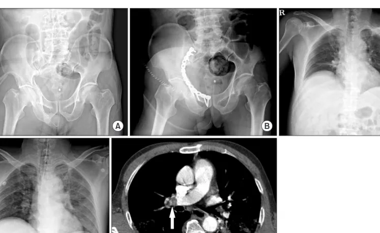
1군에서 대상 환자는 모두 31예였는데, 이 중 5예(16.1%)에서 임 상적 증상이 있는 정맥혈전색전증을 경험하였다. 심부정맥혈전 증이 1예, 폐색전증이 4예였고, 중증의 폐색전증을 보였던 경우는 2예로 중환자실에서 인공호흡기에 의존할 정도로 상태가 위중하 였다(Fig. 1). 또한 2예에서는 중증 외상으로 인해 혈역학적으로 불안정하여 정형외과 수술이 1주일 이상 지연되던 도중 폐색전
Figure 1. (A) Initial pelvis antero-posterior (AP) radiograph of a 55-year-old man with injury severity score 22; liver laceration, T12 compression fracture, right acetabular fracture and 2nd degree burn on the right arm. (B) Immediate postoperative radiograph 10 days after injury. (C) Preoperative chest AP radiograph. (D) Postoperative chest AP radiograph showing haziness at the left lower lobe with PaO2 71.5 mmHg under FiO2 50% application of external fixation. (E) Chest computed tomography image showing large thrombus (arrow) in the right main pulmonary artery. The patient was transferred to intensive care unit for ventilator care immediately.
증이 발생하여 하대정맥 필터(inferior vena cava filter)를 삽입하고 골절에 대한 수술을 진행하였다(Fig. 2). 2군에서 중증 외상 환자 는 모두 32예였는데, 이 중 임상적 증상이 있는 정맥혈전색전증은 없었다. 2군에서 대상 환자에게 수술 후 보행 시작까지 투여한 항 응고제 clexan의 사용 기간은 평균 14일(2-73일)이었다. 두 군 간 의 임상적 증상이 있는 정맥혈전색전증의 발생 빈도는 의미 있는 차이를 보였고(p=0.024), 임상 인자들에 대한 두 군 간의 의미 있 는 차이는 2군에서 ISS는 더 높고, 수상 후 48시간 동안 수혈량도 더 많은 것으로 나타났다(Table 1). 나이, 재원 기간, 중환자실 재 원 기간, 총 수혈량, ISS, 수술까지의 지연 시간에는 통계학적으로 차이를 보이지 않았다(Table 1).
고 찰
골절을 동반한 중증 외상 환자의 경우 응고 작용에 변화를 일으 켜 정맥혈전색전증에 대한 위험성이 높은 것으로 알려져 있다.5,6) Coon7)에 의하면 모든 외상 환자는 나이에 상관 없이 정맥혈전색 전증의 발생 위험도가 높다고 하였고, National Institutes of Health (NIH) 보고에 의하면 다발성 외상을 동반한 젊은 환자에서 정맥 혈전색전증의 빈도는 약 20% 정도가 된다고 하였다.8) Kudsk 등9) 은 하지 골절이 있는 경우 보행이 힘들어 침상 안정의 기간이 길 어질 경우 정맥혈전색전증의 빈도가 높다고 하였고, 무증상 정맥 혈전색전증까지 포함하면 빈도는 67%에 이른다고 하였다. 외상
Figure 2. (A) Initial left tibia antero-posterior and lateral radiographs of a 63-year-old man with injury severity score 22; hemopneumothorax right, left proximal tibia open fracture, cerebral contusion. (B) Application of external fixation. (C) Chest computed tomography image showing focal linear thrombus (arrow) in the posterior segmental branch of right pulmonary artery with PaO2 82.0 mmHg under nasal prong O2 5 L at the postoperative 6 days.
(D) Inferior vena cava filter insertion. (E) Postoperative radiographs after definitive surgery.
Table 1. Summary of Clinical Data
Group I Group II p-value
Age (yr) 43.5 (21-79) 44.1 (22-80) 0.810
Injury severity score 22.4 (17-45) 25.9 (17-50) 0.040
Hospital day 41.4 (10-156) 46.1 (12-173) 0.592
ICU day 4.9 (0-42) 8.2 (10-30) 0.312
Transfusion within 48 hours after injury (unit) 3.1 (0-22) 6.6 (0-33) 0.004
Total transfusion (unit) 11.0 (0-56) 14.4 (0-79) 0.255
Time to surgery from injury (d) 6.6 (0-37) 5.1 (0-38) 0.127
ICU, intensive care unit.
환자에 있어서 정맥혈전색전증에 대한 빈도의 보고를 살펴보면 예방 치료가 없었던 경우 Geerts 등10)은 빈도가 58%라고 보고하였 고, 예방을 하였던 경우라도 증상이 있는 심부정맥혈전증의 경우 2.5-13.4%,11-14) 증상이 있는 폐색전증의 경우 0.7-4% 정도로 보고 되어 있다.11-13,15) 정맥혈전색전증에 대한 연구에서 두 가지 항목 에 의해 빈도의 차이가 크게 보이는데, 첫째는 예방 조치 유무이 고, 둘째는 선별 검사 시행에 따른 무증상 정맥혈전색전증의 포 함 유무가 그것이다. 이번 연구에서는 선별 검사는 따로 시행하 지 않았고, 정맥혈전색전증 예방에 대한 관심의 증가로 적극적인 예방 조치를 시행한 시기를 기준으로 전후의 환자군을 비교함으 로써 정맥혈전색전증의 빈도 및 예방 효과를 후향적인 방법으로 조사하였다.
하지 골절을 동반한 중증 외상 환자에서 항혈전 스타킹만으로 예방하였을 때 정맥혈전색전증의 빈도는 16.1%였던 반면, 항응 고제 및 물리적 방법을 이용한 경우 빈도는 0%로 의미 있는 차이 를 보였다(p=0.024). 두 군 간에 나이, 중환자실 재원 기간, 재원 기 간, 총 수혈량, 수술까지 지연된 시간에는 통계학적으로 차이를 보이지 않았다. 오히려 정맥혈전색전증이 발생하지 않았던 2군 에서 ISS가 더 높고 수상 후 48시간 동안 수혈량이 더 많았던 것 으로 나타났는데, 높은 ISS와 수상 후 48시간 동안 수혈량은 정맥 혈전색전증의 위험 인자임을 감안할 때 적극적인 혈전증의 예방 의 효과가 있는 것으로 생각한다. 이번 결과를 통해 항혈전 스타 킹만으로는 정맥혈전색전증에 대한 예방이 충분하지 않다는 것 을 확인할 수 있고, 5예의 정맥혈전색전증 환자 중 폐색전증이 4 예(13.3%)로 대부분을 차지했으며 이 중 생명을 위협하는 치명적 인 폐색전증이 2예(6.7%)임을 고려할 때 정맥혈전색전증에 대한 관심을 기울여야 할 것이다. 그리고 증상이 있는 정맥혈전색전증 빈도가 16.1%임을 감안할 때 폐색전증의 위험이 있는 무증상 심 부정맥혈전증의 빈도는 이보다 더 높을 것임을 예측할 수 있다.
외상 환자에서 정맥혈전색전증의 위험 인자는 45세 이상, 3일 이상의 침상 안정, 혈전증의 병력, 척추 골절, 혼수 상태, 사지 마 비 혹은 하지 마비, 골반 골절, 하지 골절, 하지의 중요 정맥의 봉 합, 하지의 복합 상처(complex wound), 수상 후 48시간 동안 수혈 량, 장시간의 수술 시간, 수상 후 골절 고정까지의 시간이 해당된
다.10,15,16) 또한 Owings 등17)은 폐색전증에 대한 위험 인자로 장골
골절을 동반한 55세 이상의 환자, 두부 손상 및 혼수 상태, 다발 성 장관골 골절 및 골반 골절, 신경학적 손상을 동반한 척수 손상 이라고 하였다. 이와 같이 하지 혹은 골반 골절을 동반한 중증 외 상의 경우 침상 안정, 및 골절에 해당하는 위험 인자 외에 중증 외 상 환자의 특성상 많은 수혈량, 장시간의 수술 시간, 수상 후 골절 고정까지의 지연 등의 위험 인자를 동반하는 경우가 많아 예방이 필요하다 할 것이다.
연구 결과에서 보듯이 항응고제와 물리적 방법의 적극적인 조 치를 하였을 경우 정맥혈전색전증의 예방 효과는 매우 우수하였
으며, 예방에 대한 효과는 이미 잘 알려져 있다. 정맥혈전색전증 예방을 위해 사용되는 항응고제 중 저분자량 헤파린은 모든 심부 정맥혈전증에 있어 30%의 감소 효과가 있었다고 하였고, 근위부 정맥혈전증에는 58%의 감소 효과가 있었으며 저용량의 헤파린 보다 예방 효과가 더 크다고 하였다.18) 이러한 효과로 인해 저분 자량 헤파린은 정맥혈전색전증을 예방하기 위해 다발성 외상 환 자에 있어 널리 추천되어 왔다.18-22) 하지만 저분자량 헤파린을 포 함한 항응고제는 출혈에 대한 위험성을 증가시킬 수 있으므로 이 에 대한 걱정이 있어 왔고, 특히 두개내 혈종이 있거나 척수 손상, 중요 고형 장기 손상, 복합 골반 골절이 있는 경우, 주의가 필요하 다고 하였다.10) Cothren 등12)은 저분자량 헤파린의 효과 및 안전 성을 평가하는 연구에서 외상 후 혈액학적으로 안정화되고 지속 적인 출혈의 증거가 없는 시점부터 투약을 시작하여 안전하게 혈 전증을 예방할 수 있다고 하였다. The Eastern Association for the Surgery of Trauma (EAST)에서 제시한 예방 가이드라인에서는 환자의 상태에 맞게 출혈에 대한 위험성과 정맥혈전색전증 예방 효과를 비교하여 저분자량 헤파린 사용을 결정하라고 권고하였 다.23)
물리적 방법은 출혈에 대한 위험이 없다는 큰 장점이 있고, 두 가지 작용 기전으로 정맥혈전색전증을 예방하게 된다. 이는 정맥 혈류 순환 개선과 혈관벽으로부터 plasminogen activator를 유리 시켜 plasma fibrinolytic activity를 증가시킴으로써 가능하다. 일부 보고에서는 물리적인 방법이 심부정맥혈전증 예방에 효과적이라 는 보고가 있으나,24,25) 한편으로는 예방에 효과가 입증되지 않았 다는 보고도 많았다.18,26-28) 예방 효과면에서 차이를 보이는 이유 는 물리적 방법의 경우 환자들의 순응도가 낮기 때문이며, 실제 지침대로 착용을 잘 하는 환자는 19%에 불과하다고 하였다.29) 또 한 하지의 골절이나 심한 창상이 있는 경우 착용하기에는 어려운 점이 있고, 외상 환자의 35%에서 이러한 이유로 착용의 부적응증 이 된다고 하였다.28) Stannard 등13)은 외상 환자를 대상으로 수상 후 24-48시간 이후에 저분자량 헤파린을 사용한 군과 수상 직후 물리적 방법을 사용하면서 급성 출혈이 해소된 이후 수상 후 5일 뒤에 저분자량 헤파린을 지연 투여한 군을 비교하는 무작위, 전 향적 연구에서 후자의 경우에서 임상적 결과가 우수하였던 바 2 단계 접근법이 효과적이라고 권고하였는데, 예방 효과와 안정성 을 고려한 연구 결과로 이에 대한 추가적인 연구가 뒷받침되면 더욱 신뢰를 얻을 방법이라고 여겨진다.
이번 연구에서는 연구 대상의 적은 수와 단일 의료 기관에서 이루어졌다는 한계가 있으며, 추후 대규모의 전향적 무작위 연구 를 통한 정맥혈전색전증의 빈도와 예방 효과를 파악하여 골절을 동반한 중증 외상 환자에 대한 예방 가이드 라인을 제시해야 할 것이다.
결 론
중증 외상 환자에서 정맥혈전색전증에 대한 적극적인 예방 조치 를 하지 않았을 경우 임상적으로 나타나는 혈전증이 16.1%에서 나타났으며 이는 적극적인 예방을 통해 현저히 감소하는 것을 확 인할 수 있었다. 따라서 하지 골절을 동반한 중증 외상 환자에서 정맥혈전색전증에 대한 적극적인 예방법이 필요하다 할 것이다.
참고문헌
1. Cha SI, Lee SY, Kim CH, et al. Venous thromboembolism in Korean patients undergoing major orthopedic surgery: a prospective observational study using computed tomographic (CT) pulmonary angiography and indirect CT venography. J Korean Med Sci. 2010;25:28-34.
2. Lee HY, Koh SH, Oh SM, Park HC. Deep vein thrombosis after total hip arthroplasty: the incidence of DVT and Cor- relation between DVT and risk factors. Korean Soc Vasc Surg.
2005;21:40-4.
3. Lee JS, Kim TW, Suh JT. Deep vein thrombosis after total knee arthroplasty: correlation between the incidence and clinical risk factors. J Korean Knee Soc. 2010;22:270-7.
4. Park KH, Cheon SH, Lee JH, Kyung HS. Incidence of venous thromboembolism using 64 channel multidetector row com- puted tomography-indirect venography and anti-coagulation therapy after total knee arthroplasty in Korea. Knee Surg Relat Res. 2012;24:19-24.
5. Dries DJ. Activation of the clotting system and complement after trauma. New Horiz. 1996;4:276-88.
6. Seyfer AE, Seaber AV, Dombrose FA, Urbaniak JR. Coagu- lation changes in elective surgery and trauma. Ann Surg.
1981;193:210-3.
7. Coon WW. Epidemiology of venous thromboembolism. Ann Surg. 1977;186:149-64.
8. Prevention of venous thrombosis and pulmonary embolism.
Natl Inst Health Consens Dev Conf Consens Statement.
1986;6:1-8.
9. Kudsk KA, Fabian TC, Baum S, Gold RE, Mangiante E, Voeller G. Silent deep vein thrombosis in immobilized mul- tiple trauma patients. Am J Surg. 1989;158:515-9.
10. Geerts WH, Code KI, Jay RM, Chen E, Szalai JP. A prospec- tive study of venous thromboembolism after major trauma. N Engl J Med. 1994;331:1601-6.
11. Adams RC, Hamrick M, Berenguer C, Senkowski C, Ochsner
MG. Four years of an aggressive prophylaxis and screening protocol for venous thromboembolism in a large trauma population. J Trauma. 2008;65:300-6.
12. Cothren CC, Smith WR, Moore EE, Morgan SJ. Utility of once-daily dose of low-molecular-weight heparin to prevent venous thromboembolism in multisystem trauma patients.
World J Surg. 2007;31:98-104.
13. Stannard JP, Lopez-Ben RR, Volgas DA, et al. Prophylaxis against deep-vein thrombosis following trauma: a prospective, randomized comparison of mechanical and pharmacologic prophylaxis. J Bone Joint Surg Am. 2006;88:261-6.
14. Stannard JP, Singhania AK, Lopez-Ben RR, et al. Deep-vein thrombosis in high-energy skeletal trauma despite thrombo- prophylaxis. J Bone Joint Surg Br. 2005;87:965-8.
15. Abelseth G, Buckley RE, Pineo GE, Hull R, Rose MS. In- cidence of deep-vein thrombosis in patients with fractures of the lower extremity distal to the hip. J Orthop Trauma.
1996;10:230-5.
16. Hak DJ. Prevention of venous thromboembolism in trauma and long bone fractures. Curr Opin Pulm Med. 2001;7:338- 43.
17. Owings JT, Gosselin RC, Battistella FD, Anderson JT, Petrich M, Larkin EC. Whole blood D-dimer assay: an effective non- invasive method to rule out pulmonary embolism. J Trauma.
2000;48:795-9.
18. Geerts WH, Jay RM, Code KI, et al. A comparison of low- dose heparin with low-molecular-weight heparin as prophy- laxis against venous thromboembolism after major trauma. N Engl J Med. 1996;335:701-7.
19. Cipolle MD, Wojcik R, Seislove E, Wasser TE, Pasquale MD.
The role of surveillance duplex scanning in preventing venous thromboembolism in trauma patients. J Trauma. 2002;52:453- 62.
20. Greenfield LJ, Proctor MC, Rodriguez JL, Luchette FA, Ci- polle MD, Cho J. Posttrauma thromboembolism prophylaxis.
J Trauma. 1997;42:100-3.
21. Knudson MM, Morabito D, Paiement GD, Shackleford S. Use of low molecular weight heparin in preventing thromboem- bolism in trauma patients. J Trauma. 1996;41:446-59.
22. Sharma OP, Oswanski MF, Joseph RJ, et al. Venous thrombo- embolism in trauma patients. Am Surg. 2007;73:1173-80.
23. Yenna ZC, Roberts C. Thromboprophylaxis after multiple trauma: what treatment and for how long? Injury. 2009;40 Suppl 4:S90-4.
24. Spain DA, Bergamini TM, Hoffmann JF, Carrillo EH, Rich- ardson JD. Comparison of sequential compression devices and foot pumps for prophylaxis of deep venous thrombosis in high-risk trauma patients. Am Surg. 1998;64:522-5.
25. Stannard JP, Riley RS, McClenney MD, Lopez-Ben RR, Volgas DA, Alonso JE. Mechanical prophylaxis against deep-vein thrombosis after pelvic and acetabular fractures. J Bone Joint Surg Am. 2001;83:1047-51.
26. Knudson MM, Lewis FR, Clinton A, Atkinson K, Megerman J.
Prevention of venous thromboembolism in trauma patients. J Trauma. 1994;37:480-7.
27. Napolitano LM, Garlapati VS, Heard SO, et al. Asymptomatic deep venous thrombosis in the trauma patient: is an aggres- sive screening protocol justified? J Trauma. 1995;39:651-7.
28. Shackford SR, Davis JW, Hollingsworth-Fridlund P, Brewer NS, Hoyt DB, Mackersie RC. Venous thromboembolism in patients with major trauma. Am J Surg. 1990;159:365-9.
29. Cornwell EE 3rd, Chang D, Velmahos G, et al. Compliance with sequential compression device prophylaxis in at-risk trauma patients: a prospective analysis. Am Surg. 2002;68:470- 3.
Incidence and Prevention of Venous Thromboembolism in Severely Injured Patients with Lower Extremity Fracture
Ji Wan Kim, M.D., Ph.D.*, Ji Ho Choi, M.D., and Jung Jae Kim, M.D., Ph.D.
Department of Orthopaedic Surgery, Asan Medical Center, College of Medicine, University of Ulsan, Seoul,
*Haeundae Paik Hospital, College of Medicine, Inje University, Busan, Korea
Purpose: To compare the incidence of venous thromboembolism (VTE) in severely injured patients with lower extremity fracture in a
prophylactic group with that in a non-prophylactic group during a retrospective study.Materials and Methods: Severely injured patients with lower extremity fracture were enrolled in this study and were divided into
the following two groups: Group I, non-prophylactic group, and Group II, prophylactic group using anticoagulants and mechanical prophylaxis.Results: Symptomatic VTE occurred in 5 cases (16.1%) among the 31 cases in Group I; i.e., deep vein thrombosis in one case and
pulmonary embolism (PE) in four cases, including life threatening PE 2 cases. There were no patients with symptomatic VTE among the 32 cases in Group II.Conclusion: The incidence of symptomatic VTE without the use of prophylaxis was 16.1%. This rate dramatically decreased with the
use of prophylaxis. Therefore, we believe that prophylaxis with anticoagulants and mechanical device is necessary in order to prevent VTE in severely injured patients with lower extremity fracture.Key words: severely injured trauma patients, lower extremity fracture, venous thromboembolism, deep vein thrombosis, pulmonary
embolismReceived April 2, 2012 Revised June 7, 2012 Accepted July 19, 2012 Correspondence to: Jung Jae Kim, M.D., Ph.D.
Department of Orthopaedic Surgery, Asan Medical Center, College of Medicine, University of Ulsan, 88, Olympic-ro 43-gil, Songpa-gu, Seoul 138-736, Korea
TEL: +82-2-3010-3530
FAX: +82-2-488-7877 E-mail: [email protected]Copyright © 2012 by The Korean Orthopaedic Association
“This is an Open Access article distributed under the terms of the Creative Commons Attribution Non-Commercial License (http://creativecommons.org/licenses/by-nc/3.0/) which permits unrestricted non-commercial use, distribution, and reproduction in any medium, provided the original work is properly cited.”
The Journal of the Korean Orthopaedic Association Volume 47 Number 6 2012