RESEARCH ARTICLE
Received: December 28, 2017, Revised: January 26, 2018, Accepted: February 1, 2018 ISSN 2233-7679 (Online)
†
Correspondence to: Hyo-Jin Ko
Department of Dental Hygiene, Choonhae College of Health Sciences, 9 Daehak-gil, Ungchon-myeon, Ulju-gun, Ulsan 44965, Korea Tel: +82-52-270-0294, Fax: +82-52-270-0239, E-mail: [email protected]
Copyright © 2018 by Journal of Dental Hygiene Science
Quantitative Analysis of Oral Pathogenic Bacteria according to Smoking Using Real-Time PCR
Eun-Suk Jeon, Hyo-Jin Heo, and Hyo-Jin Ko †
Department of Dental Hygiene, Choonhae College of Health Sciences, Ulsan 44965, Korea
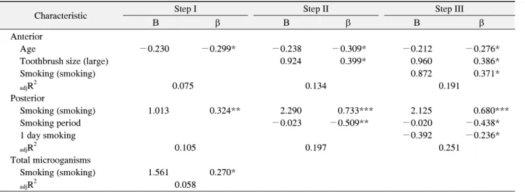
This study investigates the relationship between smoking and periodontal disease through quantitative analysis of intra-buccal oral pathogenic bacteria detected in smokers and aims to yield objective baseline data for applications in anti-smoking and dental health education programs. From April to May 2016, participants in an oral health management program within an intensive dental hygiene training course at Choonhae College of Health Sciences received an explanation of the study purposes and methods, after which male smokers aged 18∼30 years agreed to participate voluntarily. Real-time polymerase chain reaction (PCR) analysis of oral pathogenic bacteria was performed after collecting gingival sulcus fluid samples from 67 smokers. The intra-buccal oral pathogenic bacteria distributions were analyzed based on the subjects' general characteristics, smoking behaviors, and oral care behaviors. The distribution results show that pathogens in the anterior teeth are affected (in this order) by age, toothbrush size, and smoking status; older people had fewer pathogens, those who used larger toothbrushes had more pathogens, and smokers had more pathogens, compared to non-smokers (
adjR
2=19.1). In the posterior teeth, pathogens were influenced (in this order) by smoking status, smoking duration, and the number of tooth brushings per day; smokers had more pathogens than non-smokers, and those who brushed their teeth more often had fewer pathogens (
adjR
2=25.1). The overall pathogen distribution was affected only by smoking status: smokers generally had more pathogens, compared to non-smokers. Therefore, it is necessary to provide information about the risk of periodontal disease due to smoking during anti-smoking or dental health education sessions; particularly, the use of smaller toothbrushes for anterior teeth and the need for smokers in their early twenties to quit smoking for dental health should be highly emphasized.
Key Words: Oral pathogenic bacteria, Real-time polymerase chain reaction, Smokers
Introduction
Periodontal disease, a quintessential oral disease, involves the chronic infection of periodontal cells found in periodontal ligaments, the gums, cement, and alveolar bones, resulting in the destruction of cells that support the teeth. Periodontal diseases occur in response to various causes, such as microbes, dental prostheses, tooth loss, smoking, nutritional disorders, and genetic factors, and can be divided into inflammation of the gums, which involve infections of the gum only, and periodontitis, which involves the destruction of both the attached cells and alveolar bones 1) . Microbes found in dental plaques are the core causes of such inflammation.
The oral cavity contains 300∼500 types of microbes, and in the normal gums, aerobic, inactive, gram-positive oral micrococcal species comprise the majority of these entities; however, the contraction of periodontal disease causes a shift to anaerobic, active gram-negative microbes 2) . Socransky et al. 3) asserted that periodontal diseases increase in the presence of anaerobic gram-negative mic- robes, and Greenstein and Polson 4) asserted that the focal points of periodontal disease are an increase in the population of active microbes and reduction in micro- coccal strains. In other words, changes in the composition of bacterial species and other microorganisms are associated with the occurrence of periodontal diseases.
The key microbial species that cause periodontal diseases
include Aggregatibacter actinomycetemcomitans, Porphy- romonas gingivalis, and Prevotella intermedia. Of these, A. actinomycetemcomitans causes invasive periodontitis and facilitates periodontal cell destruction, P. gingivalis is associated with gum inflammation and alveolar bone destructions, and P. intermedia is known to facilitate hormone-related and complex periodontitis 5) .
According to the 2016 statistics from the Organization for Economic Co-operation and Development (OECD), the percentage of daily smokers older than 15 years was 20.0% in the Korean population; this was higher than the OECD average of 18.6% 6) . The 2012 Korean National Oral Health Survey 7) reported a continuous increase in periodontal diseases among Korea adults, and found that smoking, dietary habits, stress, and the usage of oral hygiene items influenced oral health 8,9) . Moreover, smoking is a known cause of general diseases such as lung cancer and cardiovascular diseases and is known to be related to epithelial attachment, alveolar bone loss, gum recession, and dental pocket formation 8,10,11) . Barbour et al. 12) reported that smokers have a 2.5 to 6-fold increased risk of periodontal diseases relative to non-smokers, while Won et al. 13) reported an increased prevalence of perio- dontal diseases among smokers, compared to non- smokers. Reibel 14) also reported a higher prevalence of periodontal diseases among smokers than among non-smokers after comparing periodontal disease rates between the two groups, and also reported an increased likelihood of failure of dental treatments because of smoking.
To prevent periodontal diseases that destroy cells around the teeth and the alveolar bones, it is important to verify the composition of microorganisms within the oral cavity and quantify the results. Real-time polymerase chain reaction (PCR) is the most widely used technique for quantitatively observing the volume of microbes within the oral cavity, and involves the real-time quan- tification of acid levels throughout the PCR process to accurately quantify the substrates in each sample 15) .
Research regarding the effects of smoking on periodontal diseases and associated correlations is ongoing 8,10-14) ; however, quantitative analyses of oral pathogenic bacteria according to oral cavity sections are lacking, particularly
in terms of the associations of these characteristics with smoking behavior and oral health management.
Therefore, this study aimed to quantitatively analyze the intra-buccal oral pathogenic bacteria of smokers, identify the relationship between smoking and oral diseases, and provide objective baseline data that could be applied in anti-smoking and dental health education programs.
Accordingly, this study sought to identify quantitative differences in oral pathogenic bacteria among different sections of the oral cavity based on smoking behavior and oral health management behavior with the intent to determine the influential factors.
Materials and Methods
1. Subjects
From April to May 2016, participants in an oral health management program within the intensive dental hygiene training course at C College, Ulsan Metropolitan City, received an explanation of the study purpose and method.
A total of 67 male subjects between the ages of 18 and 30 years, including non-smokers, were enrolled after agreeing to participate voluntarily.
2. Research method
To ensure the ethical protection of the subjects,
approval was obtained from the Institutional Review
Board of Choonhae College of Health Sciences prior to
the study (IRB approval no. CH-201604-12). The subjects
received oral and written explanations of the study
purpose and method, the commitment to confidentiality
regarding study participation, the voluntary nature of
participation, ability to opt in or out, and the potential
advantages and disadvantages of participation; sub-
sequently, agreement to participate was obtained from the
subjects. A self-administered survey addressing sex, age,
and general diseases was conducted, after which gingival
sulcus fluid samples were collected from two anterior
teeth and posterior teeth to compare quantitative
differences in the oral pathogenic bacteria distributions by
section. DNA was extracted from these samples, and
multiplex real-time PCR was used to generate data.
1) Collection of gingival sulcus fluid
To collect DNA from oral pathogenic bacteria, samples were collected from the subjects’ oral cavities by inserting 4 sterilized no. 30 endodontic paper points (Sure Dent, Seongnam, Korea) for 10 seconds into the labial and lingual sections of the upper right central incisor and lower left central incisor among the anterior teeth, and by inserting 4 sterilized no. 90 endodontic paper points (Sure Dent) for 10 seconds in the buccal and lingual sections of the upper left first molar tooth and lower right first molar tooth. After removal, the paper points were frozen in 1.5-ml micro tubes containing 1 μl of phosphate-buffered saline (Gibco BRL, Grand Island, NY, USA) (two per each of the four teeth) and sent to the DNA lab where they were stored at −20 o C until use.
2) DNA extraction of oral pathogenic bacteria and quantitative analysis
An Exgene Clinic SV mini-kit (Biokorea, Cheongju, Korea) was used to extract DNA from oral microor- ganisms according to the manufacturer’s protocol. For the quantitative analysis of oral pathogenic bacteria, a Cytoperio analysis system (Cytogen, Seoul, Korea) was used to expand the 16s rRNA and thus collect data of all microorganisms existing within the oral cavity.
Real-time PCR reactions were set so that the total volume was 20 μl; each reaction included 2 μl of total extracted DNA, 10 pmol of each primer set, probe, and buffer solution, and 1 unit of Hot-start Taq DNA Polyme- rase (GeneAll, Seoul, Korea). The reactions were placed in 96-well plates and quantitatively analyzed using an ABI 7500 Fast Real-Time PCR System (Life Technologies Korea, Seoul, Korea). The real-time PCR conditions were as follows: denaturalization for 15 minutes at 95 o C, 15 seconds at 95 o C, and 45 cycles of 15 seconds at 55 o C and 20 seconds at 72 o C. The cycle threshold (Ct) analysis, which began in the exponential phase, used known DNA concentrations to measure the number of microbes in each sample according to the standard curve. In this study, the total volume of oral pathogenic bacteria is presented by section, and large numbers have been transformed into natural logs to simplify the comparison of differences.
3. Statistical analysis
The collected data were analyzed using IBM SPSS Statistics (ver. 21.0; IBM Co., Armonk, NY, USA) and the statistical significance level was at a p<0.05. The subjects’ general characteristics, smoking behavior, and oral health management behavior were analyzed using a frequency analysis, and a Student’s t-test and one-way ANOVA were conducted to verify quantitative differences in oral pathogenic bacteria based on general charac- teristics, smoking behavior, and oral health management behavior. Moreover, a multiple regression analysis was conducted to identify the factors influencing quantitative differences in intra-buccal oral pathogenic bacteria.
Results
1. Oral pathogenic bacteria and general characteristics In this study, 49.3% of the subjects were 18∼22 years, and 50.7% were 23∼30 years; furthermore, 67.2% were students, representing the largest subset of the population.
Regarding residence, 50.7%, 35.8%, and 13.4% of the subjects resided in Ulsan, Busan, and other regions, respectively. As noted, this study analyzed oral pathogenic bacteria by dental section, as well as the relationship of these pathogens with general characteristics. Among subjects aged 18∼22 years, 20.02±1.68 and 20.21±1.66 oral pathogenic bacteria were detected from the anterior and posterior teeth, with an overall number of 20.97±1.53 pathogens; this was higher than the numbers detected in subjects aged 23∼30 years, in whom 18.64±1.35 and 19.31±1.35 were detected from the anterior and posterior teeth, with an overall number of 19.83±1.25 pathogens (p
<0.05). However, no statistically significant differences were observed with respect to occupation or place of residence (Table 1).
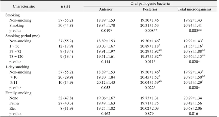
2. Oral pathogenic bacteria and smoking behavior The analysis of smoking behavior indicated that 55.2%
of the subjects were non-smokers and 44.8% were smokers; of the former, the largest subset, 17.9%, had smoked for 1∼36 months. Furthermore, 29.9% reported smoking fewer than 10 cigarettes per day, whereas 14.9%
reported smoking more than 11 cigarettes per day.
Table 1. Oral Pathogenic Bacteria according to General Characteristics
Characteristic n (%) Oral pathogenic bacteria
Anterior Posterior Total microoganisms
Age (y)
18∼22 33 (49.3) 20.02±1.68 20.21±1.66 20.97±1.53
23∼30 34 (50.7) 18.64±1.35 19.31±1.35 19.83±1.25
p-value <0.001*** 0.017* 0.001**
Occupation
Inoccupation 9 (13.4) 18.71±1.38 19.72±1.20 20.13±1.10
Student 45 (67.2) 19.32±1.74 19.70±1.67 20.35±1.61
Soldier 4 (6.0) 18.91±1.60 19.76±1.21 20.13±1.33
Office workers 9 (13.4) 20.09±1.48 20.04±1.68 20.90±1.43
p-value 0.339 0.951 0.698
Residence
Busan 24 (35.8) 19.08±1.53 19.72±1.46 20.22±1.42
Ulsan 34 (50.7) 19.30±1.75 19.73±1.61 20.39±1.53
Etc. 9 (13.4) 20.00±1.67 19.92±1.84 20.81±1.70
p-value 0.375 0.942 0.633
Values are presented as n (%) or mean±standard deviation.
Bacteria levels were expressed as log scale of concentration.
*p<0.05, **p<0.01, ***p<0.001 statistically significant by t-test.
Table 2. Oral Pathogenic Bacteria Caused by Smoking Behavior
Characteristic n (%) Oral pathogenic bacteria
Anterior Posterior Total microoganisms
Smoking
Non-smoking 37 (55.2) 18.89±1.53 19.30±1.46 19.92±1.43
Smoking 30 (44.8) 19.84±1.70 20.31±1.53 20.94±1.41
p-value 0.019* 0.008** 0.005**
Smoking period (mo)
Non-smoking 37 (55.2) 18.89±1.53 19.30±1.46
a19.92±1.43
a1∼36 12 (17.9) 20.03±1.67 20.89±1.18
b21.35±1.16
b37∼72 9 (13.4) 19.91±1.97 20.29±1.92
a,b20.88±1.88
a,b73∼120 9 (13.4) 19.51±1.61 19.57±1.32
a,b20.46±1.15
a,bp-value 0.114 0.011* 0.020*
1-day smoking
Non-smoking 37 (55.2) 18.89±1.53 19.30±1.46
a19.92±1.43
a≤10 20 (29.9) 19.70±1.84 20.45±1.52
b20.93±1.50
a,b≥11 10 (14.9) 20.12±1.43 20.04±1.59
a,b20.95±1.29
bp-value 0.053 0.022* 0.020*
Family smoking
None 32 (47.8) 19.06±1.67 19.73±1.31 20.29±1.34
Father 27 (40.3) 19.49±1.63 19.71±1.75 20.42±1.56
Etc. 8 (11.9) 19.75±1.82 20.02±2.03 20.68±2.06
p-value 0.462 0.879 0.816
Values are presented as n (%) or mean±standard deviation.
Bacteria levels were expressed as log scale of concentration.
*p<0.05, **p<0.01, statistically significant by t-test.
*p<0.05, statistically significant by one way ANOVA.
a,b
Values are significantly different using the Scheffe method.
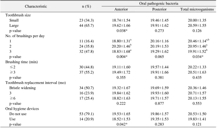
Table 3. Oral Pathogenic Bacteria according to Oral Health Behavior
Characteristic n (%) Oral pathogenic bacteria
Anterior Posterior Total microoganisms
Toothbrush size
Small 23 (34.3) 18.74±1.54 19.46±1.45 20.00±1.35
Large 44 (65.7) 19.62±1.66 19.91±1.62 20.59±1.55
p-value 0.038* 0.273 0.126
No. of brushings per day
1 11 (16.4) 18.80±1.31
a20.16±1.16 20.46±1.14
a,b2 24 (35.8) 20.20±1.46
b20.19±1.53 20.95±1.46
a3 32 (47.8) 18.83±1.68
a19.29±1.62 19.91±1.52
bp-value 0.004* 0.065 0.034*
Brushing time (min)
≤2 30 (44.8) 19.11±1.60 19.57±1.44 20.22±1.33
≥3 37 (55.2) 19.49±1.72 19.91±1.66 20.51±1.63
p-value 0.355 0.381 0.435
Toothbrush replacement interval (mo)
Bristle widening 34 (50.7) 19.32±1.67 19.69±1.59 20.36±1.46
3 16 (23.9) 19.84±1.62 19.93±1.60 20.71±1.57
≥6 17 (25.4) 18.82±1.63 19.71±1.57 20.13±1.55
p-value 0.222 0.877 0.553
Oral hygiene devices
Do not use 53 (79.1) 19.53±1.65 19.86±1.57 20.53±1.50
Use 14 (20.9) 18.52±1.53 19.35±1.53 19.83±1.41
p-value 0.042* 0.283 0.121
Values are presented as n (%) or mean±standard deviation.
Bacteria levels were expressed as log scale of concentration.
*p<0.05, statistically significant by one way ANOVA.
a,b