31
and Safety
Available online at http://www.foodhygiene.or.kr
http://dx.doi.org/10.13103/JFHS.2014.29.1.031
Real-time RT-PCR을 이용한 Feline Calicivirus 불활성화의 정량적 분석
정혜미·김광엽*
충북대학교 식품생명공학과
Quantitative Analysis of Feline Calicivirus Inactivation using Real-time RT-PCR
Hye Mi Jeong and Kwang Yup Kim*
Dept. of Food Science and Biotechnology, Chungbuk National University, Chungbuk 361-763, Korea (Received September 3, 2013/Revised December 20, 2013/Accepted February 25, 2014)
ABSTRACT - Norovirus causes acute gastroenteritis in all age groups and its food poisoning outbreaks are rap- idly increasing in Korea. Reverse transcription-polymerase chain reaction (RT-PCR) is most widely used for the rapid detection of foodborne viruses due to high sensitivity. However, the false positive results of RT-PCR obtained against already inactivated viruses could be a serious drawbacks in food safety area. In this study, we investigated a method to yield true positive RT-PCR results only with alive viruses. To decompose the RNA genes from dead viruses, the enzymatic treatments composed of proteinse K and Ribonuclease A were applied to the sanitized and inactivated virus particles. Another aim of this study was to quantify the efficiencies of several major sanitizing treatments using real- time RT-PCR. Feline calicivirus (FCV) that belongs to the same Caliciviridae family with norovirus was used as a surrogate model for norovirus. The initial level of virus in control suspension was approximately 104PFU/mL. Most of inactivated viruses treated with the enzymatic treatment for 30 min at 37oC were not detected in RT-PCR, Quanti- fication results to verify the inactivation efficiencies of sanitizing treatments using real-time RT-PCR showed no false positive in most cases. We could successfully develope a numerical quantification process for the inactivated viruses after major sanitizing treatments using real-time RT-PCR. The results obtained in this study could provide a novel basis of rapid virus quantification in food safety area.
Key words : Norovirus, Feline Calicivirus, Inactivation, Enzymatic treatment, Real-time RT-PCR
노로바이러스(Norovirus)는 Caliciviridae과에 속하는 소형 구형 정이십면체 RNA 바이러스로서, GI부터 GV까지 그 유전자 다양성이 크며, 이 중 GI, GII, GIV 그룹이 주로 사 람에게 감염을 일으키는 인체 서식종으로 보고되어 있다1,2,3). 노로바이러스는 분변 및 구강 경로를 통하여 감염이 되 며, 감염 시 24~48시간의 잠복기를 거쳐 구토, 설사, 어지 럼증 등의 증세를 동반하게 된다4,5). 세균성 식중독과는 다 르게 10~100개의 작은 입자로도 감염을 유발시킬 수 있 고, 인체 감염 시 많은 양의 바이러스를 배출하는 특징을 지니고 있다6).
최근 전세계적으로 노로바이러스에 의한 식중독 사고가 많이 보고되고 있으며, 미국 질병통제예방센터(Centers for Disease Control and Prevention, CDC)에 의하면 2006~2010
년 노로바이러스에 의한 식중독 발생률은 49%로 가장 높 은 비중을 차지하고 있으며, 세균 40%, 화학물질 6%, 기 타 4%, 기생충 1% 순으로 보고되어 있다7).
국내에서는 식품의약품안전처(Ministry of Food and Drug Safety, MFDS)에서 발표한 한국의 원인균별 식중독 발생 현황 중 노로바이러스에 의한 식중독 발생건수는 2010년 271건 중 31건(11%), 2011년 249건 중 31건(12%), 2012 년 266건 중 50건(19%)으로 세균성 식중독 건수보다 높 은 수치를 보이며 매년 지속적인 증가추세를 보이고 있다8). 노로바이러스에 관한 연구들로는 오염원이 될 수 있는 식 수, 어패류, 과일, 채소 등에서 BDC방법 등을 이용하여 효 율적으로 검출하는 방법9), 분리·농축방법10) 및 UV11), 열처 리12), 염소13), 에탄올14)등의 물리, 화학적 위생 처리를 통하 여 불활성화 시키는 방법 등에 관한 연구들이 진행되고 있다.
일반적으로 노로바이러스를 검출하기 위해서는 reverse transcription-polymerase chain reaction (RT-PCR)을 이용한 유전적 분석법이 사용되는데, 이 방법은 결과를 얻기까지 많은 시간과 노동력이 소요되고, 위양성의 결과를 얻을 수
*Correspondence to: Kwang Yup Kim, Dept. of Food Science and Biotechnology, Chungbuk National University, Chungbuk 361- 763, Korea
Tel: 82-43-261-2568, Fax: 82-43-271-4412 E-mail: [email protected]
도 있으며, 최종 증폭산물을 agarose gel 상으로 확인하기 때문에 정확한 정량분석을 할 수 없다는 한계점을 지닌 다15). 이에 반하여 Real-time RT-PCR법은 형광기술을 이 용하여 증폭과정을 실시간으로 모니터링 하는 방법으로서 RT-PCR법보다 신속하고 민감하게 바이러스의 정성은 물 론 정량검출까지 가능하다16,17).
본 연구에서는 바이러스 현탁액에 물리, 화학적 위생처 리를 한 후 RT-PCR법과 Real-time RT-PCR법을 진행하여 살균효능을 비교분석코자 하였다. 이 때 불활성화 된 바 이러스들이 PCR에 의해 증폭되어 위양성의 결과가 나오 는 것을 방지하고자 PCR 이전에 복합효소(Proteinase K, Ribonuclease A)처리의 전처리과정을 적용하였다18). 또한 복합효소처리 후 RT-PCR법을 이용하여 정성분석에 그친 선행연구18)를 한 단계 더 발전시켜 증폭산물을 전기영동 으로 확인할 필요없이 신속하게 높은 민감도로 확인할 수 있는 Real-time RT-PCR법으로 정량분석함으로써 물리, 화 학적 살균효능도 비교분석하였다.
노로바이러스는 인간의 장을 숙주로 하기 때문에 동물 실험이나 세포배양의 방법으로는 증식이 어렵고, 단일가 닥의 RNA로 구성되어 있어 불안정한 특성을 지니기 때 문에 본 연구에서는 노로바이러스와 동일한 Calicoviridae 과에 속하고 유전학, 형태학, 생화학적 특성이 매우 유사 한 Feline calicivirus (FCV)를 노로바이러스의 대체모델로 서 사용하였다19,20,21,22).
재료 및 방법
세포배양
FCV VR-782는 American Type Culture Collection (ATCC, Manassas, VA, USA)으로부터 구매하였고, Crandell-Reese feline kidney (CRFK) 세포는 한국세포주은행(KCLB 10094, Seoul, Korea)에서 제공받았다.
CRFK 세포는 10% fetal bovine serum (FBS, GIBCO, Grand island, NY), 1% Penicillin-streptomycin (GIBCO, Grand island, NY), 1% non-essential amino acids (NEAA, GIBCO, Grand island, NT, USA)가 포함된 Dulbecco's Modified Eagle Medium (DMEM, GIBCO, Grand island, NY, USA)을 사용하여 37oC, 5% CO2 incubator (MCO- 15AC, SANYO Electric Biomedical Co., Ltd, Japan)에서 배양하였다. 75 cm2-Tissue flask (Corning, NY, USA)에 90% 이상 세포가 자라면 25 cm2-Tissue flask (Corning, NY, USA)에 계대하여 활성화 시켰다9,10).
바이러스
CRFK 세포가 단층(monolayer)을 형성하면 FCV를 접종 하여 37oC, 5% CO2 incubator에서 1시간 배양하였다. 배 양 후 DMEM liquid, 5% FBS, 1% NEAA, 1% Penicillin-
streptomycin이 함유된 maintenance medium을 분주하여 37oC, 5% CO2 incubator에서 3~4일간 배양하였다. 세포병 변효과(cytopathic effect)가 나타나 부착된 세포가 떨어지 면 액체질소로 급속 동결 후 37oC 항온수조로 해동하는 과정을 3반복 후, 2,000 rpm에서 8분 동안 원심분리 후 상 등액을 취하여 1 mL씩 cryogenic vials (Nalgene, Rochester, NY, USA)에 나누어 담았다. 이렇게 만들어진 바이러스 stock은 액체질소(−80oC)에서 보관하여 사용하였다9,23,24).
Plaque assay
실험에 사용한 바이러스 stock은 Dulbecco's Phosphate Buffered Saline, pH 7.4 (DPBS, GIBCO, Grand island, NY)로 10−2 부터 10−6까지 십진희석하여 25 cm2-Tissue flask (Corning, NY, USA)에 배양하여 단층을 형성한 CRFK 세 포에 0.5 mL씩 접종 후 37oC, 5% CO2 incubator에서 1시 간 흡착시켰다. 흡착 후 DMEM liquid, 10% FBS, 1%
NEAA, 1% Penicillin-streptomycin가 함유된 2× overlay medium에 1.5% agarose gel (JUNSEI, Tokyo, Japan)을 1:1로 혼합하여 5 mL씩 각각의 흡착시킨 flask에 분주하였 다. Plaque가 형성되면 18.5% formaldehyde를 20분간 방 치하여 세포를 고정시키고 흐르는 물로 세포층을 제거 후 crystal violet (JUNSEI, Tokyo)으로 5분간 염색하였다. 염 색액을 흐르는 물로 제거하여 건조시킨 후 plaque 수를 측 정하여 바이러스 stock 1 mL당 plaque forming units (PFU) 를 계산하였다9,25,26).
Standard curve 산출
Standard curve는 바이러스 stock을 100 부터 10−5까지 십 진희석하여 CFX 96 Real-time System software (Bio-Rad, Hercules, CA, USA)을 이용하여 분석하였고, 바이러스의 particle 수는 plaque assay를 통해 정량화하였다. 농도가 다른 바이러스의 Ct값으로부터 얻어진 standard curve는 물리, 화학적 위생처리 후 회수된 바이러스의 농도를 결 정하는데 사용하였다10).
물리, 화학적 위생처리를 통한 FCV의 불활성화 물리적 위생처리
물리적 위생처리 방법으로 UV(254 nm, TUV 36 T5 SP, Phillips)와 열처리(63, 72oC)방법을 선정하여 실험을 진행 하였다.
FCV 현탁액에 UV 및 열처리 후 바로 분석에 들어갔으 며, 모든 실험은 3반복으로 수행하였다.
UV등(germicidal lamp; TUV 36 T5 SP, 40 W, Phillips)은 5, 15, 30분 점등하여 FCV 현탁액에 조사하였다. 재현성 있는 결과를 얻고자 실험시작 전 30분 동안 UV등을 예열 시킨 후 진행하였다18).
열처리는 유전자증폭기(Eppendorf Mastercycler Gradient, Hamburg, Germany)를 이용하였으며, FCV 현탁액을 0.2 mL PCR tube에 100 µL씩 분주 후 63oC와 72oC에서 각각 2, 5, 10분 동안 열처리하였다. 열처리 온도는 바이러스의 외 피 단백질이 완전히 파괴되지 않을 정도인 72oC 이하에서 수행하였다12,18).
화학적 위생처리
화학적 위생처리 방법으로는 염소(SIGMA, Louis, MO, USA), 에탄올(MERCK, Darmstadt, Germany), 과초산계열 의 제품 bactzero (Omegachem, Korea)를 선정하여 실험을 진행하였다.
FCV 현탁액에 염소, 에탄올, 과초산계열 제품 처리 후 바로 분석에 들어갔으며, 모든 실험은 3반복으로 수행하였다.
염소의 농도에 따른 살균효과를 조사하기 위하여 10, 25, 50, 100, 150, 200 ppm의 염소수를 제조하여 FCV 현 탁액에 0.5 mL씩을 5분 동안 처리 후 염소작용의 활성을 중화시키기 위해 0.12% sodium thiosulfate(Na2S2CO3, Sigma, Louis, MO)를 0.5 mL씩 처리하였다18).
에탄올의 농도에 따른 살균효과를 조사하기 위하여 50, 70, 90% 에탄올을 제조하여 사용하였다. FCV 현탁액에 0.5 mL씩을 5분 동안 처리 후 에탄올에 의한 살균효능을 중화시키고자 3% beef extract (BD, NJ, USA)-0.05 mol/L glycine buffer (Duchefa, Haarlem, Netherlands) (pH 8.5)를 0.5 mL씩 처리하였다14,27).
과초산계열 제품 농도에 따른 살균효과를 조사하기 위 하여 100, 500, 1,000 ppm의 농도로 제조하여 FCV 현탁 액에 0.5 mL씩을 5분 동안 처리 하였다. 중화액으로는 1%
sodium thiosulfate (Na2S2CO3, Sigma, Louis, MO) + 1%
Tween 80 (JUNSEI, Tokyo)을 사용하였다27,28).
화학적 위생처리의 대조군으로는 0.5 mL의 증류수를 사 용하였다.
복합효소처리
물리, 화학적 위생처리에 의해 불활성화 된 FCV 현탁 액에 Proteinase K (PK, Sigma P-4850, Louis, MO) 20 U 과 Ribonuclease A (RNase A, Sigma R-4642, Louis, MO) 100 ng을 첨가하여 37oC에서 30분 동안 배양하였다. PK 와 RNase A는 불활성화 된 바이러스들이 위양성의 결과 로 검출되는 것을 방지하여 감염성 있는 바이러스만을 검 출함으로써 PCR의 검출효율을 높이는데 사용되었다. 이 들 효소는 −80oC의 deep freezer (Ultra-low temperature freezer, MDF-192, SANYO Electric Biomedical Co., Ltd., Osaka, Japan)에 넣어 보관하며 매 실험에 사용하였다18,29).
Viral RNA 추출
FCV의 Viral RNA는 QIAamp viral RNA mini kit
(QIAGEN, Hilden, Germany)의 매뉴얼 순서(lysis → RNA banding→ washing → RNA elution)에 따라 추출하였다. 추 출한 RNA는 즉시 RT-PCR과 Real-time RT-PCR법으로 분
석하였다10,18).
RT-PCR
RT-PCR을 위한 primer는 FCV의 capsid protein gene을 이용한 673 b.p.(base pair)의 calcapF(5'-TTCGGCCTTTTG TGTTCC-3')와 calcapR(5'-TTGAGAATTGAACACATCAA TAGATC-3')을 사용하였다. RT-PCR은 Accupower RT-PCR premix (Bioneer, Daejeon, Korea)를 사용하여 전처리를 하 고 유전자증폭기(Eppendorf Mastercycler Gradient, Hamburg, Germany)를 이용하여 수행하였다. 이 때, 추출된 RNA 13 µL에 reverse primer인 Calcap R을 0.4 µL 넣어 70oC에서 5분간 반응시킨 뒤 forward primer인 calcapF를 0.4 µL 넣 고 DEPC-DW (Bioneer)를 6.2 µL 넣어 최종부피를 20 µL 로 조정하였다. 이렇게 만들어진 혼합액은 RT-PCR PreMix 에 넣고 42oC에서 60분(cDNA 합성단계), 94oC에서 5분간 (RTase 불활성화 단계) 방치 후 다시 95oC에서 5분간 방 치(초기 단백질 변성단계)하고, 91oC에서 1분, 56oC에서 1 분, 72oC에서 1분의 과정을 40회 반복하고 최종적으로 72oC에서 방치하여 RT-PCR을 수행하였다. RT-PCR의 최 종산물은 0.5 µg/mL(w/v) ethidium bromide (Bioneer)가 포 함된 2% agarose (SeaKem LE, Rockland, ME, USA)로 전 기영동하여 확인하였다9,30,31).
Real-time RT-PCR
Real-time RT-PCR을 위한 primer와 probe는 선행연구를 참고하여 FCV strain F-9(GenBank accession number M86379)의 viral polymerase를 포함한 ORF1 (open reading frames)을 중점으로 하는 Forward primer(5'-GTAAAAGAA ATTTGAGACAAT-3'), Reverse primer(5'-TACTGAAGWT CGCGYCT-3'), Fluorogenic probe(5'-FAM-CAAACTCTG AGCTTCGTGCTTAAA-TAMRA-3')를 사용하였다. 모든 유 전자증폭 반응은 iScript One-Step RT-PCR kit for Probes (Bio-Rad, CA, USA)와 200 nM probe, 500 nM primer를 사용하여 총 부피가 50 µL가 되도록 하였고, CFX96 Real- time System (Bio-Rad, CA)을 이용하여 수행하였다. 반응 조건은 50oC, 10분(cDNA 합성), 95oC, 5분(reverse tran- scription 불활성화) 진행 후 95oC, 15초(변성), 56oC, 60초 (annealing), 72oC, 30초(extension) 세 단계를 40회 반복하 였다. 대조군으로는 RNase-free water를 사용하였다10,32).
불활성화 효율 계산
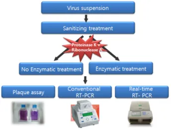
Fig. 1과 같은 과정으로 FCV에 물리, 화학적 위생처리 후 복합효소 처리구와 무처리구를 Real-time RT-PCR법으 로 3반복하여 실험한 결과의 평균을 아래식에 대입하여
물리, 화학적 위생처리의 살균효능(%)을 비교분석하였다.
% inactivation efficacy =
Vsample treatment : Ct value of inactivated virus Vcontrol : Ct value of intact virus
결과 및 고찰
Plaque assay
Plaque assay는 1952년 Renato Dubecco에 의해 개발된 바이러스 정량법으로, plaque란 바이러스 감염에 의해 세 포변성을 일으킨 단층세포부위로 plaque 주위의 감염되지 않은 정상세포는 vital dye로 염색되어 구별할 수 있다25). Plaque assay를 통하여 실험에 사용한 FCV의 농도를 정 량한 결과 9.53 × 10⁵PFU/mL로 나타났으며(Fig. 2), RT- PCR법에 의해 재현성있는 양성결과를 얻을 수 있도록 물 리, 화학적 위생처리에 사용된 바이러스의 초기농도는 104PFU/mL로 조정하였다18,29).
Standard curve
Standard curve는 FCV stock을 9.53 × 100 부터 9.53 × 10⁵ 까지 십진희석하여 CFX 96 Real-time System software (Bio-Rad, CA)로 분석하여 작성하였다. FCV에 대한 검출 한계는 9.53 × 10²PFU/mL로 확인되었고, 100 부터 10−5까 지 십진희석한 FCV의 선형분석 결과 상관계수 0.993, 기 울기는 −4.663으로 나타났다(Fig. 3). Standard curve에 물 리, 화학적 위생처리를 3반복 실험한 Ct값의 평균을 대입 하여 바이러스의 농도를 산출하였다.
물리적 위생처리
UV는 물뿐만 아니라 식품표면 및 식품접촉표면을 살균 하는데 많이 사용되는 방법으로서 UV에너지는 바이러스
의 외피단백질보다 RNA에 많이 흡수되어 photodimer와 photohydrate와 같은 photoproduct를 만들며 바이러스를 불 활성화시키는 작용기전을 가지고 있다18,28).
UV양과 FCV의 감염능의 상관관계를 연구한 Nuanualsuwan S. 등11)의 연구에서는 실험에 사용된 바이러스 FCV, HAV, PV1, MS2, φX174를 불활성화 시키기 위해 필요한 UV양 은 각각 47.85, 36.50, 24.10, 23.04, 15.48 mW·s/cm2 으 로 FCV가 다른 바이러스들에 비해 UV에 대한 저항성이 큰 것으로 나타났다고 보고하고 있다.
본 실험에서는 254 nm 파장의 UV를 FCV 현탁액에 각 각 5, 15, 30분 조사 후 RT-PCR을 실시한 정성분석결과, UV 조사 후 복합효소 무처리구는 Fig. 4 의 lane 2, 4, 6 과 같이 양성결과가 나온 반면, UV 조사 후 복합효소 처 리구는 lane 3, 5, 7과 같이 음성결과가 도출되었다(Fig. 4).
Real-time RT-PCR법을 통하여 살균 효능을 계산한 정량 분석결과, 5, 15, 30분 조사 후 복합효소 무처리구의 불활 성화 효율은 각각 23.29, 24.48, 40.51%, 복합효소 처리구 의 불활성화 효율은 각각 39.95, 47.44, 55.74%로 복합효소 처리구가 무처리구에 비해 높은 살균효능을 보였다(Table 1).
100 Vsample treatment–--- 100Vcontrol ×
Fig. 1. Outline of experimental design. Fig. 2. Images of FCV plaques on CRFK cells. CRFK cell line propagated in 25 cm2-Tissue flasks. Flask containing a full and adherent cell monolayer exhibiting visible plaques.
Fig. 3. Standard curve by serial dilutions of FCV. 1: 9.53 × 10⁵, 2:
9.53× 10⁴, 3: 9.53× 10³, 4: 9.53× 10²PFU/mL.
이는 복합효소 처리가 불활성화 된 바이러스만을 분해 하여 불활성화 되지 않은 바이러스만을 증폭시킨 선행연
구 결과11,18)와 일치하며, UV조사 시간이 증가하고 복합효
소를 처리하였을 경우 불활성화 효율이 증가한다는 것을
확인할 수 있었다.
열처리는 식품조리 및 가공에 흔히 사용되는 방법으로 서 바이러스의 coat protein에 작용하여 불활성화 시키는 작용기전을 가지며, 열처리 온도가 높아질수록 그 효과도 상승한다고 보고되고 있다11,12,18).
Fig. 4. RT-PCR results of UV-inactivated FCV. M: 100 b.p. size marker, lane 1: control (0 min), lane 2: 5 min, lane 3: 5 min + enzymatic treatment, lane 4: 15 min, lane 5: 15 min + enzymatic treatment, lane 6: 30 min, lane 7: 30 min + enzymatic treatment, lane 8: negative control (RNA extraction), lane 9: negative control (conventional RT-PCR).
Table 1. Quantitative analysis of UV-inactivated FCV
Treatment time (min) Concentration of FCV (PFU/suspension) 1)Inactivation efficacy (%)
No enzymatic treatment 2)Enzymatic treatment No enzymatic treatment 2)Enzymatic treatment
Control 2,480.42 ± 2,230.12 -
5 1,997.24 ± 2008.53 1,595.13 ± 1535.45 23.28 ± 13.52b3) 39.95 ± 17.45ab
15 1,963.09 ± 1962.52 1,460.94 ± 1595.86 24.48 ± 12.11b 47.44 ± 12.29ab
30 1,430.83 ± 1322.33 1,382.29 ± 1671.43 40.51 ± 18.85ab 55.74 ± 19.61a
1) Inactivation efficacy(%) = 100− (sanitizing treatment/control) × 100
2) Enzymatic treatment : proteinase K+RNase A treatments for 30 min at 37oC
3) Values are the mean ± standard deviation of triplicate experiments. Means with different letter in the same column are significantly different from the others at p < 0.05.
Table 2. Quantitative analysis of thermally inactivated FCV
Heat (oC) Treatment time (min)
Concentration of FCV (PFU/suspension) 1)Inactivation efficacy (%)
No enzymatic treatment 2)Enzymatic treatment No enzymatic treatment Enzymatic treatment
Control 0 11,813.76 ± 3,582.55 -
63
2 616.97 ± 308.93 352.99 ± 47.90 93.99 ± 4.60b3) 96.27 ± 1.44ab
5 421.54 ± 63.63 286.15 ± 92.03 96.12 ± 1.68ab 97.04 ± 1.48ab
10 416.94 ± 183.08 258.70 ± 23.64 96.53 ± 1.17ab 97.30 ± 1.01ab
72
2 592.82 ± 76.15 255.42 ± 63.09 94.79 ± 1.00ab 97.35 ± 1.19ab
5 421.37 ± 61.67 261.71 ± 45.99 96.12 ± 1.66ab 97.28 ± 1.11ab
10 395.08 ± 2.82 212.33 ± 55.39 96.45 ± 1.05ab 97.80 ± 1.01a
1) Inactivation efficacy(%) = 100− (sanitizing treatment/control) × 100
2) Enzymatic treatment : proteinase K+RNase A treatments for 30 min at 37oC
3) Values are the mean ± standard deviation of triplicate experiments. Means with different letter in the same column are significantly differ- ent from the others at p < 0.05.
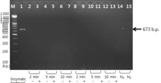
Fig. 5. RT-PCR results of thermally inactivated FCV. M: 100 b.p.
size marker, lane 1: control (0 min), lane 2: 63oC, 2 min, lane 3:
63oC, 2 min + enzymatic treatment, lane 4: 63oC, 5 min, lane 5: 63oC, 5 min + enzymatic treatment, lane 6: 63oC, 10 min, lane 7: 63oC, 10 min + enzymatic treatment, lane 8: 72oC, 2 min, lane 9: 72oC, 2 min + enzymatic treatment, lane 10: 72oC, 5 min, lane 11: 72oC, 5 min + enzymatic treatment, lane 12: 72oC, 10 min, lane 13: 72oC, 10 min + enzymatic treatment, lane 14: negative control (RNA extraction), lane 15: negative control (conventional RT-PCR).
열처리 온도에 따른 살균 효능을 조사하기 위하여 63, 72oC에서 각각 2, 5, 10분 처리 후 RT-PCR법에 의해 FCV 를 검출한 정성분석결과, 복합효소 처리구 및 무처리구 모 두 음성결과가 나와 검출되지 않았다(Fig. 5).
Real-time RT-PCR법을 통하여 살균 효능을 계산한 정량 분석결과, 63oC에서 2, 5, 10분 처리 후 복합효소 처리구 는 각각 96.27, 97.04, 97.30%, 복합효소 무처리구는 93.99, 96.12, 96.53%로 나타났으며, 63oC에서 10분 열처리 후 복 합효소를 처리하였을 때 가장 높은 살균효능을 보였다.
72oC에서 2, 5, 10분 처리 후 복합효소 처리구는 97.35, 97.28, 97.80%, 복합효소 무처리구는 94.79, 96.12, 96.45%
로 나타났으며, 72oC에서 10분 열처리 후 복합효소를 처 리하였을 때 가장 높은 살균효능을 보였다(Table 2). 이러 한 결과를 통하여 동일한 온도에서 처리시간이 길어지고 복합효소를 처리하였을 때 불활성화효율이 증가하는 것을 확인 할 수 있었다.
화학적 위생처리
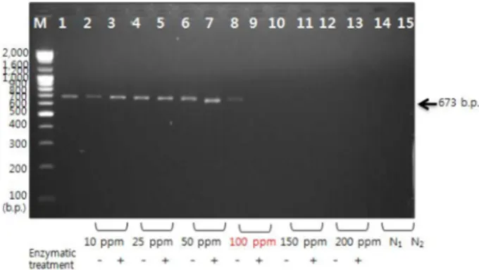
FCV에 염소수를 5분 동안 각각 10, 25, 50 ppm 처리 후 RT-PCR을 실시한 정성분석결과, 복합효소 처리구 및 무처리구 모두 양성결과가 나왔다. 100 ppm을 처리하였 을 때 복합효소 무처리구는 Fig. 6의 lane 8에서 음성, 복 합효소 처리구는 lane 9에서 양성결과가 나왔다. 150 ppm 처리부터는 복합효소 처리구 및 무처리구는 모두 음성결 과가 나온 것을 통해 FCV를 불활성화 시킬 수 있는 염소 수의 최소농도는 150 ppm인 것을 확인하였다.
Real-time RT-PCR법을 통하여 살균 효능을 계산한 정량 분석결과, 10, 25, 50, 100, 150, 200 ppm 처리 후 복합효 소 무처리구의 경우 각각 30.02, 38.55, 45.36, 85.27, 97.61, 98.15%, 복합효소 처리구의 경우 55.98, 59.31, 77.63, 92.91, 98.52, 99.47%로 나타나 복합효소 처리 시 불활성화 효율 이 증가하였으며, 염소수의 농도가 높아질수록 살균효능 도 증가되는 양상을 보였다(Table 3).
Kim 등33)의 연구에서는 병원성 세균에 대하여 100 ppm 의 염소계 용액을 5분 동안 처리한 결과 4 log 이상 감소 하였으며, Nuanualsuwan S 등18)의 연구에서는 염소계 용 액은 바이러스의 coat protein과 RNA에 모두 작용하여 불 활성화 시켰다고 보고한 것을 통해 염소계 용액은 세균뿐 만 아니라 바이러스를 불활성화 시키는데 효과적으로 작 용한다는 것을 확인할 수 있었다.
에탄올을 50, 70, 90% 처리 후 RT-PCR을 실시한 정성 분석결과, 50, 70% 에탄올 처리 후 복합효소 처리구 및 무처리구는 모두 양성, 90% 에탄올 처리 후 복합효소 처 리구는 음성, 복합효소 무처리구는 양성의 결과가 도출된 것을 통해 FCV를 불활성화 시킬 수 있는 에탄올의 최소 농도는 90%인 것을 확인하였다(Fig. 7).
Real-time RT-PCR법을 통하여 살균 효능을 계산한 정량 분석결과, 50, 70, 90% 에탄올 처리 후 복합효소 무처리
Table 3. Quantitative analysis of sodium hypochlorite-inactivated FCV Concentration of
chlorine (ppm)
Treatment time (min)
Concentration of FCV (PFU/suspension) 1)Inactivation efficacy (%)
No enzymatic treatment 2)Enzymatic treatment No enzymatic treatment Enzymatic treatment 0 (control)
5
1,070.83 ± 976.31 -
10 819.42 ± 855.12 602.87 ± 693.63 30.02 ± 10.81e3) 53.03 ± 17.32cd
25 618.78 ± 506.27 362.72 ± 239.64 38.55 ± 6.45de 60.05 ± 11.89c
50 504.74 ± 333.50 262.78 ± 267.32 45.36 ± 12.40cde 77.63 ± 7.48b
100 457.44 ± 465.51 231.13 ± 301.40 85.27 ± 11.94ab 92.91 ± 7.41ab
150 77.36 ± 117.03 41.89 ± 55.63 97.61 ± 2.82a 98.52 ± 1.38a
200 44.03 ± 56.51 8.79 ± 8.11 98.15 ± 1.60a 99.47 ± 0.60a
1) Inactivation efficacy(%) = 100− (sanitizing treatment/control) × 100
2) Enzymatic treatment : proteinase K+RNase A treatments for 30 min at 37oC
3) Values are the mean ± standard deviation of triplicate experiments. Means with different letter in the same column are significantly different from the others at p < 0.05.
Fig. 6. RT-PCR results of sodium hypochlorite-inactivated FCV.
M: 100 b.p. size marker, lane 1: control (0 ppm), lane 2: 10 ppm, lane 3: 10 ppm + enzymatic treatment, lane 4: 25 ppm, lane 5: 25 ppm + enzymatic treatment, lane 6: 50 ppm, lane 7: 50 ppm + enzymatic treatment, lane 8: 100 ppm, lane 9: 100 ppm + enzymatic treatment, lane 10: 150 ppm, lane 11: 150 ppm + enzymatic treat- ment, lane 12: 200 ppm, lane 13: 200 ppm + enzymatic treatment, lane 14: negative control (RNA extraction), lane 15: negative control (conventional RT-PCR).
구는 각각 43.48, 44.11, 48.31%, 복합효소 처리구는 각각 70.73, 79.07, 86.25%로 나타나 복합효소 처리 시 불활성 화 효율이 증가하였다(Table 4).
이는 Malik 등14)의 연구에서 FCV에 에탄올을 처리하였 을 때, 낮은 농도의 에탄올에 비해 높은 농도의 에탄올이 바이러스를 불활성화 시키는데 더 효과적이었다는 결과와 일치한다.
과초산계열 제품 bactzero를 FCV에 100, 500, 1,000 ppm
처리 후 RT-PCR을 실시한 정성분석결과, 100 ppm 처리 시 복합효소 처리구 및 무처리구 모두 양성, 500 ppm 처리 시 복합효소 처리구는 음성, 무처리구는 양성, 1,000 ppm 처 리 시 복합효소 처리구 및 무처리구 모두 음성결과가 도출 되었으며(Fig. 8), 이로써 FCV를 불활성화 시킬 수 있는 과 초산의 최소농도는 500 ppm임을 확인하였다.
Real-time RT-PCR법을 통하여 살균 효능을 계산한 정량
Table 4. Quantitative analysis of ethanol-inactivated FCV Concentration of
EtOH (%)
Treatment time (min)
Concentration of FCV (PFU/suspension) 1)Inactivation efficacy (%)
No enzymatic treatment Enzymatic treatment No enzymatic treatment 2)Enzymatic treatment
0 (control)
5
3,145.66 ± 2301.06 -
50 1,427.51 ± 925.73 1,213.30 ± 102.68 43.48 ± 9.67c 70.73 ± 6.31b3)
70 1,608.20 ± 1096.55 876.03 ± 250.19 44.11 ± 11.08c 79.07 ± 6.94ab
90 1,469.36 ± 1036.98 560.78 ± 310.84 48.31 ± 3.63c 86.25 ± 8.77a
1) Inactivation efficacy(%) = 100− (sanitizing treatment/control) × 100
2) Enzymatic treatment : proteinase K+RNase A treatments for 30 min at 37oC
3) Values are the mean ± standard deviation of triplicate experiments. Means with different letter in the same column are significantly different from the others at p < 0.05.
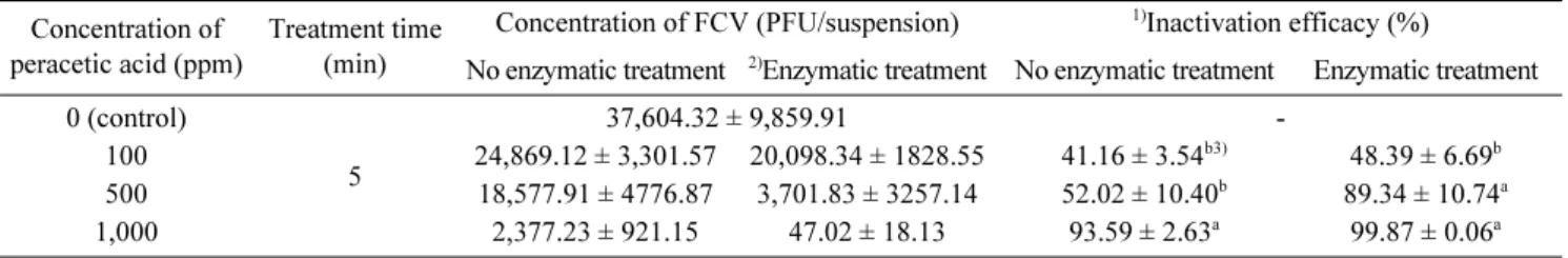
Table 5. Quantitative analysis of peracetic acid-inactivated FCV Concentration of
peracetic acid (ppm)
Treatment time (min)
Concentration of FCV (PFU/suspension) 1)Inactivation efficacy (%)
No enzymatic treatment 2)Enzymatic treatment No enzymatic treatment Enzymatic treatment 0 (control)
5
37,604.32 ± 9,859.91 -
100 24,869.12 ± 3,301.57 20,098.34 ± 1828.55 41.16 ± 3.54b3) 48.39 ± 6.69b
500 18,577.91 ± 4776.87 3,701.83 ± 3257.14 52.02 ± 10.40b 89.34 ± 10.74a
1,000 2,377.23 ± 921.15 47.02 ± 18.13 93.59 ± 2.63a 99.87 ± 0.06a
1) Inactivation efficacy(%) = 100− (sanitizing treatment/control) × 100
2) Enzymatic treatment : proteinase K+RNase A treatments for 30 min at 37oC
3) Values are the mean ± standard deviation of triplicate experiments. Means with different letter in the same column are significantly different from the others at p < 0.05.
Fig. 7. RT-PCR results of ethanol-inactivated FCV. M: 100 b.p.
size marker, lane 1: control (0%), lane 2: 50%, lane 3: 50% + enzymatic treatment, lane 4: 70%, lane 5: 70% + enzymatic treat- ment, lane 6: 90%, lane 7: 90% + enzymatic treatment, lane 8:
negative control (RNA extraction), lane 9: negative control (con- ventional RT-PCR).
Fig. 8. RT-PCR results of peracetic acid-inactivated FCV. M: 100 b.p. size marker, lane 1: control (0 ppm), lane 2: 100 ppm, lane 3:
100 ppm + enzymatic treatment, lane 4: 500 ppm, lane 5: 500 ppm + enzymatic treatment, lane 6: 1,000 ppm, lane 7: 1,000 ppm + enzymatic treatment, lane 8: negative control (RNA extraction), lane 9: negative control (conventional RT-PCR).
분석결과, 100, 500, 1,000 ppm 처리 시 복합효소 무처리 구는 41.16, 58.01, 93.59%, 복합효소 처리구는 48.39, 95.53, 99.87%로 나타났으며, 과초산의 농도가 높아질수록 살균효능이 증가함을 알 수 있었다(Table 5).
Wutzler P 등27)의 연구에서는 0.2% 과초산이 첨가된 80%
에탄올을 아데노바이러스 등에 처리하였을 때 바이러스를 100% 불활성화 시킬 수 있었으며, 과초산의 바이러스에 대한 불활성화 기작은 아직 확실히 밝혀진 바가 없으나, 과초산이 바이러스의 외벽과 capsid 단백질의 S-H그룹과 N-H그룹을 산화시켜 기능을 제대로 하지 못하도록 불활 성화시킴으로써 에탄올에 의한 불활성화 효율에 상승효과 를 더한 것으로 보고하였다. 따라서 물리, 화학적 위생처 리 시 효과와 이러한 효과를 증진시킬 수 있는 방법에 관 한 연구도 지속적으로 이루어져야 할 것으로 사료된다.
요 약
본 연구에서는 FCV 현탁액에 물리, 화학적 위생처리 후 복합효소처리라는 전처리과정을 적용한 뒤 real-time RT- PCR법을 이용하여 살균효능을 분석하였다. RT-PCR 이전 에 37oC에서 30분 동안 PK와 RNase A를 처리함으로써 UV, 열, 염소, 에탄올, 과초산계열 제품에 의해 불활성화 된 바이러스들은 음성 결과를 나타내었고, real-time RT- PCR법을 통해 살균 효능을 정량분석한 결과, 복합효소처 리를 했을 경우 무처리구보다 더 높은 살균 효능을 보이 는 것을 확인할 수 있었다.
이로써 Nuanualsuwan S. 등11,18,29)의 선행연구에서와 같 이 PK와 RNase A로 전처리하는 단계를 통하여 물리, 화 학적 위생처리에 의해 손상되지 않은 바이러스가 RT-PCR 법에 의해 증폭되는 것을 방지함으로써 Real-time PCR법 에 대한 검출 감도를 높일 수 있음을 확인하였다. 또한, FCV를 검출하기 위해 사용된 RT-PCR과 real-time RT-PCR 두 방법 중에서도 real-time RT-PCR법이 가장 신속하면서 도 민감도 높은 결과로 도출되었다. 따라서, 유전자 분석 이전에 복합효소처리는 물리, 화학적 위생처리에 의해 불 활성화 된 바이러스의 RNA가 transcription 또는 증폭되는 것을 방지하기 위한 수단으로 real-time RT-PCR법과 결합 됨으로써 노로바이러스를 비롯한 식중독 바이러스를 검출 하는데 효과적으로 적용될 것으로 판단된다. 또한 식품현 장에서 전기영동 과정없이 신속하게 살아있는 바이러스만 을 수치적으로 정량화함으로써 식품안전에도 기여할 것으 로 사료된다.
감사의 글
본 연구는 2012년도 충북대학교 학술연구지원사업의 연 구비 지원에 의하여 수행되었습니다.
참고문헌
1. Dean O Cliver and Hans P Riemann: Foodborne Deseases.
2nd Ed. Academin press. San Diego. Califronia. USA. pp.
161-175 (2002).
2. Woo, G.J., Hwang, I.G., Kwak, H.S., Kim, M.G., Park, J.S., Lee, G.Y. and Koh, Y.H.: Apply of detection method and eval- uation for foodborne virus. The Annual Report of KFDA, 8-1, 569-575 (2004).
3. Jung, W.Y., Eom, J.H., Kim, B.J., Yun, M.H., Ju, I.S., Kim, C.S., Kim, M.R., Byun, J.A., Park, Y.G., Son, S.H., Lee, E.M., Jung, R.S., Na, M.A., Yuk, D.Y., Gang, J.Y.
and H대, O.S.: Investigation of norovirus occurrence and influence of environmental factors in food service in- stitiutions of chungcheong area. J. Fd Hyg. Safety, 25, 153-161 (2010).
4. Sarbelio M, Espinosa M, Farkas T, Jiang X.: Human calicivi- ruses and pediatric gastoenteritis. Semin. Pediatr. Infect. Dis, 15, 237-245 (2004).
5. Koopmans M, Durzer E.: Foodborne viruses: An emerging problem. Int. J. Food Microbiol. 90, 23-41 (2004).
6. Blackburn, B. G., G. F. Craun, J. S. Yoder, V. Hill, R. L. Cal- deron, N. Chen, S. H. Lee, D. A. Levy, and M. J. Beach.: Sur- veillance for waterborne-disease outbreaks associated with drinking water-United States. MMWR Surveillance Summa- ries, 53, 23-45 (2004).
7. Centers for Disease Control and Prevention. Available at:
http://www.cdc.gov
8. Ministry of Food and Drug Safety. Present Condition of Food- borne. Available at: http://www.mfds.go.kr
9. Lee, S.Y., Jang, K.I., Woo, G.J., Kwak, H.S. and Kim, K.Y.:
Development of protocol for the effective detection of feline calicivirus as norovirus surrogate in oyster and lettuce.
KOREAN J. FOOD SCI. TECHNOL, 39, 71-76 (2007).
10. Cho, M.G., Jeong, H.M., Ahn, J.B. and Kim, K.Y.: Detection of feline calicivirus as norovirus surrogate in food and water sources using filtration and real-time RT-PCR. Food Sci.
Biotechnol, 20, 1475-1480 (2011).
11. Nuanualsuwan S., Mariam T., Himathongkham S., and Cliver D.
O.:. Ultraviolet inactivation of feline calicivirus, human enteric viruses and coliphages. Photochemistry and phtobiology, 76, 406-410 (2002).
12. Hewitt J., Rivera-Aban M., and Greening G. E.: Evalution of murine norovirus as a surrogate human norovirus and hepati- tis A virus in heat inactivation studies. Journal of Applied microbiology. 107, 65-71 (2009).
13. Shin, G.A. and Mark D. Sobsey.: Inactivation of norovirus by chlorine disinfection of water. water research 42, 4562-4568 (2008).
14. Malik, Maherchandani, and Goyal.: Comparative efficacy of ethanol and isopropanol against feline calicivirus, a norovi- rus surrogate. Association for professionals in infection con- trol and epidemiology. 34, 31-35 (2006).
15. Lee J. H.: Real-time PCR in the viral hepatitis. Korean Soci- ety of Gastroenterolohy. 2, 198-206 (2003).
16. Yang C, Jinag Y, Huang K, Zhu C, Yin Y.: Application of real-time PCR for quantitative detection of Campylobacter jejuni in poultry, milk, and environmental water. FEMS Immunol Med Mic. 38, 265-271(2003).
17. Manish M. Patel, Aron J. Halla, Jan Vinje, and Umesh D.:
Parashara. Noroviruses: A comprehensive review. Journal of Clinical Virology 44, 1-8 (2009).
18. Suphachai Nuanualsuwan and Dean O. Cliver.: Pretreatment to avoid positive RT-PCR result with inactivatied viruses.
Journal of Virological Methods. 104, 217-225 (2002).
19. Kim, K.R., Kim, Y.M., Lee, E.W., Lee, D.S., Lee, M.S.:
Screening of antiviral activity from natural plants against feline calicivirus. Journal of Life Science, 19, 928-933 (2009).
20. Duizer, E., K. J. Schwab, F. H. Neill, R. L. Atmar, M. P. G.
Koopman, and M. K. Estes.: Laboratory efforts to cultivate noroviruses. J. Gen. Virol. 85, 79-87 (2004).
21. Clarke I. N., Lambden P. R., and Caul E. O.: Human enteric RNA viruses: caliciviruses and astroviruses. In: Mahy BWJ, Collier L, eds. Topley and Wilson’s microbiology and micro- bial infections. London: Arnold, 511-535 (1998).
22. Koopmans M. and Durzer E.: Foodborne viruses: an emerg- ing problem. Int. J. Food Microbiol. 90, 23-41 (2004).
23. Park, K.J., Ha, H.C., Kim, H.S., Chiba, K., Yeo, I.H. and Lee, S.Y.: The neuroprotective and neurotrophic effects of korean gardenia(Gardenia jasminoides ellis) in PC12h cells.
Food Sci. Biotechnol. 15, 735-738 (2006).
24. Chi, H.Y., Lee, C.H., Kim, K.H., Kim, S.I. and Chung, I.M.:
Induction of apoptotic cell death by red pericarp rice(Jak- wangchalbyeo) extracts. Food Sci. Biotechnol. 15, 534-542 (2006).
25. Kang, J.O.: Quantitation of virus. Korean J Clin Microbiol, 4, 1-4 (2001).
26. Bidawid, S., Malika, N., Adegbunrinb, O., Sattarb, S.A. and Farber, J.M.: A feline kidney cell line-based plaque assay for feline calicivirus, a surrogate for norwalk virus. J Virol.
Method. 107, 163-167 (2003).
27. Wutzler P. and Sauerbrei A.: Virucidal efficacy of a combi- nation of 0.2% peracetic acid and 80% (v/v) ethanol (PAA- ethanol) as a potential hand disinfectant. Journal of Hospital infection. 46, 304-308 (2000).
28. Guerreiro-Tanomaru J. M., Morqental R. D., Faria-Junior N.
B., Berbert F. L., and Tanomaru-Filho M.: Antibacterial effec- tiveness of peracetic acid and conventional endodontic irrig- ants. Braz Dent J. 22, 285-287 (2011).
29. Suphachai Nuanualsuwan and Dean O. Cliver.: Capsid Func- tions of Inactivated Human Picornaviruses and Feline Cali- civirus. Applied and environmental micobiology. 69, 350-357 (2003).
30. Bidavid S., Farber J.M., Sattar S.A.: Rapid concentration and detection of hepatitis A virus from lettuce and strawberry. J.
Virol. Methods. 88, 175-185 (2000).
31. Sykes J.E., Allen J.L., Studdert P., and Browning G.F.: Detec- tion of feline calicivirus, feline herpesvirus I and Chlamydia psittaci mucosal swabs by multiplex RT-PCR/PCR. Vet.
Microbiol. 81, 95-108 (2001).
32. Abd-Eldaim M. M., Wilkes R. P., Thomas K. V., and Kennedy M. A.: Development and validation of a Taqman real-time reverse transcription-PCR for rapid eetection of feline calicivirus. Arch. Virol. 154, 555-560 (2009).
33. Kim, H.I., Jeon, D.H., Yoon, H.J., Choi, H.C., Eom, M.O., Sung, J.H., Park, N.Y., Won, S.A., Lim, N.Y. and Lee, Y.J.:
Evaluation of the efficacy of sanitizers on food contact surface using a surface test method. J Fd Hyg Safety. 23, 291-296 (2008).