Ⅰ. 서 론
치주질환은 구강 내에 존재하는 치주질환 원인균 및 이 세균 들이 생성하는 독소들에 의해서 치주조직이 파괴되는 염증성 질환이다. 구강 내에는 많은 세균이 존재하고 있으나 이들 모든 세균이 치주질환에 관여하지는 않는다. 국소적 공격형 치주염 과 관련이 있는 세균은Actinobacillus actinomycetemcomi- tans1), 성인의 치주질환에 중요한 역할을 하는 원인균은 red complex로 분 류 되 는 Porphyromonas gingivalis, Tannerella forsythia(Bacteroides forsythus), Treponema denticola와 orange complex의 하나인 Fusobacterium nu- cleatum 등으로 알려져 있다2). 이러한 치주질환 원인균을 발견 하기 위한 전통적인 세균배양방법은 치주질환 여부와 관계없이
발견되지 않는 경우도 있었다3,4). 그러나 1990년대 중반부터는 분자생물학의 발전으로 25-100개의 세균만 있어도 검출이 가 능한 중합효소연쇄반응(Polymerase chain reaction, PCR)이 사용되고 있다. 중합효소연쇄반응은 세균 특이성이 있는 primer를 이용하여 적은 수의 세균이 있을지라도 쉽게 검출할 수 있는 유용한 방법으로 이를 이용하여 구강내 치태나 타액으 로부터 직접 세균을 검출할 수 있게 되었고, 치주질환 원인균에 대한 연구가 많이 이루어지고 있다5-8). 하지만 중합효소연쇄반 응은 중합반응의 최종 단계를 알아내는 것으로 존재여부를 조 사할 수 있지만 정량적 계산은 어렵다. 최근에는 real-time PCR이 이용되고 있는데 이는 지수적 증식 단계를 조사함으로 써 원인균의 존재여부를 알 수 있을 뿐 아니라 정량적 분석이 가능하게 되었다. 이러한 정량적 분석은 치주낭내 세균의 조성
Real-time PCR을 이용한 치주질환 원인균의 정량적 분석
김선미∙양규호∙최남기∙강미선*∙오종석*
전남대학교 치의학전문대학원 소아치과학교실 및 치의학연구소 및 2단계 BK21, *의과대학 미생물학교실
Real-time PCR 기법을 이용하여 구강내 치태에서 특정 세균의 존재여부를 알 수 있을 뿐 아니라 정량적 분석이 가능하게 되었다. 이 연구는 real-time PCR 기법을 이용하여 8-18세의 청소년에서 치주질환 원인균의 조성을 알아보고자 전신질환이 없는 어린이 및 청소년 65명을 대상으로 치은연하의 치태를 채취하여 5종의 치주질환 원인균의 출현율과 그 양을 측정하고, 또한 함께 측정한 치태지수 및 치은지수와의 상관관계를 조사하여 다음과 같은 결론을 얻었다.
1. 치태지수의 평균은 1.33이었으며, 치은지수의 평균은 0.97이었다. 치태지수는 나이가 증가할수록 낮아졌다(p<0.05).
2. 치주질환 원인균의 출현율은P. gingivalis 61.5%, T. forsythia 53.8%, T. denticola 29.2%, A. actinomycetem- comitans 15.4%, F. nucleatum은 100%였다. P. gingivalis, T. denticola는 10세 이후 출현율이 증가하였고 A.
actinomycetemcomitans는 8-10세 군에서 출현율이 높았다(p<0.05).
3. 정량적 분석에서 치태지수와 치은지수는F. nucleatum의 양과 유의한 상관성이 있었으며, 치태지수는 치은지수와 유 의한 상관성이 있었다. T. forsythia는 A. actinomycetemcomitans, T. denticola양과 강한 상관성을 보였으며, T.
denticola양과A. actinomycetemcomitans도 강한 상관관계를 가지고 있었다.
주요어 : 치주질환 원인균, Real-time PCR, 치태지수, 치은지수 국문초록
교신저자 : 김 선 미
광주광역시 동구 학동 8번지 / 전남대학교병원 소아치과학교실 / Tel: 062-220-5476 / E-mail: [email protected]
원고접수일: 2008년 3월 11일 / 원고최종수정일: 2008년 4월 21일 / 원고채택일: 2008년 4월 28일
을 알아내는데 중요한 역할을 할 수 있다. 또한 치주질환을 진 단하고 적절히 치료하는데 치주질환 원인균의 양을 비교 조사 하는 것은 치주질환에 있어서 세균의 역할을 이해하는데 중요 하다고 할 수 있다. 전통적 중합효소연쇄반응과 real-time PCR을 이용한 연구에서 타액과 치은연하 치태에서 치주세균의 출현율은 높은 일치성을 보였고, real-time PCR은 40분 정도 시간이 단축되었다고 보고하고 있다9). 치주상태와 P. gingi- valis의 관계에 대해 real-time PCR을 이용한 연구에서 치주 낭의 깊이와 세균의 양은 상관성이 있었으며, 치주치료 후 이 세균의 양이 유의하게 감소함이 보고되었다10). 이들 보고들을 통해 치주상태의 평가에 real-time PCR을 이용한 세균의 절대 적 양 및 상대적 양을 측정하는 것이 필요하다고 할 수 있다.
건강한 정상의 어린이에서 치주질환으로 치주조직이 파괴되 는 경우는 드물다11). 하지만 치주질환의 병력이 있던 어린이는 결국 청소년기를 지나 성인이 되어서 다시 치주질환을 가지게 될 가능성이 높다12,13). 이는 유치열에 존재했던 치주질환 원인 균들이 치주조직에 잔존하여 새롭게 영구치열이 형성될 때 재 집락을 이루기 때문이라고 추정되고 있다14). 어린이 및 청소년 의 치은연하 치태에서 치주질환 원인균의 양을 측정하는 것은 추후 치주질환의 예측 및 진단, 그리고 치료효과의 평가에 기여 할 수 있다.
따라서 이 연구는 어린이 및 청소년의 구강내 치태에서 P.
gingivalis, T. forsythia, T. denticola, A. actinomycetem- comitans, F. nucleatum 등 5종의 치주질환 원인균을 real- time PCR을 이용하여 출현율과 양을 측정하고, 측정한 치태지 수 및 치은염과의 상관관계를 조사하여 상관성을 평가해보고자 하였다.
Ⅱ. 연구 대상 및 방법 1. 연구 대상
본 연구대상으로는 초,중,고교에 재학 중이며 전신질환이 없 는 학생 65(남자 32명, 여자 33명)명을 조사대상으로 하였다.
연구대상의 평균 연령은 12.6세였으며 연령 범주는 혼합치열 기, 영구치열기 등을 고려하여 4개의 범주로 구분하였다(Table 1).
2. 연구 방법
1) 치태지수와 치은지수의 검사
구강위생상태를 측정하기 위해서 치태지수 (Plaque index of Silness and Loe¨)15)를 사용하였으며 측정부위는 상악 좌우 제1대구치의 협측, 상악 우측 중절치와 하악 좌측 중절치의 순 측, 하악 좌우 제1대구치의 설측이었다. 제 1대구치가 없는 경 우는 인접 제 2유구치나 제 2 소구치를 검사하였다. 치태침착 정도는 0-3점 (0점: 치태가 없음, 1점: 치은변연을 따라 탐침 시 묻어나는 정도, 2점: 눈에 보이는 상태, 3점: 치태가 많은 경우)으로 구분하였다. 치주조직의 염증 상태를 측정하기 위해 치은지수 (Gingival index of Loe¨)16)를 사용하였으며, 조사 치 아는 치태지수와 같다. 탐침부위는 해당면의 근심, 중앙, 원심 세 부위였다. 치은염의 정도는 0-3점 (0점: 염증소견이 없음, 1 점: 약간의 발적과 부종, 2점: 탐침후 출혈을 동반한 발적과 부 종, 3점: 자연출혈을 동반한 염증)으로 구분하였다.
2) 치주질환 원인균의 검사 (1) 치태 채취
치태시료를 얻기 위해 대상자의 하악 우측 제1대구치 또는 제2유구치의 근심협측부위에서 멸균된 이쑤시개를 사용하여 조 심스럽게 치은부위의 치태를 채취하였다. 채취한 치태는 멸균 된 0.2 ml phosphate-buffered saline (PBS)이 들어있는 1.5 ml tube에 수집한 후 DNA 추출을 위해 실험실로 옮겨 4
℃에 보관하였다.
(2) 치태내 치주세균의 DNA 추출
시료를 원심분리 (12,000 rpm, 2 min)한 후 다시 0.1 ml cell lysis buffer (1.0% Triton X-100, 20 mM Tris-HCl, 2 mM ethylenediaminetetraacetic acid, pH 8.0)로 현탁하고 100℃에서 10분간 끓여서 DNA를 추출한다.
(3) real-time PCR
PCR 조건의 표준화를 위해 5종류의 혐기성세균을 각각의 배 지에 혐기적으로 배양한 후 표준균주의 세균수를 측정한 후 시 료와 같은 방법으로 1 ml를 튜브에 취하여 원심한 후 PBS로 2 회 세척하고 PBS 0.1 ml로 suspension하여 10배 농축시킨 후 DNA를 추출한 다음 102~108 까지 10배씩 연속적으로 희 석하여 real-time PCR 정량에 필요한 표준곡선을 작성하였다.
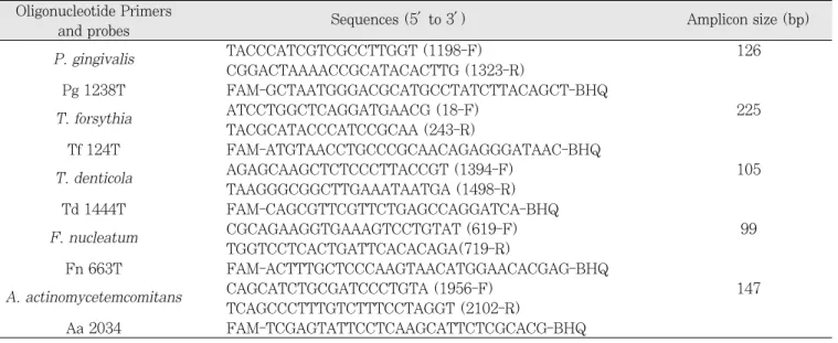
5종 세균의 sense primer와 antisense primer (400 nM) 각 각 1 ㎕, dual labelled probe (5‘-FAM, BHQ-3')가 이용되 었다17)(Table 2). template DNA 3 ㎕에 2배의 QuantiTect Probe(Qiagen GmbH, Hilden, Germany) 12.5 ㎕, PCR Master Mix(HotStarTaq polymerase, QuantiTec Probe PCR buffer, dNTP mix including dUTP, 발색제 ROXTM, 8mM MgCl), 3차 멸균수를 6.5 ㎕ 첨가하여 최종 용량 25 ㎕ Table 1.
The distribution of the study subjects
Age(year) Male Female
8-10 10 7
11-13 8 14
14-16 10 9
17-18 4 3
Total 32 33
3000™ Real-time Thermal Cycler (Corbett research, Sydney, Australia)를 이용하여 중합효소연쇄반응을 시행하였 다. 이 때 사용한 real-time PCR의 조건은 최초 변성을 위해 95℃에서 15분간, 이후 45번의 중합효소연쇄반응 cycle은 95
℃에서 15초, 58℃에서 1분간 시행하였다. 데이터 분석은 Rotor-Gene analysis software version 6.0을 이용하였다.
Cycle threshold (CT)는 알려진 숫자의 DNA농도의 대수기에 서 시작하며, 세균수에 대한 표준곡선에 의해서 시료당 세균수 가 측정이 되었다(Fig. 1, 2).
3) 통계 분석
위 데이터는 SPSS 12.0을 이용하여 통계학적 평가를 시행하 였다. 치태지수와 치은지수를 나이군에 대해서 비교하기 위해 ANOVA를 시행하였고, 사후검정으로 Tukey’s studentized range test를 시행하였다. 치주질환 원인균의 연령에 따른 출 현율을 비교하기 위해 카이제곱 검정을 시행하였다. 그리고 치 태지수, 치은지수와 치주질환 원인균의 상관성을 조사하기위해 Pearson 상관계수를 계산하였다.
Table 2.
Oligonucleotide primers and probes used for real-time PCR Oligonucleotide Primers
Sequences (5′to 3′ ) Amplicon size (bp)
and probes
P. gingivalis TACCCATCGTCGCCTTGGT (1198-F) 126
CGGACTAAAACCGCATACACTTG (1323-R)
Pg 1238T FAM-GCTAATGGGACGCATGCCTATCTTACAGCT-BHQ
T. forsythia ATCCTGGCTCAGGATGAACG (18-F) 225
TACGCATACCCATCCGCAA (243-R)
Tf 124T FAM-ATGTAACCTGCCCGCAACAGAGGGATAAC-BHQ
T. denticola AGAGCAAGCTCTCCCTTACCGT (1394-F) 105
TAAGGGCGGCTTGAAATAATGA (1498-R) Td 1444T FAM-CAGCGTTCGTTCTGAGCCAGGATCA-BHQ
F. nucleatum CGCAGAAGGTGAAAGTCCTGTAT (619-F) 99
TGGTCCTCACTGATTCACACAGA(719-R)
Fn 663T FAM-ACTTTGCTCCCAAGTAACATGGAACACGAG-BHQ
A. actinomycetemcomitans CAGCATCTGCGATCCCTGTA (1956-F) 147
TCAGCCCTTTGTCTTTCCTAGGT (2102-R)
Aa 2034 FAM-TCGAGTATTCCTCAAGCATTCTCGCACG-BHQ
P. gingivalis: Porphyromonas gingivalis, T. forsythia: Tannerella forsythia, T. denticola: Treponema denticola, F. nucleatum:
Fusobacterium nucleatum, A. actinomycetemcomitans: Actinobacillus actinomycetemcomitans
Fig. 1. Amplification of profiles for
P. gingivalis-specific PCR products.
Fig. 2. Standard curve was generated from the amplication
plot of P. gingivalis. A suspension of P. gingivalis cells was
serially diluted and subjected to real-time PCR.
Ⅲ. 연구 성적 1. 치태지수와 치은지수(Table 3)
치태지수의 전체 평균은 1.33이었다. 나이가 증가할수록 치 태지수가 낮아지는 양상이었다(p<0.05). 치은지수의 전체 평균 은 0.97이었다.
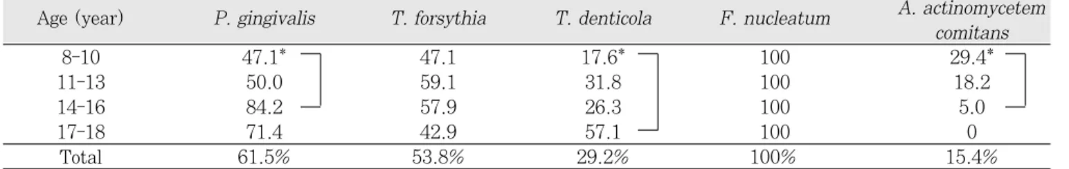
2. 치주질환 원인균의 출현율
real-time PCR을 이용한 치주질환 원인균의 출현율은 P.
gingivalis 61.5%, T. forsythia 53.8%, T. denticola 29.2%, A. actinomycetemcomitans 15.4%를 나타내었다.
각 균의 연령별 비교를 보면 F. nucleatum은 모든 연령에서 100%의 출현율을 보였다. P. gingivalis, T. denticola는 10 세 이후 출현율이 증가하는 양상을 보였고 A. actino- mycetemcomitans는 어린 연령군에서 더 많이 출현하였다 (p<0.05) (Table 4).
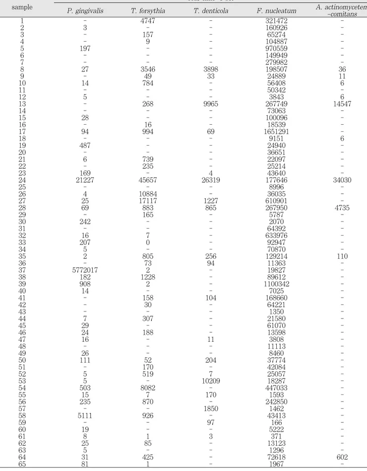
3. 치주질환 원인균의 정량적 조사
real-time PCR을 이용한 치주질환 원인균의 양적 검사에서 F. nucleatum은 모든 대상자에서 많은 수의 세균이 분포하였 으며 다른 세균보다 더 많은 양이 관찰되었다. A. actino-
mycetemcomitans는 출현율도 적을 뿐 아니라 양도 적게 분포 하였다. 어린 나이군 부터 치주질환 원인균들이 다양하게 분포 하고 있었으나, 나이가 증가함에 따라 조사한 5종의 치주질환 원인균의 양은 유의한 차이를 보이지 않았다(p>0.05). 한편 P. gingivalis와 T. forsythia가 함께 출현하는 비율은 33%였 으며, T. forsythia와 T. denticola가 함께 출현하는 경우는 20%, P. gingivalis와 T. denticola가 함께 출현하는 경우는 18%, red complex로 분류되는 세 종류의 세균이 모두 출현한 경우가 12%였다(Table 5).
4. 치주질환 세균의 양과 치태지수, 치은 지수와의 상관성
치태의 침착정도를 나타내는 치태지수는 F. nucleatum의 양과 유의한 상관성이 있었으며(상관계수 0.46), 치은염을 나 타내는 치은지수 또한F. nucleatum의 양과 유의한 상관성이 있었다(상관계수 0.3). 치태지수는 치은지수와 유의한 상관성 이 있었다(상관계수 0.7). T. forsythia는 A. actinomycetem- comitans, T. denticola양과 강한 상관성을 보였으며(상관계 수 0.81, 0.79), T. denticola양과 A. actinomectemcomi- tans도 강한 상관관계를 가지고 있었다(상관계수 0.92). 연령 별로는 11-13세군에서P. gingivalis가 T. forsythia, T. den- ticola A. actinomycetemcomitans의 양과 강한 상관관계를 보이고 있었다(상관계수 0.94, 0.99, 0.99)(p<0.05).
Table 3.
Plaque index and gingival index of study population
Age (year) Plaque index Gingival index
8-10 1.62 0.98
11-13 1.45 1.01
14-16 1.11 1.00
17-18 0.88 0.74
Total 1.33 0.97
Table 4.
Prevalence of periodontopathic bacteria found in subjects(%)
Age (year) P. gingivalis T. forsythia T. denticola F. nucleatum A. actinomycetem comitans
8-10 47.1* 47.1 17.6* 100 29.4*
11-13 50.0 59.1 31.8 100 18.2
14-16 84.2 57.9 26.3 100 5.0
17-18 71.4 42.9 57.1 100 0
Total 61.5% 53.8% 29.2% 100%` 15.4%
* p<0.05
P. gingivalis: Porphyromonas gingivalis, T. forsythia: Tannerella forsythia, T. denticola: Treponema denticola, F. nucleatum:
Fusobacterium nucleatum, A. actinomycetemcomitans: Actinobacillus actinomycetemcomitans
Table 5.
Amount of periodontopathic bacteria in the subgingival plaque real-time- PCR sample
P. gingivalis T. forsythia T. denticola F. nucleatum A. actinomycetem -comitans
1 - 4747 - 321472 -
2 3 - - 160926 -
3 - 157 - 65274 -
4 - 9 - 104887 -
5 197 - - 970559 -
6 - - - 149949 -
7 - - - 279982 -
8 27 3546 3898 198507 36
9 - 49 33 24889 11
10 14 784 - 56408 6
11 - - - 50342 -
12 5 - - 3843 6
13 - 268 9965 267749 14547
14 - - - 73063 -
15 28 - - 100096 -
16 - 16 - 18539 -
17 94 994 69 1651291 -
18 - - - 9151 6
19 487 - - 24940 -
20 - - - 36651 -
21 6 739 - 22097 -
22 - 235 - 25214 -
23 169 - 4 43640 -
24 21227 45657 26319 177646 34030
25 - - - 8996 -
26 4 10884 - 36035 -
27 25 17117 1227 610901 -
28 69 883 865 267950 4735
29 - 165 - 5787 -
30 242 - - 2070 -
31 - - - 64392 -
32 16 7 - 633976 -
33 207 0 - 92947 -
34 5 - - 70870 -
35 2 805 256 129214 110
36 - 73 94 11363 -
37 5772017 2 - 19827 -
38 182 1228 - 89612 -
39 908 2 - 1100342 -
40 14 - - 7025 -
41 - 158 104 168660 -
42 - 30 - 64221 -
43 - - - 1350 -
44 7 307 - 21580 -
45 29 - - 61070 -
46 24 188 - 13598 -
47 16 - 11 3808 -
48 - - - 11113 -
49 26 - - 8460 -
50 111 52 204 37774 -
51 - 170 - 42084 -
52 5 519 7 25057 -
53 5 - 10209 18287 -
54 503 8082 - 447033 -
55 15 7 170 1593 -
56 235 870 - 242850 -
57 - - 1850 1462 -
58 5111 926 - 43413 -
59 - - 97 166 -
60 19 - - 5222 -
61 8 1 3 371 -
62 25 85 - 13123 -
63 5 - - 1296 -
64 31 425 - 72618 602
65 81 1 - 1967 -
P. gingivalis: Porphyromonas gingivalis, T. forsythia: Tannerella forsythia, T. denticola: Treponema denticola, F. nucleatum:
Fusobacterium nucleatum, A. actinomycetemcomitans: Actinobacillus actinomycetemcomitans
Ⅳ. 총괄 및 고찰
건강한 치주조직에 비하여 치주병소에서 흔히 나타나는 미생 물들이 치주질환 병원균으로 제시되어 왔으며, 이러한 병원균 이 군집한 부위에서는 치주파괴의 위험이 증가한다. 유년형 치 주염 환자나 성인형 치주염 환자의 병소에서 발견되는 세균은 같은 사람의 건강한 부위에서나 건강한 사람에서는 낮은 비율 로 나타났다18,19). 따라서 치주질환의 적절한 치료를 위해서는 치태내 특정 병원균을 알고 이를 제거하거나 감소시키는 것이 필요하다.
치은연하 치태는 그람양성균과 그람음성 구균, 간균, 사상균 으로 구성되어 있으며, 나선균과 다양한 편모를 가진 세균들이 치태의 하방성장을 주도한다20,21). 1996년 세계치주학회에서는 A. actinomycetemcomitans, P. gingivalis, T. forsythia를 파괴적 치주질환을 야기하는 치주병원균으로 규정하였다22).
A. actinomycetemcomitans는 그람음성, 호이산화탄소성, 끝이 둥근 간균으로 건강한 상태의 치주에서보다 국소적 유년 형 치주염, 사춘기전 치주염에서 증가함이 보고되었다1,23,24). P.
gingivalis는 그람음성, 혐기성, 비운동성의 간균으로 건강한 사람이나 치은염 환자에서는 드물고 파괴성 질환에서 더 많이 나타나고 특히 치주질환이 진행된 환자에서 더 많이 발견되었
다2,25). T. forsythia는 그람음성, 혐기성, 방추형의 다형성 간균
으로 건강한 부위나 치은염부위보다 파괴성 치주질환 또는 치 주농양 부위에 많이 보이고 비활동성 병소보다는 활동성 병소 에서 더 많이 발견되었다26,27). T. denticola는 그람 음성, 혐기 성의 활발한 운동성을 가진 나선균으로 건강한 사람이나 치은 염 환자보다는 급성괴사성궤양성치주염에서 그리고 심한 치주 염환자에서 더 많이 나타난다28,29). F. nucleatum은 그람 음성, 혐기성, 방추형의 간균으로 치은연하 치태에서 가장 흔히 발견 되는 세균으로 치주염을 가진 사람에게서 널리 퍼져 있다30).
PCR에 의한 치주질환 원인균의 출현율을 조사한 연구를 살 펴보면 Ashimoto 등은 치은염이 있는 어린이에서 P. gingi- valis, T. forsythia, T. denticola, 그리고 A. actino- mycetemcomitans가 8-14%로 출현한다고 하였다31). 한편 Kimura 등은 2-13세 정상 어린이에서 치주질환의 임상증상 없이A. actinomycetemcomitans 50%, T. forsythia 20%가 발견되었고P. gingivalis와 T. denticola는 발견되지 않았다고 하였다32). Umeda 등33)은 치주세균검사를 시행하여 어린이에서 P. gingivalis 8.9%, T. forsythia 42.9%, T. denticola 48.2%, A. actinomycetemcomitans가 1.8%의 빈도를 보인 다고 보고하였다. 국내의 정34)은 2-12세의 어린이 치주질환 원 인균을 조사하여P. gingivalis 95.7%, T. forsythia 44.3%, T. denticola 65.7%, A. actinomycetemcomitans가 20%의 빈도를 보인다고 보고하였다. 김 등35)은 다운증후군 대상자에서 P. gingivalis 63%, T. forsythia 74.1%, T. denticola
치주질환의 진단과 추적조사에 기여하는 치주질환 원인균의 정성 및 정량적 분석이 최근에 real-time PCR을 사용하여 이 루어지고 있다36-38). 전통적 중합효소연쇄반응과 real-time PCR 두 방법을 비교한 연구에서 Sakamoto 등은 타액과 치은 연하 치태에서 P. gingivalis, T. forsythia, T. denticola, A.
actinomycetemcomitans등의 치주세균의 출현율에 있어서 높 은 일치성을 보였다고 하였다9). 한편 Yoshida 등은 치주질환이 있는 환자의 타액과 치은연하 치태에서 A. actinomycetem- comitans와 P. gingivalis의 수를 검사함으로써 전통적 중합효 소연쇄반응과 real-time PCR sensitivity를 비교하였는데, real-time PCR이 더 민감했다고 하였다37). 본 연구의 8-18세 대상자의 real-timePCR을 이용한 연구에서는 P. gingivalis 61.5%, T. forsythia 53.8%, T. denticola 29.2%, A.
actinomycetemcomitans 15.4%를 나타내어 정등의 보고보다 낮았으나 다른 연구자들보다는 높은 출현율을 보였는데 대상자 들의 연령이 증가된 것과 real-time PCR이 더 민감한 방법이 라는 주장도 그 이유의 하나로 설명할 수 있다. Suzuki 등36)은 T. forsythia와 F. nucleatum의 치주낭내 출현율과 조성을 평 가했는데, 두 균 모두 깊은 치주낭에서 잘 자라며, 두 균이 서로 공존관계를 가진다고 보고하였다. 본 연구에서도P. gingivalis 와T. forsythia가 함께 출현하는 비율은 33%였으며, T. for- sythia와 T. denticola가 함께 출현하는 경우는 20%, P. gin- givalis와 T. denticola가 함께 출현하는 경우는 18%, red complex로 분류되는 세 종류의 세균이 모두 출현한 경우가 12%로 나타나 치주질환 원인균들이 서로 공존하는 양상을 보 였다. Kawada 등10)은 치주상태와 P. gingivalis의 관계에 대 해 평가하였는데, 치주낭의 깊이가 1mm 깊어질수록 세균의 수 는 10배씩 증가하였다고 하였으며, 또한 치주낭의 깊이가 증가 할수록P. gingivalis균의 상대적 양이 4.69% 증가하며, 치주 치료 후 이 세균의 양이 유의하게 감소하였다고 보고하였다. 이 를 통해 치주치료를 평가하는 데 있어 치주상태와 연관하여 세 균의 절대적 양 및 상대적 양을 측정하는 것이 중요하다고 주장 하였다. 본 연구에서는 치은지수와 F. nucleatum간에 유의한 상관성을 보였고 치태지수와 치은지수간에 상관성을 보였는데 이는 F. nucleatum의 양이 많을수록, 그리고 치태가 많이 축 적되어있을 수록 치은염이 증가함을 의미하였다. Asai 등39)은 T. denticola와 치주낭의 깊이가 상관성이 있음을 보고하였고, Sawamoto 등40)도 타액에서 P. gingivalis와 T. forsythia는 건강한 사람보다 치주환자에서 더 많았으며, 초기 치주치료 후 P. gingivalis의 수, 치주낭 깊이, 탐침후 출혈이 감소함을 보고 하였다. T. forsythia는 A. actinomycetemcomitans, T.
denticola양과 강한 상관성을 보였으며, T. denticola양과 A.
actinomectemcomitans도 강한 상관관계를 보이고 있어 이러 한 치주질환 원인균의 공존은 추후 치주질환을 가지게 될 가능 성을 높일 수 있다고 추정할 수 있다.
이상의 연구인 real-time PCR을 이용한 치주질환 원인균의
균들이 다양하게 분포하고 있음을 알 수 있었다. 또한 나이가 증가함에 따라 치주질환 원인균의 출현율은 증가하는 양상이 었으며, 연령에 따른 치주질환 원인균의 양에는 유의한 차이가 없었지만 치주질환 원인균이 서로 상관성를 가지고 공존하고 있음을 발견하였다. 또한F. nucleatum의 양이 많을수록, 그 리고 치태가 많이 축적되어있을 수록 치은염이 증가함을 나타 내고 있어, 추후 청소년에서도 정확한 치주건강 상태를 평가함 에 있어 real-time PCR을 이용한 치주질환 원인균의 주기적인 정성 및 정량적 검사가 도움이 되리라 사료된다.
Ⅴ. 결 론
최근 real-time PCR 기법을 이용하여 구강내 치태에서 특정 세균의 존재여부를 알 수 있을 뿐 아니라 정량적 분석이 가능하 게 되었다. 이 연구는 real-time PCR 기법을 이용하여 8-18세 의 청소년에서 치주질환 원인균의 조성을 알아보고자 전신질환 이 없는 어린이 및 청소년 65(남자 32명, 여자 33명)명을 대상 으로 치은연하의 치태를 채취하여P. gingivalis, T. forsythia, T. denticola, A. actinomycetemcomitans, F. nucleatum 등 5종의 치주질환 원인균의 출현율과 그 양을 측정하고, 또한 함께 측정한 치태지수 및 치은지수와의 상관관계를 조사하여 다음과 같은 결론을 얻었다.
1. 치태지수의 평균은 1.33이었으며, 치은지수의 평균은 0.97이었다. 치태지수는 나이가 증가할수록 낮아졌다 (p<0.05).
2. 치주질환 원인균의 출현율은 P. gingivalis 61.5%, T.
forsythia 53.8%, T. denticola 29.2%, A. actino- mycetemcomitans 15.4%, F. nucleatum은 100%의 출현율을 보였다. P. gingivalis, T. denticola는 10세 이 후 출현율이 증가하는 양상을 보였고 A. actino- mycetemcomitans는 어린 연령군인 8-10세군에서 출현 율이 높았다(p<0.05).
3. 정량적 분석에서F. nucleatum은 모든 대상자에서 다른 세균보다 많은 양이 관찰되었다. 연령에 따른 치주질환 원 인균의 양에는 유의한 차이가 없었지만 치주질환 원인균 이 서로 상관성을 가지고 공존하고 있었다.
4. 치태지수와 치은지수는F. nucleatum의 양과 유의한 상 관성이 있었으며, 치태지수는 치은지수와 유의한 상관성 이 있었다. T. forsythia는 A. actinomycetemcomitans, T. denticola양과 강한 상관성을 보였으며(상관계수 0.81, 0.79), T. denticola양과 A. actinomycetem- comitans도 강한 상관관계를 가지고 있었다(상관계수 0.92).
청소년의 구강 내 치태에는 치주질환을 유발하는 세균들이 다양하게 공존하고 있었으며, 치은염을 나타내는 치은지수는 치태지수와F. nucleatum의 양과 유의한 상관성이 있었다.
참고문헌
1. Mandell RL : A longitudinal microbiological investi- gation of Actinobacillus actinomycetemcomitans and Eikenella corrodens in juvenile periodontitis. Infect Immun, 45:778-780, 1984.
2. Haffajee AD, Socransky SS : Microbial etiological agents of destructive periodontal diseases.
Periodontol, 5:78-111, 1994.
3. Frisken KW, Tagg JR, Laws AJ, et al. : Suspected periodontopathic microorganisms and their oral habitats in young children. Oral Microbiol Immunol, 2:60-64, 1987.
4. Delaney JE, Kornman KS : Microbiology of subgingi- val plaque from children with localized prepubertal periodontitis. Oral Microbiol Immunol, 2:71-76, 1987.
5. Slots J, Ashimoto A, Flynn MJ, et al. : Detection of putative periodontal pathogens in subgingival speci- mens by 16S ribosomal DNA amplification with the polymerase chain reaction. Clin Infect Dis, 20(2):
304-307, 1995.
6. Conrads G, Mutters R, Fischer J, et al. : Reaction and dot-blot hybridization to monitor the distribu- tion of oral pathogens within plaque samples of peri- odontally healthy individuals. J Periodontol, 67:994- 1003, 1996.
7. Watanabe K, Frommel TO : Porphyromonas gingi- valis, Actinobacillus actinomycetemcomitans and Treponema denticola detection in oral plaque sam- ples using the polymerase chain reaction. J Clin Periodontol, 23:212-219, 1996.
8. Okada M, Hayashi F, Nagasaka N. : PCR detection of 5 putative periodontal pathogens in dental plaque samples from children 2 to 12 years of age. J Clin Periodontol, 28:576-582, 2001.
9. Sakamoto M, Takeuchi Y, Umeda M, et al. : Rapid detection and quantification of five periodontopathic bacteria by real-time PCR. Microbiol Immunol, 45:39-44, 2001.
10. Kawada M, Yoshida A, Suzuki N, et al. : Prevalence of Porphyromonas gingivalis in relation to periodon- tal status assessed by real-time PCR. Oral Microbiol Immunol, 19:289-292, 2004.
11. Ko¨no¨nen E : Development of oral bacterial flora in young children. Ann Med, 32:107-112, 2000.
12. Sweeny EA, Alcoforado GAP, Nyman S, et al. : Prevalence and microbiology of localized prepubertal periodontitis. Oral Microbiol Immunol, 2:65-70, 1987.
13. Sjo¨din B, Crossmer CG, Unell L, et al. : A retro- spective radiographic study of alveolar bone loss in the primary dentition in patients with localized juve- nile periodontitis. J Clin Periodontol, 16:125-127, 1989.
14. Watanabe K, Frommel TO : Detection of Porphyromonas gingivalis in oral plaque samples by use of the polymerase chain reaction. J Dent Res, 72:1040-1044, 1993.
15. Silness J, Lo¨e H : Periodontal disease in pregnancy.
Correlation between oral hygiene and periodontal condition. Acta Odontol Scand, 22:112-135, 1964.
16. Lo¨e H : The gingival index, the plaque index and the retention index system. J Dent Res, 38:610- 616, 1927.
17. Kato H, Yoshida A, Awano S, et al. : Quantitative detection of volatile sulfur compound producing mi- croorganism in oral specimens using real-time PCR.
Oral Dis, 11:67-71, 2005.
18. Slots J : The predominant cultivable organisms in juvenile periodontitis. Scand J Dent Res, 84:1-10, 1976.
19. Slots J : The predominant cultivable microflora of advanced periodontitis. Scand J Dent Res, 85:114- 121, 1997.
20. Schroeder HE, De Boever J : The structure of micro- bial dental plaque. In: McHugh, W.D., ed. Dental plaque. Edinburgh: Livingstone, 49-74, 1970.
21. Listgarten MA : Structure of the microbial flora as- sociated with periodontal health and disease in man.
A light and electron microscopic study. J Periodontol, 47:1-18, 1976.
22. Consensus report. Anonymous. Periodontal diseases:
Pathogenesis and microbial factors. Ann Periodontol, 1:926-932, 1996.
23. Slots J, Reynolds HS, Genco RJ : Actinobacillus actinomycetemcomitans in human periodontal dis- ease: a cross-sectional microbiological investigation.
Infect Immun, 29:1013-1020, 1980.
24. Newman MG, Socransky SS, Savitt ED, et al. : Studies of the microbiology of periodontosis. J Periodontol, 47:373-379, 1976.
momycetemcomitans, Porphyromons gingivalis and Prevotella intermedia in progressing adult periodon- titis. J Periodontol, 71:948-954, 2000.
26. Papapanou PN, Neiderud AM, Papadimitriou A, et al. : "Checkerboard" assessments of periodontal mi- crobiota and their antibody responses: a case control study. J Periodontol, 71:885-987, 2000.
27. Lai CH, Listgarten MA, Shirakawa M, et al. : Bacteroides forsythus in adult gingivitis and peri- odontitis. Oral Microbiol and Immunol, 2:152-157, 1987.
28. Listgarten MA : Electron microscopic observations of the bacterial flora of acute necrotizing ulcerative gin- givitis. J Periodontol, 36:328-339, 1965.
29. Simonson LG, Robinson PJ, Pranger RJ, et al. : Quantitative relationship of Treponema denticola to severity of periodontal disease. Infect Immun, 56:726-728, 1988.
30. Dzink JL, Tanner ACR, Haffajee AD, et al. : Gram- negative species associated with active destructive periodontal lesions. J Clin Periodontol, 12:648-659, 1985.
31. Ashimoto A, Chen C, Bakker I, et al. : Polymerase chain reaction detection of 8 putative periodontal pathogens in subgingival plaque of gingivitis and ad- vanced periodontitis lesions. Oral Microbiol Immunol, 11:266-273, 1996.
32. Kimura S, Ooshima T, Takiguchi M, et al. : Periodontopathic bacterial infection in childhood. J Periodontol, 73:20-26, 2002.
33. Umeda M, Miwa Z, Takeuchi Y, et al. : The distrib- ution of periodontopathic bacteria among Japanese children and their parents. J Periodontol Res, 39:398-404, 2004.
34. 정우성 : 어린이의 치은연하 치태내 치주질환 원인균의 출 현율 조사. 경희대학교 대학원, 2004.
35. 김선미, 양규호, 최남기, 등 : 다운증후군 환자의 치주질환 원인균의 출현율. 대한소아치과학회지, 32(4):717-725, 2005.
36. Suzuki N, Yoshida A, Saito T, et al. : Quantitative microbiological study of subgingival plaque by real- time PCR shows correlation between levels of Tannerella forsythensis and Fusobacterium spp. J Clin Microbiol, 42:2255-2257, 2004.
37. Yoshida A, Suzuki N, Nakano Y, et al. : Development of a 5' fluorogenic nuclease-based real-
Actinobacillus actinomycetemcomitans and Porphyromonas gingivalis. J Clin Microbiol, 41:863- 866, 2003.
38. Lyons SR, Griffen AL, Leys EJ : Quantitative real- time PCR for Porphyromonas gingivalis and total bacteria. J Clin Microbiol, 38:2362-2365, 2000.
39. Asai Y, Jinno T, Igarashi H, et al. : Detection and
quantification of oral treponemes in subgingival plaque by real-time PCR. J Clin Microbiol, 40:3334- 3340, 2002.
40. Sawamoto Y. Sugano N, Tanaka H, et al : Detection of periodontopathic bacteria and an oxidative stress marker in saliva from periodontitis patients. Oral Microbiol Immunol, 20:216-220, 2005.
Abstract
QUANTITATIVE DETECTION OF PERIODONTOPATHIC BACTERIA USING REAL-TIME PCR
Seon-Mi Kim, Kyu-Ho Yang, Nam-Ki Choi, Mi-Sun Kang
*, Jong-Suk Oh
*Department of Pediatric Dentistry, School of Dentistry, Chonnam National University and Dental Research Institute and second stage of BK 21,
*
Department of Microbiology, College of Medicine, Chonnam National University
The detection of special bacteria of oral plaque and quantitative analysis are possible through the real-time PCR technique. The aim of this study was to investigate the prevalence and the amount of five periodontopathic bacteria using real-time PCR, and evaluate the relationship among amount of bacteria, plaque index and gingi- val index. Sixty five subjects ranging from 8 to 18 years old were enrolled. Plaque index and gingival index were examined and five periodontopathic bacteria were surveyed in subgingival plaque samples. The results were an- alyzed by ANOVA and Pearson's correlation.
Results: The prevalence of P. gingivalis was 61.5%, T. forsythia was 53.8%, T. denticola was 29.2%, A.
actinomycetemcomitans was 15.4% and F. nucleatum was 100%. Plaque index and gingival index have a signif- icant relationship with the amount of F. nucleatum. The amount of T. forsythia has a strong relationship with the amount of T. denticola and A. actinomycetemcomitans(p<0.05).