INTRODUCTION
Human metapneumovirus (hMPV), which was identified recently in the Netherlands in respiratory samples from chil- dren with acute respiratory symptoms, is enveloped non-seg- mented RNA virus belonging to genus Metapneumovirus, family Paramyxoviridae (1, 2). hMPV has been emerged as an important etiologic agent of upper and lower respiratory tract infections in individuals of all age, especially in young children (3-8). The clinical manifestations associated with hMPV ranges from upper respiratory tract infection includ- ing cough, rhinorrhea, to lower respiratory tract diseases such as severe bronchiolitis and pneumonia, similar to those of human respiratory syncytial virus (RSV) (1, 9). Although the prevalence of hMPV infection by using reverse transcriptase PCR (RT-PCR) has been reported in several countries includ- ing U.S.A., Canada, Australia, England, Japan, and Hong Kong (3, 4, 8, 10-12), it is still unclear in Korean children.
The purpose of our study was to determine the frequency of hMPV in hospitalized children with acute respiratory tract disease in Korea.
MATERIALS AND METHODS Respiratory samples and study subjects
Between December 2003 and February 2005, we collected nasal aspirates from 381 children under 15 yr (median age=19 months; range 0-144 months) who was hospitalized with res- piratory infections in the Department of Pediatrics, Sanggye- Paik Hospital, Inje University, Seoul, Korea. The specimens were immediately transferred to the laboratory and each sam- ple analyzed for RSV, adenovirus, influenza virus A and B, and parainfluenza virus by immunofluorescent assay using fluo- rescein-labelled monoclonal antibodies specific for each virus (IMAGEN; Dako Diagnostics Ltd., Cambridgeshire, U.K.).
The specimens were stored at -70℃until further testing. An informed consent was obtained at admission from patient’s parents. The Ethics Committee of Inje University College of Medicine, approved the study.
RT-PCR
RNA was extracted using the QIAmp Viral RNA Mini Kit (Qiagen GmbH, Hilden, Germany). To synthesize cDNA,
Ju Young Chung, Tae Hee Han*, Byung Eui Kim, Chang Keun Kim, Sang Woo Kim, Eung-Soo Hwang�
Department of Pediatrics and Diagnostic Laboratory Medicine*, Sanggyepaik Hospital, Inje University College of Medicine, Seoul; Department of Microbiology and Immunology�, Seoul National University College of Medicine, Seoul, Korea
Address for correspondence Sang Woo Kim, M.D.
Department of Pediatrics, Sanggyepaik Hospital, Inje University College of Medicine, 761-1 Sanggye 7-dong, Nowon-gu, Seoul 139-707, Korea
Tel : +82.2-950-1077, Fax : +82.2-950-1955 E-mail : [email protected]
*This study was supported by the grants of Inje Uni- versity 2004.
838
Human Metapneumovirus Infection in Hospitalized Children with Acute Respiratory Disease in Korea
Human metapneumovirus (hMPV) is a recently isolated virus, mostly associated with acute lower respiratory infection in children, of which symptoms are similar to those of respiratory syncytial virus (RSV) infection. The aim of our study was to determine the frequency of hMPV in hospitalized children with acute respiratory tract disease in Korea. Nasal aspirates from hospitalized children with respiratory infections under 15 yr old between December 2003 and February 2005 were included in the study.
Each sample was analyzed for RSV, adenovirus, influenza virus A and B, and para- influenza virus by indirect fluorescent assay (IFA). F-gene sequences were used for PCR for the detection and sequencing of hMPV. In total 381 samples, negative samples in which any viral pathogen could not be identified by IFA were 231 cases.
hMPV was detected using reverse transcriptase-PCR (RT-PCR) in 28 of 231 (12.1%) children who were not infected with another respiratory viruses. The hMPV-infect- ed children were diagnosed as having pneumonia, bronchiolitis, bronchial asthma exacerbation, croup, and upper respiratory tract infection. Most of the RT-PCR posi- tive samples for hMPV were collected in winter season. These results suggest that hMPV may be a responsible pathogen causing acute respiratory tract infection in Korean children.
Key Words : Metapneumovirus; Child; Respiratory Tract Infections; Korea
Received : 18 October 2005 Accepted : 29 March 2006
0.2 g of of RNA was incubated in a solution containing 5 M random hexamer (Bioneer, Daejeon, Korea), 1 mM of each dNTP, 2 units of RNase inhibitor, and 10 units of reverse transcriptase (Bioneer, Daejeon, Korea) in a final volume of 20 L at 42℃for 60 min. The cDNA (1 L) was subjected to PCR analysis to detect the hMPV F-gene. Published F-gene sequences of hMPV were used for PCR for the detection and sequencing of hMPV (13). The forward primer sequence was 5′-GCAACAATTGAA CTGATCTTCAGGAAAC-3′(AY- 304360-2; nucleotides 627 to 749) and the reverse primer sequence was 5′-GCAACATTGAACTGATCTTCAGGA- AAC -3′(AY304360-2; nucleotides 1350 to 1376). When the first PCR was negative, a nested PCR was performed using forward primer [5′-ACATGCCAACATCTGCAG- GA CAAATAAAAC-3′(AY304360-2; nucleotides 698 to 727)] and the reverse primer [5′-ACAT GCTGTTCACC- TTCAACTTTGC-3′(AY304360-2; nucleotides 1285 to 1307)]. Amplification was performed in a 20 L reaction mixture containing the following: 0.2 M of each primer;
200 M dATP, dCTP, dTTP, and dGTP; 10 mM Tris-HCl (pH 9.0), 1.5 mM MgCl2, 40 mM KCl; 1 unit of Taq poly- merase (Bioneer, Daejeon, Korea); and 1 L of cDNA sam- ple (or the first round PCR product). Amplification was pre- formed in a DNA thermal cycler (iCycler Thermal Cycler, Bio-Rad Laboratories, Hercules, CA, U.S.A.). The amplifi- cation conditions were as follows: 95℃for 15 min followed by 35 cycles of amplification (95℃for 1 min, 55℃for 1 min, 72℃extension for 1 min) and a final extension at 72℃ for 3 min. The amplified DNA fragments were size-separat- ed on a 2% agarose gel with ethidium bromide, and visual- ized with UV light (Fig. 1). To examine the sensitivity and specificity of RT-PCR, ten of hMPV-positive RNA samples (0.1 g) and ten of RSV-positive samples were used. The amplified products were detected to 10 pg of RNA and no product was detected from RSV positive samples. cDNA from a clinical specimen was used as the positive control.
Phylogenic tree analysis
The PCR products were purified using Accuprep PCR Pu- rification kit (Bioneer, Daejeon, Korea) and subsequently se- quenced directly on both strands using BigDye Terminator v3.1 Cycle Sequencing kit (Applied Biosystems, Foster, CA, U.S.A.) with ABI prism 377 analyzer (Applied Biosystems).
Phylogenic trees were constructed using MEGA version 3.0 (14). Sequences are available from GenBank under accession No. DQ092710-DQ092737.
RESULTS Detection of hMPV by RT-PCR
In total 381 samples, negative samples in which any viral pathogen could not be identified by IFA were 231 cases (60.6
%). Respiratory syncytial virus was detected in 129/381 chil- dren (33.8%). In addition, parainfluenza virus (n=9), influen- za A (n=9), influenza B (n=2), and adenovirus (n=1) were identified (Fig. 1).
hMPV was detected using RT-PCR in 28 of 381 children (7.3%) and in 12.1% of children who were not infected with other common respiratory viruses. Of the 28 RT-PCR positive
750 bp 610 bp 1 2 3 4 5 6 7 8 9 10 11 12 13 14 15 16 17
Fig. 1.Human metapneumovirus F gene amplificaion using nested polymerase chain reaction. Lane 9 shows a DNA ladder marker.
Lanes 1 and 10 contain positive control (750 bp for first round PCR, 610 bp for second round PCR) Lanes 2 and 11 contain negative control. Lanes 3-8 and lanes 12-17 contain samples 1-6. Sample (lane 4 and 8) and (lane 13 and 17) show positive reactions and the other samples show negative reaction.
KR04-694 87 33 20
73
98
99 82
23
77 79
70
48
96 80
99
99 51
KR05-81 KR04-708
KR05-127 KR04-586 KR04-531 KR04-602 KR04-534 KR04-577 KR04-71 KR04-693
KR05-120 KR05-54 KR05-119 NL-00-17
KR05-106 KR04-467 KR04-600 KR05-47
KR04-456 KR04-550 KR04-570
KR04-625 KR04-701 KR05-39 KR05-80
KR05-95 NL-00-1 CA N99-81
NL-94-1 CA N98-75
KR04-86 KR04-73 NL-99-1 0.02
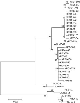
Fig. 2.Phylogenetic analysis of the F genes of hMPV isolated in Korean children, which was reconstructed using neighbour-join- ing method. Bootstrap proportions (500 replicates) are plotted at the branches of phylogram to show support values. GenBank accession numbers are DQ092710 to DQ092737.
samples for hMPV, 25 cases (89.2%) were collected between November 2004 and February 2005 (Table 1).
Clinical findings of hMPV positive patients
Clinical manifestations of the 28 children with hMPV infec- tion are presented in Table 2. The median age was 15 months
(range 7-86 months) in the 28 patients. Symptoms included cough (92.8%), fever (60.2%), sputum (42.8%), rhinorrhea (39.2%), dyspnea (17.8%), diarrhea (7.2%), and vomiting (7.2%). Expiratory wheezing and crackles were observed in 21.4% and 25% of the patients, respectively. Abnormal infil- trates of chest radiograph were noted in 10 (35.7%) of the 28 patients. The hMPV-infected children were diagnosed as having pneumonia (35.7%), bronchiolitis (28.5%), bronchial asthma exacerbation (14.3%), croup (10.7%), and upper res- piratory tract infection (7.1%). Six of 28 patients had a his- tory of reactive airway disease. A 27-month-old girl who was admitted with respiratory distress expired of cardiorespiratory failure 2 days after admission without known bacteremia. Only one patient of the 28 patients had underlying medical prob- lem, chronic lung disease of prematurity.
Phylogenetic analyses of hMPV strains
Amplified products corresponding to part of the hMPV F gene (750 bp) were visualized on 1.5% gels. The PCR prod- ucts from each positive specimen were sequenced and were
*RSV, respiratory syncytial virus; hMPV, human metapneumovirus.
Dec 2003- Feb 2004
Mar- May 2004
Jun- Aug 2004
Sep- Nov 2004
Dec 2004- Feb 2005 No. of tested samples (No. of positive)
Common respiratory viruses
RSV 27 (2) 20 (0) 48 (6) 191 (99) 95 (22) Influenza A 27 (1) 20 (0) 48 (0) 191 (1) 95 (7) Influenza B 27 (0) 20 (0) 48 (2) 191 (0) 95 (0) Parainfluenza 27 (0) 20 (0) 48 (3) 191 (3) 95 (3) Adenovirus 27 (1) 20 (0) 48 (0) 191 (0) 95 (0)
hMPV 5 (0) 18 (3) 33 (0) 120 (8) 54 (17)
Table 1.Virus detection in nasal swabs in hospitalized children with acute respiratory disease
Case
No. Sex Age
(months) Symptoms Diagnosis Duration of hospi-
talization (days)
Underlying condition
Chest radiography findings
1 F 5 Cough, Rh, sputum Bronchiolitis 4 None Hyperaeration
2 F 26 Fever, cough, stridor Croup 4 None WNL
3 M 12 Fever, cough, sputum Pneumonia 5 None WNL
4 F 52 Cough, Rh, wheezing Pneumonia 7 RAD RLL
5 F 52 Cough, wheezing B.A. 3 RAD WNL
6 F 10 Fever, cough, sputum Bronchiolitis 3 None WNL
7 M 11 Cough Bronchiolitis 10 None WNL
8 M 8 Fever, Rh, sore throat URI 5 None WNL
9 M 15 Fever, Rh, cough, sputum Bronchiolitis 7 None LLL
10 F 24 Fever, Rh, cough Bronchiolitis 6 None Hyperaeration
11 F 14 Cough, Rh, wheezing Pneumonia 10 None WNL
12 M 32 Fever, Rh, cough, sputum Bronchiolitis 6 None RML
13 F 3 Fever, cough B.A. 5 None WNL
14 M 8 Cough, Rh, stridor Croup 7 RAD WNL
15 M 22 Fever, cough Pneumonia 8 None WNL
16 M 37 Fever, cough, sputum, Croup 7 RAD Interstitial
wheezing, stridor, Rh
17 M 22 Fever, cough Bronchiolitis 7 None WNL
18 M 4 Cough B.A. 18 RDS WNL
19 M 79 Cough, wheezing B.A. 4 RAD WNL
20 M 6 Wheezing Pneumonia 15 RAD Interstitial
21 M 86 Fever, cough, sputum Pneumonia 5 None Interstital
22 F 27 Fever, cough, sputum ARDS, expired 2 None BUL, BLL
23 F 22 Fever, cough URI 8 None WNL
24 M 26 Fever, cough, sputum Pneumonia 6 None Interstitial
25 F 9 Cough, diarrhea Bronchiolitis 7 None Hyperaeration
26 F 16 Fever, cough, sputum, Rh Pneumonia 5 None Interstitial
27 M 70 Fever, cough, sputum, Rh Pneumonia 6 None BLL
28 M 6 Cough, sputum Bronchiolitis 5 None Hyperaeration
Table 2.Clinical manifestations of hMPV infection
URI, upper respiratory tract infection; B.A., bronchial asthma; RAD, reactive airway disease; ARDS, acute respiratory distress syndrome; RDS, respira- tory distress syndrome; Rh, rhinorrhea; WNL, within normal limit; BUL, both upper lobe infiltration; BLL, both lowe lobe infiltration; RML, right middle lobe infiltration, Interstitial, interstitial infiltration.
consistent with hMPV. The 28 hMPV strains detected in this study were classified into two distinct F lineages, 26 strains belonged to genogroup A (A2) and two strains to genogroup B (Fig. 2).
DISCUSSION
This study shows that hMPV is an important etiologic agents among Korean children hospitalized with acute res- piratory tract infections. The prevalence of hMPV detected in nasopharyngeal samples from children with respiratory infections of unknown etiology has varied from 1.5% to 21%
(1, 11, 15-18), but it has been unclear in Korea due to lack of published reports. The difference of prevalence in several studies may be explained by yearly variation in incidence, different group of patients and primers used in PCR assays for hMPV (15-18). Because we studied hospitalized children only, actual prevalence of hMPV infections may be higher.
Some researcher suggested that hMPV cause lower respira- tory tract infection in healthy children at a relatively high frequency (19).
It seems that hMPV shows a seasonal variation, with spo- radic epidemics. In a study, hMPV was the most common virus isolates during the winter season 2002-2003 in children hospitalized for acute respiratory tract infection, but no spe- cimen was found positive for hMPV in the following season (18). Maggi et al. (7) reported that the incidence of hMPV infection in infants varied from season to season over 3 yr peri- od. The peak time of hMPV transmission in the Netherlands was in December, in Canada in April, and in Hong Kong in spring and early summer (1, 4, 12). Some reported that sig- nificant hMPV activity occurred in every month of the year with the peak incidence in the winter and spring seasons (19, 20). In our study, we detected 25 hMPV positive samples from November to February during the winter season of 2004- 2005, with a peak in November. In the spring season of 2004, we also detected three hMPV positive samples. But, the exact prevalence of hMPV during the winter season of the 2003- 2004 in this study is uncertain due to small number of avail- able respiratory specimens.
The clinical features of children with hMPV positive sam- ples observed in our study were similar to those of previous reports (8, 20). Exacerbation of asthma was observed in 14%
(4/28) of hMPV only positive patients in this study. Like other viruses, such as hRSV and rhinoviruses, which have been sug- gested as important triggers of asthma exacerbation in chil- dren, an association between hMPV and asthma exacerbation has been implicated. In a Finnish study, the positive rate of hMPV was 32% (10/31) in hospitalized children with acute wheezing during the period of peak of hMPV infection (21).
Although it may be possible that asthmatic bronchitis is trig- gered by hMPV, it is not certain to conclude definitely the association between and bronchial asthma (22). hMPV has
been suggested as a common and frequent etiologic agent of bronchiolitis in young children (8, 23, 24). Some reported that co-infection with RSV and hMPV may be a possible cause of increasing clinical severity in hospitalized children (25, 26), but others reported different results (7). The limi- tations of this study are relatively short study period, small number of samples in the first half of the 2004, which is not sufficient to know the exact epidemiologic characteristics. The data of combined infections of hMPV with other common respiratory viruses are also lacking.
RT-PCR is more useful method than cell culture, which has characteristics of slow growth and mild cytopathic effect of hMPV, and is now used commonly in epidemiologic stud- ies in various populations (27, 28). The strains of hMPV can be divided into two major genetic lineages (1, 3). Variabili- ty of hMPV genes may affect the sensitivity of study due to limited available sequence information. We have performed the analysis of F-gene as van den Hoogen et al. described (13), which is known to have highly conserved sequence and allow the differentiation of all four sublineages in several studies (5, 8, 9, 13). Phylogenetic analysis of F gene sequences of our strains showed highly nucleotide identity with viruses belonging to genogroup A during the winter season of 2004- 2005, all belonged to A2. Otherwise, three hMPV positive samples detected in spring of 2004 belonged to genogroup B2 in two, genogroup A1 in one. An Italian study reported striking variation in the overall circulation rates and co-circu- lation of multiple strains in the same area in different years (18).
This study suggests that hMPV may be an important etio- logic agent of respiratory tract infection requiring hospital- ization in children and two distinct groups of hMPV are co- circulating in Korea.
ACKNOWLEGEMENTS
The authors owe thanks to Miss Kim SJ at Research Cen- ter of Sanggyepaik Hosiptal for her technical assistance.
REFERENCES
1. van den Hoogen BG, de Jong JC, Groen J, Kuiken T, de Groot R, Fouchier RA, Osterhaus AD. A newly discovered human pneumovirus isolated from young children with respiratory tract disease. Nat Med 2001; 7: 719-24.
2. van den Hoogen BG, Bestebroer TM, Osterhaus AD, Fouchier RA.
Analysis of the genomic sequence of a human metapneumovirus. Vi- rology 2002; 295: 119-32.
3. Peret TC, Boivin G, Li Y, Couillard M, Humphrey C, Osterhaus AD, Erdman DD, Anderson LJ. Characterization of human metapneu- movirus isolated from patients in North America. J Infect Dis 2002;
185: 1660-3.
4. Boivin G, De Serres G, Cote S, Gilca R, Abed Y, Rochette L, Berg- eron MG, Dery P. Human metapneumovirus infections in hospital- ized children. Emerg Infect Dis 2003; 9: 634-40.
5. Esper F, Boucher D, Weibel C, Martinello RA, Kahn JS. Human metapneumovirus infection in the United States: clinical manifesta- tions associated with a newly emerging respiratory infection in chil- dren. Pediatrics 2003; 111: 1407-10.
6. Freymouth F, Vabret A, Legrand L, Eterradossi N, Lafay-Delaire F, Brouard J, Guillois B. Presence of the new human metapneumovirus in French children with bronchiolitis. Pediatr Infect Dis J 2003; 22:
92-4.
7. Maggi F, Pifferi M, Vatteroni M, Fornai C, Tempestini E, Anzilotti S, Lanini L, Andreoli E, Ragazzo V, Pistello M, Specter S, Bendinelli M. Human metapneumovirus associated with respiratory tract infec- tions in a 3-year study of nasal swabs from infants in Italy. J Clin Mic- robiol 2003; 41: 2987-91.
8. Ebihara T, Endo R, Kikuta H, Ishiguro N, Ishiko H, Hara M, Taka- hashi Y, Kobayashi K. Human metapneumovirus infection in Japanese children. J Clin Microbiol 2004; 42: 126-32.
9. Biovin G, Abed Y, Pelletier G, Ruel L, Moisan D, Cote S, Peret TC, Erdman DD, Anderson LJ. Virological features and clinical mani- festations associated with the human metapneumovirus, a new para- myxovirus responsible for acute respiratory tract infections in all age groups. J Infect Dis 2002; 186: 1330-4.
10. Stockton J, Stephenson I, Fleming D, Zambon M. Human metapneu- movirus as a cause of community acquired respiratory illness. Emerg Infect Dis 2002; 8: 897-901.
11. MacKay IM, Jacob KC, Woolhouse D, Waller K, Syrmis MW, Whiley DM, Siebert DJ, Nissen M, Sloots TP. Molecular assay for detection of human metapneumovirus. J Clin Microbiol 2003; 41: 100-5.
12. Peiris JS, Tang WH, Chan KH, Khong PL, Guan Y, Lau YL, Chiu SS. Children with respiratory disease associated with metapneumo- virus in Hong Kong. Emerg Infect Dis 2003; 9: 628-33.
13. van den Hoogen BG, Herfst S, Sprong L, Cane PA, Forleo-Neto E, de Swart RL, Osterhaus AD, Fouchier RA. Antigenic and genetic variability of human metapneumoviruses. Emerg Infect Dis 2004;
10: 658-66.
14. Kumar S, Tamura K, Nei M. MEGA 3: Integrated software for molec- ular evolutionary genetics analysis and sequence alignments. Bioin- formatics 2004; 5: 150-63.
15. von Linstow ML, Henrik Larsen H, Eugen-Olsen J, Koch A, Nord- mann Winther T, Meyer AM, Westh H, Lundgren B, Melbye M, Hogh B. Human metapneumovirus and respiratory syncytial in hos- pitalized Danish children with acute respiratory tract infection. Scan J Infect Dis 2004; 36: 578-84.
16. Galiano M, Videla C, Puch SS, Martinez A, Echavarria M, Carballal
G. Evidence of human metapneumovirus in children in Argentina. J Med Virol 2004; 72: 299-303.
17. Dollner H, Risnes K, Radtke A, Nordbo SA. Outbreak of human metapneumovirus infection in Norwegian children. Pediatr Infect Dis J 2004; 23: 436-40.
18. Gerna G, Campanini G, Rovida F, Sarasini A, Lilleri D, Paolucci S, Marchi A, Baldanti F, Revello MG. Changing circulation rate of human metapneumovirus strains and types among hospitalized pedi- atric patients during three consecutive winter-spring seasons. Arch Virol 2005; 150: 2365-75.
19. Williams JV, Harris PA, Tollefson SJ, Halburnt-Rush LL, Pingster- haus JM, Edwards KM, Wright PF, Crowe JE Jr. Human metapneu- movirus and lower respiratory tract disease in otherwise healthy in- fants and children. N Engl J Med 2004; 350: 443-50.
20. Robinson JL, Lee BE, Bastien N, Li Y. Seasonality and clinical fea- tures of human metapneumovirus infection in children in Northern Alberta. J Med Virol 2005; 76: 98-105.
21. Jartti T, van den Hoogen B, Garofalo RP, Osterhaus AD, Ruuskanen O. Metapneumovirus and acute wheezing in children. Lancet 2002;
360: 1393-4.
22. Rawlinson WD, Waliuzzaman Z, Carter JW, Belessis YC, Gilbert KM, Morton JR. Asthma exacerbation in children associated with rhinovirus but not human metapneumovirus infection. J Infect Dis 2003; 187: 1314-8.
23. Viazov S, Ratjen F, Scheidhauer R, Fielder M, Roggendorf M. High prevalence of human metapneumovirus infection in young children and genetic heterogeneity of the viral isolates. J Clin Microbiol 2003;
41: 3043-5.
24. Bouscambert-Duchamp M, Lina B, Trompette A, Moret H, Motte J, Andreoletti L. Detection of human metapneumovirus RNA sequences in nasopharyngeal aspirates of young French children with acute bronchiolitis by real time reverse transcriptase PCR and phylogenetic analysis. J Clin Microbiol 2005; 43: 1411-4.
25. Cuevas LE, Nasser AM, Dove W, Gurgel RQ, Greensill J, Hart CA.
Human metapneumovirus and respiratory syncytial virus, Brazil.
Emerg Infect Dis 2003; 9: 1626-8.
26. Konig B, Konig W, Arnold R, Werchau H, Ihorst G, Forster J. Pros- pective study of human metapneumovirus infection in children less than 3 years of age. J Clin Microbiol 2004; 42: 4632-5.
27. Falsey AR, Erdman D, Anderson LJ, Walsh EE. Human metapneu- movirus infections in young and elderly adults. J Infect Dis 2003; 187:
785-90.
28. Carr MJ, McCormack GP, Crowley B. Human metapneumovirus associated respiratory tract infections in the Republic of Ireland dur- ing the influenza season of 2003-4. Clin Microbiol Infect 2005; 11:
366-71.