INTRODUCTION
Uncontrolled cell proliferation is the hallmark of cancer, and tumor cells have acquired damage to genes that directly regulate their cell cycles. In the cell cycle, the period from the late G1 to the S phase is the most important for cell proliferation. The decision to divide occurs as cell passes a restriction point late in G1, after which they commit to the autonomous program that carries them through to division.
The restriction point is regulated largely by retinoblastoma gene product (pRb). In pRb phosphorylation, two major G1 cyclins have important roles. Cylin D1 forms a complex with cyclin-dependent kinase (cdk) 4 or cdk6, and works for the phosphorylation of pRb. Cyclin E binds to cdk2 and also phosphorylates pRb (1). Cyclin D1 gene is amplified in human carcinomas of esophagus (2) and breast (3). Deran- ged cyclin E mRNA expression has been reported in carci- nomas of the breast (4). Rb gene is deleted or mutated in a wide range of human cancers (5).
To prevent unrestrained proliferation, there are a number of cyclin-dependent kinase inhibitors (CKIs) which bind with specific cdks, preventing the cdks from binding to cyclin and hence blocking a cascade of events which ulti- mately leads to cell proliferation. Two subtypes of CKIs are the INK (inhibitor of kinase) family, which includes p16 (INK4a), p15 (INK4b), p18 (INK4c) and p19 (INK4d), and the CIP/KIP (cdk interacting protein/kinase inhibitory protein) family, which includes p21 (WAF1/CIP1), p27 (KIP1) and p57 (KIP2) (6). After exposure to DNA damag- ing agent, wild-type p53 induced p21 protein expression allows apoptosis, thus, induction of p21 protein seems to occur with functional p53. In contrast, recent study has demonstrated that p21 can be induced by a p53-indepen- dent pathway (7). p16 is implicated in carcinogenesis as a result of frequent gene alterations such as deletion, muta- tion, and aberrant DNA methylation in human cancers (8).
Unlike p16 gene, mutation of the p21 or p27 gene is an extremely rare event in human carcinomas (9-11).
Yoon-La Choi, Seong Hoe Park*, Ja-June Jang*, Cheol Keun Park
Department of Diagnostic Pathology, Samsung Medical Center, Sungkyunkwan University School of Medicine, Seoul; Department of Pathology*, Seoul National University School of Medicine, Seoul, Korea
Address for correspondence Cheol Keun Park, M.D.
Department of Diagnostic Pathology, Samsung Medical Center, 50 Ilwon-dong, Kangnam-gu, Seoul 135-710, Korea
Tel : + 82.2-3410-2766, Fax : +82.2-3410-0025 E-mail : [email protected]
*This work was supported by the Samsung grant,
#SBRI C-A0-033-1.
424
Expression of the G1-S Modulators in Hepatitis B Virus-Related
Hepatocellular Carcinoma and Dysplastic Nodule: Association of Cyclin D1 and p53 Proteins with the Progression of Hepatocellular Carcinoma
Deranged expression of cell cycle modulators has been reported to contribute to the development and progression of hepatocellular carcinoma (HCC). However, their expression patterns remain poorly understood in hepatitis B virus (HBV)- related HCC, which constitutes about 65-70% of HCC in Korea. The aims of this study were to evaluate the expressions of G1-S modulators in HBV-related HCCs and dysplastic nodules (DNs), and to correlate with the histopathologic features of HCCs. Immunohistochemical expressions of cyclin D1, cyclin E, p53, p27, p21, p16, Rb, and PCNA proteins were investigated in 80 HCCs and 22 DNs. Cyclin D1 overexpression showed positive relationships with advanced tumor stage, poor differentiation, larger tumor size, microvascular invasion, intrahepatic meta- stasis, no tumor capsule formation, infiltrative growth, aberrant p53 expression, and high PCNA labeling index (LI) of HCC (p<0.05). Aberrant p53 expression showed positive relationship with poor differentiation of HCC (p<0.01). Expression of cyclin D1 or p53 was not observed in DNs. The p27 LI and p16 LI were lower in HCCs with intrahepatic metastasis (p<0.05). Cyclin D1 overexpression and aber- rant p53 expression could be associated with the progression of HBV-related HCC, and might have a less crucial role in the DN-HCC sequence. In addition, elevated expression of p27 and p16 proteins might have inhibitory action to the intrahepatic metastasis of HBV-related HCC.
Key Words : Carcinoma, Hepatocellular; Dysplastic Nodule; Hepatitis B Virus; Protein cyclin D1; Pro- tein p53; Protein p27; Protein p16
Received : 14 February 2001 Accepted : 11 April 2001
p53 is considered to be a stress response gene, and its product (the p53 protein) induces cell cycle arrest or apop- tosis in response to DNA damage. Mutation of p53 gene is the most common genetic abnormality identified in human carcinomas and may result in a biologically altered protein that is detectable by immunohistochemical method (12).
Proliferating cell nuclear antigen (PCNA) is an auxiliary protein for DNA polymerase-delta, and accumulates in the nucleus from the late G1 to the S phase. PCNA can be used as an immunohistochemical marker of proliferating cells (13).
Hepatocellular carcinoma (HCC) is one of the most com- mon human malignancies, especially in sub-Saharan Africa and Southeast Asia including Korea (14). Hepatitis B and C viruses, alcohol and aflatoxin B1 are known risk factors for HCC. It is probable that the ongoing process of hepatocyte necrosis and liver cell renewal coupled with inflammation, which is characteristic of chronic viral hepatitis, causes not only cirrhosis but also progressive genomic errors in hepato- cytes as well as unregulated growth and repair mechanism leading to hepatocyte dysplasia and, in some cases, HCC (15). Investigation of the cell proliferating status in HCC is important for evaluating the biological aggressiveness of HCC, because distant and lymphatic metastases are not very common. Recently, both genetic alterations and over- expressions of cell cycle modulators in HCC have been investigated, but there were small numbers of cases with hepatitis B virus (HBV)-related HCC (16-18). HBV-related HCC constitutes about 65-70% of HCC in Korea (19). It is necessary to study on the cell cycle modulators for a large number of cases with HBV-related HCC for the prevention and treatment of HCC.
Dysplastic nodule (DN) has recently been described as possible precancerous lesion in multistep hepatocarcinogen- esis (20). Sequential development of low grade DN (LDN), high grade DN (HDN) and early HCC is very similar to multistep tumorigenesis in colon (21). In colorectal carcino- genesis, p53 mutation occurs in about 20% of adenomas and seems to be a rate-limiting step that makes an impor- tant contribution to tumor progression (22). p53 mutation might be an unusual event in DN (23). No crucial events or pivotal alterations in the molecular basis of the hepatocar- cinogenesis have yet to be elucidated.
The aims of this study were to evaluate the expressions of cyclin D1, cyclin E, p53, p27, p21, p16, Rb, and PCNA proteins in HBV-related HCCs and DNs with a correlation with the histopathologic features of HCCs.
MATERIALS AND METHODS Tissue specimens and histopathology
Eighty HCCs (Edmondson grade I, 20; grade II, 24;
grade III, 36 cases) and 22 DNs (LDN, 10; HDN, 12 cases)
were derived from 72 and 18 surgically resected livers, respectively, at Samsung Medical Center from 1996 to 1999. All patients were of Korean origin. The patients were diagnosed as HCC or liver cirrhosis, and undergone surgery without transcatheter arterial chemoembolization or lipi- odol injection. All patients were seropositive for hepatitis B surface antigen (HBsAg), showed positive immunoreactivi- ty for HBsAg in nontumorous liver, and had no serum anti- body against hepatitis C virus (HCV). Seventy two patients with HCC were 58 men and 14 women with a mean age at diagnosis of 55.2 yr (range, 31-79 yr). Eighteen patients with DN were 16 men and 2 women with a mean age at diagnosis of 56.1 yr (range, 33-79 yr). Tissue were fixed in 10% formalin for 12 hr and embedded in paraffin. Sections of 4- m thickness were stained with hematoxylin and eosin. HCCs were graded histologically according to the Edmondson and Steiner (24), and DNs were classified according to the International Working Party (20) into two groups, i.e., LDN and HDN. Among the patients with HCC, 67 patients had single HCC, 2 patients had two HCCs, and 3 patients had three HCCs. Among the patients with DN, 15 patients had single DN, 2 patients had two DNs, and 1 patient had three DNs. Among the patients with HCC, 56 patients showed cirrhosis in nontumorous liver, and 16 patients showed chronic hepatitis. All patients with DN showed cirrhosis in nontumorous liver, and five patients with DN had no HCC. The histopathologic fea- tures of HCCs were examined: tumor stage, tumor size, his- tological differentiation, portal vein invasion, microvascular invasion, intrahepatic metastasis, tumor capsule formation, and growth pattern. The tumor stage was determined according to the TNM atlas (UICC) (25). Portal vein inva- sion was defined as tumor thrombus in the lobar or seg- mental branches of the portal veins. Microvascular invasion was considered as present when tumor was seen in a vessel, that is, at least 1 or more endothelial cells or the tunica media of the vessel were recognized to surround a neoplastic cell group. Intrahepatic metastasis and tumor capsule for- mation conformed to the Classification of Primary Liver Cancer of the Liver Cancer Study Group of Japan (26). The growth pattern was divided as infiltrative and expansive, and infiltrative pattern was defined as the presence of one or more foci of microscopic parenchymal invasion. Blocks of normal liver were prepared from 10 patients with metastat- ic colonic carcinoma of the liver as control cases.
Immunohistochemistry
Representative tissue sections including both HCC or DN and adjacent nontumorous liver were selected and sec- tioned in 4- m thickness. Immunohistochemical study was performed using the streptavidin-biotin complex method and TechMateTM 1,000 automated staining system (Dako Chemmate, Glostrup, Denmark). Primary antibodies used
and working dilutions employed were as follows: cyclin D1 (clone P2D11F11, monoclonal, 1:10), cyclin E (clone 13A3, monoclonal, 1:20), and Rb (clone 1F8, monoclonal, 1:20) from Novocastra (London, UK), p21 (clone SX118, mono- clonal, 1:25) and PCNA (clone PC10, monoclonal, 1:1000) from Dako (Glostrup, Denmark), p53 (clone BP53.12, mono- clonal, 1:80), p27 (clone 57, monoclonal, 1:200), and p16 (rabbit polyclonal, 1:20) from Zymed (San Francisco, CA, U.S.A.), Transduction (Lexington, KY, U.S.A.), and Santa Cruz (Santa Cruz, CA, U.S.A.), respectively. Deparaffinized sections were treated with 3% hydrogen peroxide in metha- nol for 10 min to block endogenous peroxidase. Sections were processed in 0.05 M citrate buffer (pH 6.0) and heated in a microwave oven for 10 min for antigen retrieval. Sec- tions were then incubated with the primary antibody for 60 min at room temperature. Each section was treated with biotinylated secondary antibody and streptavidin-peroxi- dase complex (Dako, Glostrup, Denmark), sequentially.
DAB (3,3′-diaminobenzidine tetrahydrochloride) was used as a chromogen, and Mayer’s hematoxylin counterstain was applied. Negative controls were run simultaneously with an omission of primary antibody.
Immunohistochemical evaluation
For assessment of the positivity of immunostaining for each section, only nuclear staining was regarded as positive.
We regarded the G1 cyclins and p53 as positive when ≥
5% of tumor cell nuclei showed positive immunostaining.
We counted positive cells for p27, p21, p16, Rb and PCNA by monitoring at least 1,000 cells in HCC or DN and non- tumorous liver from more than five high power fields where positive cells were present at a relatively uniform density.
Then, we calculated the labeling index (LI) as a percentage for each protein.
Statistical analyses
Values were expressed as mean±SD. Testing for associa- tions between the expression of proteins and histopatholog- ic parameters was performed by Chi-square analysis. When appropriate, the unpaired Student t-test was used to test for statistical differences between groups. p-values of less than 0.05 were considered statistically significant.
RESULTS
Lymphocytes in nontumorous liver were positive for all proteins, which were regarded as internal positive controls for each section.
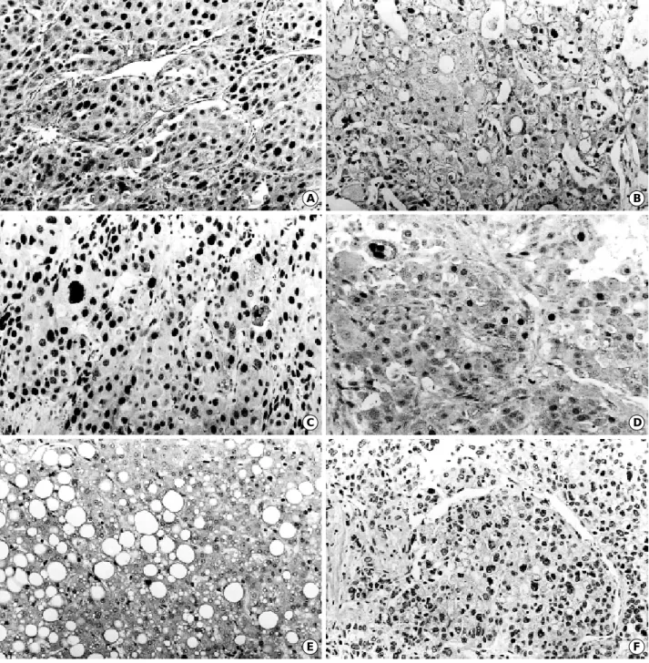
Cyclin D1 protein was overexpressed in 17 of the 80 cases (21.3%) of HCC (Fig. 1A). Overexpression of cyclin D1 protein was significantly associated with advanced tumor stage (p=0.032), larger tumor size (p=0.021), poorer histo- logical grade of differentiation (p<0.0001), microvascular invasion (p=0.023), intrahepatic metastasis (p=0.049), no tumor capsule formation (p=0.003), infiltrative growth
p value Categories Cyclin D1 positive cases
Tumor stage I 2/27 (7.4%) 0.032
II 3/21 (14.3%) III 7/20 (35.0%) IV 5/12 (41.7%)
Tumor size �2 cm 2/32 (6.3%) 0.021
2-5 cm 7/26 (26.9%)
>5 cm 8/22 (36.4%)
Differentiation Ed I 0/20 (0%) <0.001 Ed II 2/24 (8.3%)
Ed III 15/36 (41.7%)
Portal vein + 3/10 (30.0%) NS
invasion - 14/70 (20.0%)
Microvascular + 12/37 (32.4%) 0.023
invasion - 5/43 (11.6%)
Intrahepatic + 5/11 (45.5%) 0.049
metastasis - 12/69 (17.4%)
Capsule + 3/37 (8.1%) 0.003
formation - 14/38 (36.8%)
Growth pattern Infiltrative 10/27 (37.0%) 0.016 Expansive 7/53 (13.2%)
p53 expression Positive 9/18 (50.0%) 0.002 Negative 8/62 (12.9%)
Table 1.Relationship between the overexpression of cyclin D1 protein and histopathologic features of hepatocellular carcinomas
Ed, Edmondson grade; NS, not significant
p value
Categories p53 positive cases
Tumor stage I 2/27 (7.4%) NS
II 5/21 (23.8%) III 7/20 (35.0%) IV 4/12 (33.3%)
Tumor size �2 cm 2/32 (6.3%) NS
2-5 cm 9/26 (34.6%)
>5 cm 7/22 (31.8%)
Differentiation Ed I 0/20 (0%) 0.003
Ed II 4/24 (16.7%) Ed III 14/36 (38.9%)
Portal vein + 3/10 (30.0%) NS
invasion - 15/70 (21.4%)
Microvascular + 11/37 (29.7%) NS
invasion - 7/43 (16.3%)
Intrahepatic + 3/11 (27.3%) NS
metastasis - 15/69 (21.7%)
Capsule + 8/37 (21.6%) NS
formation - 10/38 (26.3%)
Growth pattern Infiltrative 8/27 (29.6%) NS Expansive 10/53 (18.9%)
Table 2.Relationship between the aberrant expression of p53 protein and histopathologic features of hepatocellular carcinomas
Ed, Edmondson grade; NS, not significant
(p=0.016), and aberrant expression of p53 protein (p=0.002) (Table 1). The average LI of PCNA was significantly higher in cases with overexpression of cyclin D1 protein (cyclin D1 positive: 76.0±16.0, cyclin D1 negative: 56.3±27.0, p=
0.018).
Only one case (1.3%) of stage III HCC showed overex- pression of cyclin E protein (Fig. 1B).
Aberrant expression of p53 protein was observed in 18 of the 80 cases (22.5%) of HCC (Fig. 1C). There was positive
correlation between the aberrant expression of p53 protein and poorer histological grade of differentiation (p=0.003) (Table 2). The average LI of PCNA was higher in cases with aberrant p53 protein expression, but there was no statistical significance (p=0.076).
Expression of cyclin D1, cyclin E, and p53 proteins was not observed in DNs, nontumorous livers, and normal con- trols.
p27 protein was expressed in 64 cases (80%) of HCC
E F
C D
A B
Fig. 1. Representative examples of immunohistochemical staining of the G1-S modulators in HCCs (A-D, F, G, I) and dysplastic nodules (E, H, J). Overexpression of cyclin D1 (A) and cyclin E (B), and aberrant expression of p53 (C) in HCCs Ed III. Nuclear immunoreactivity for p27 in HCC Ed II (D) and high grade dysplastic nodule (E). Nuclear immunoreactivity for p21 in HCC Ed III (F). (Fig. 1 continued next)
(Fig. 1D) and its average LI was 45.3±38.4. The p27 LI in HCC was significantly lower in cases with intrahepatic metastasis (p=0.040) (Table 3). p27 protein was expressed in 6 of the 12 cases (50%) of HDN (Fig. 1E) and 6 of the 10 cases (60%) of LDN. The average LIs of HDN, LDN, and nontumorous liver were 7.0±11.9, 3.0±4.1, and 8.0
±18.2, respectively (Table 5), and were significantly lower than that of HCC (p<0.001).
p21 protein was expressed in 18 cases (22.5%) of HCC (Fig. 1F) and its average LI was 0.6±1.8. No correlation could be established between the p21 LI and histopatholog- ic features of HCCs, including aberrant expression of p53 protein (Table 3). p21 protein was not expressed in DNs and nontumorous livers (Table 5).
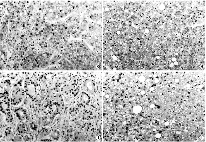
p16 protein was expressed in 58 cases (72.5%) of HCC (Fig. 1G) and its average LI was 35.2±37.4. p21 LI in HCC was significantly lower in cases with intrahepatic metastasis (p=0.012) (Table 3), and there was no association with the pRb LI. p16 protein was expressed in 6 cases (50%) of HDN (Fig. 1H) and 6 cases (60%) of LDN. The average LIs of HDN, LDN, and nontumorous liver were 9.5±16.1, 1.8±2.1, and 9.0±23.8, respectively (Table 5),
and were significantly lower than that of HCC (p<0.001).
Rb protein was expressed in 62 cases (77.5%) of HCC (Fig. 1I) and its average LI was 34.6±35.6. There was no statistically significant correlation between the pRb LI and histopathologic features of HCCs (Table 4). Rb protein was expressed in 7 cases (58.3%) of HDN (Fig. 1J) and 5 cases (50%) of LDN. The average LIs of HDN, LDN, and nontu- morous liver were 17.5±21.4, 1.3±2.0, and 5.9±13.1, respectively (Table 5), and were significantly lower than that of HCC (p<0.05).
The average PCNA LIs of nontumorous liver, LDN, HDN, and HCC were 2.3±1.2, 3.8±2.6, 10.0±9.7, and 33.3±21.7 (Edmondson I, 10.1±7.1; Edmondson II, 23.6
±12.4; Edmondson III, 43.3±21.2), respectively (Table 5). The PCNA LI significantly increased from LDN to HDN, and from HDN to HCC (p<0.001). The PCNA LI in HCC was significantly higher in more poorly differentiat- ed lesions (p=0.000).
There were no significant differences between the average LI of p27, p21, p16, and Rb proteins in nontumorous liver and those in normal controls.
I J
G H
Fig. 1. (Continued from the previous page) Nuclear immunoreactivity for p16 in HCC Ed II (G) and high grade dysplastic nodule (H). Nucle- ar immunoreactivity for Rb in HCC Ed II (I) and high grade dysplastic nodule (J). (×200). Ed: Edmondson grade.
DISCUSSION
Hepatitis B virus is an important etiologic factor that contributes to HCC, but there have been few studies for a large number of cases with HBV-related HCC. Nishida et al. (27) demonstrated that amplification and overexpression
of cyclin D1 gene in both HBV- and HCV-related HCCs at an advanced stage with rapid tumor growth. However, a recent study reported that downregulation of cyclin D1 mRNA was significantly associated with large HCC and did not correlate with serum HBsAg (18). Ito et al. (16) reported that cyclin D1 protein overexpression occurred more frequently in HCCs with poor differentiation (p=
0.0612) and intrahepatic metastasis (p=0.0675), and did not correlate with serum HBsAg or HCV antibody. In this study, overexpression of cyclin D1 protein showed positive relationship with advanced tumor stage, poor differentia- tion, larger tumor size, microvascular invasion, intrahepatic metastasis, no tumor capsule formation, and infiltrative growth of HBV-related HCCs. But cyclin D1 protein over- expression was not observed in DNs. Our findings strongly suggest that cyclin D1 protein overexpression occurs after the initial stage of hepatocellular carcinogenesis, and reflects the progression and aggressiveness of HBV-related HCCs.
Cyclin D1 alterations might be an unusual event in DNs, and might have a less crucial role in the DN-HCC sequence.
p value
Categories Rb LI
Tumor stage I 24.0±26.7 NS
II 43.9±44.2
III 34.2±38.2
IV 39.7±35.9
Tumor size �2 cm 27.8±29.1 NS
2-5 cm 39.2±33.3
>5 cm 40.3±46.2
Differentiation Ed I 26.2±29.7 NS
Ed II 39.7±38.3 Ed III 35.8±32.2
Portal vein + 46.0±48.5 NS
invasion - 33.1±33.5
Microvascular + 37.9±39.1 NS
invasion - 32.2±29.4
Intrahepatic + 15.8±11.1 NS
metastasis - 37.2±42.0
Capsule + 36.7±38.8 NS
formation - 33.6±35.9
Growth pattern Infiltrative 33.3±29.9 NS Expansive 35.3±40.1
Table 4.Relationship between the expression of Rb protein and histopathologic features of hepatocellular carcinomas
LI, labeling index; Ed, Edmondson grade; NS, not significant
HCC 45.3 0.6 35.2 34.6 33.3
HDN 7.0 0 9.5 17.5 10.0
LDN 3.0 0 1.8 1.3 3.8
p27 LI p21 LI p16 LI Rb LI PCNA LI Table 5.Average labeling indices of p27, p21, p16, Rb, and PCNA proteins in hepatocellular carcinomas and dysplastic nodules
HCC, hepatocellular carcinoma; HDN, high grade dysplastic nodule;
LDN, low grade dysplastic nodule; LI, labeling index
p value p value
p value
Categories p27 LI p21 LI p16 LI
Tumor stage I 29.4±21.2 NS 0.2±0.8 NS 31.3±28.9 NS
II 61.3±63.0 0.1±0.3 42.8±39.5
III 50.2±44.3 0.5±0.8 40.0±42.2
IV 38.3±33.2 2.1±1.9 23.0±29.1
Tumor size �2 cm 37.2±39.1 NS 0.2±0.3 NS 37.1±34.3 NS
2-5 cm 45.6±44.9 0.5±0.8 30.7±33.2
>5 cm 54.3±52.2 1.1±2.1 38.3±35.5
Differentiation Ed I 38.5±41.5 NS 0.1±0.7 NS 35.5±37.2 NS
Ed II 53.1±55.1 0.3±0.4 37.9±40.9
Ed III 43.8±33.3 1.0±0.7 33.3±31.6
Portal vein + 38.0±40.4 NS 2.1±3.1 NS 6.0±11.1 NS
invasion - 46.6±37.8 0.3±0.9 36.9±32.3
Microvascular + 49.2±47.2 NS 0.2±0.5 NS 37.0±39.0 NS
invasion - 42.1±39.2 1.0±0.4 33.8±31.7
Intrahepatic + 15.8±19.5 0.040 0.6±0.7 NS 9.2±12.1 0.012
metastasis - 50.5±47.2 0.6±0.6 39.8±34.1
Capsule + 47.6±42.9 NS 0.9±1.1 NS 39.2±30.2 NS
formation - 46.3±44.2 0.2±0.3 34.2±33.6
Growth pattern Infiltrative 42.0±40.8 NS 0.3±0.4 NS 29.0±34.3 NS
Expansive 47.0±39.6 1.2±2.1 38.6±39.1
Table 3.Relationship between the expression of p27, p21 and p16 proteins, and histopathologic features of hepatocellular carcinomas
LI, labeling index; Ed, Edmondson grade; NS, not significant
We also found that overexpression of cyclin D1 protein was significantly associated with aberrant expression of p53 pro- tein, which suggests that overexpression of cyclin D1 and p53 proteins can lead to deregulation of cell cycle and con- tribute to the progression of HBV-related HCC. The PCNA LI was higher in HCCs with overexpression of cyclin D1 protein, which suggests that cyclin D1 protein is related to the dedifferentiation and proliferative activity of HBV-relat- ed HCCs.
In this study, the histological differentiation of HCCs cor- related with the aberrant p53 protein expression, and there was no p53 protein expression in DNs. We suggest that p53 alterations may be an unusual event in the precancer- ous lesions of multistep hepatocarcinogenesis, and might have a less crucial role in the DN-HCC sequence. p53 alter- ations seem to be a relatively late occurring event and relat- ed to the aggressiveness of HCC (16). And it is suggested that p53 alterations play a less important role in the pro- gression of HBV-related HCCs than cyclin D1 alterations.
Peng et al. (18) reported that overexpression of cyclin E mRNA was observed in 56.3% of HCC, was significantly associated with poorly differentiated and invasive HCCs, and did not correlate with serum HBsAg. Ito et al. (16) reported that cyclin E protein overexpression was observed in 35.5% of HCC, and had positive relationship with stage of HCC. In the present study, cyclin E protein overexpres- sion was observed in 1.3% of HCC. Since the histopatho- logic features do not differ in HCCs from various geograph- ic areas, the discrepancy in frequency of cyclin E alterations may indicate that these changes do not represent a primary oncogenic event but are a late event in tumor progression.
Alternatively, the existence of a different genetic back- ground for the etiology of HCCs in different human popu- lations could be postulated.
p27 protein is expressed in high rate in normal tissue, but shows decrease or absence of expression in tumor tissue, indicating an unfavorable factor in human cancers (28-30).
In this study, the average LI of p27 protein was higher in HCC than in nontumorous liver or DN, unlike other can- cers. Our finding is consistent with the recent report (16).
We found that the p27 LI was significantly lower in HCCs with intrahepatic metastasis (p=0.040). This study offers the evidence for a potential role of p27 dysfunction in the process of HCC metastasis. Ito et al. (16) reported that the decreased expression of p27 protein in HCC was strikingly related to the biological aggressiveness, including portal vein invasion, poor differentiation, and larger size of HCCs as well as its prognosis, and that the p27 LI decreased with borderline significance in cases with intrahepatic metastasis (p=0.078). A previous study demonstrated that the lack or decreased expression of this protein is regulated at posttrans- lational levels of p27 gene by a ubiquitin-proteosome path- way (31), instead of common gene mutations or aberrant DNA methylation. The decreased p27 protein expression in
advanced HCC may be derived by the same mechanism.
Recent studies demonstrated that p21 is an important cell cycle regulatory factor in continuously proliferating cells. Little p21 protein or mRNA is detected in normal human liver tissue, but hepatocyte p21 expression is upreg- ulated in response to hepatic injury (32, 33). The absence of p21 protein expression in nontumorous liver in this study indicates that the level of p21 protein in hepatocytes is below the immunohistochemically detectable threshold, which might be due to the low proliferative activity in non- tumorous liver. Down-regulation of p21 expression through degradation by a ubiquitin-dependent proteolytic pathway has recently been reported (34). Our results showed that the average LI of p21 protein was very low in HCC, which sug- gests that post-transcriptional regulation might be one mechanism that down-regulate p21 expression in HCC. In this study, there was no correlation between the p21 protein expression and histopathologic features of HCCs, which is consistent with the recent reports (35, 36).
A recent study demonstrated that epigenetic change due to extensive CpG methylation is the main cause of inactiva- tion of p16(INK4a) in HCC (37). In this study, the average LI of p16 protein was higher in HCC than in nontumorous liver, which is consistent with the recent report (16). We demonstrated that the p16 LI was significantly lower in HCCs with intrahepatic metastasis (p=0.012). This study offers the evidence for a potential role of p16 dysfunction in the process of HCC metastasis. Hui et al. (38) suggested that p16 protein loss might contribute to the progression of HCC, because it occurred approximately twice as often in advanced rather than in early stage HCC.
The loss of pRb expression in HCC is mainly caused by loss of heterozygosity of the Rb gene (39). In this study, the average LI of pRb was higher in HCC than in nontumorous liver, which is consistent with the recent report (16). Hui et al. (40) suggested that elevated and absent pRb is closely associated with tumor progression and developing metasta- tic disease rather than tumor initiation in HCC. We could not determine any correlation between the pRb expression and histopathologic features of HCCs, which is consistent with previous reports (16, 41).
The PCNA LI, in this study, was significantly higher in more poorly differentiated HCCs, which is consistent with previous reports (42, 43). Our finding suggests that cell proliferative activity of HCC cells correlates with their degree of dedifferentiation. We also found that the PCNA LI increased from LDN, to HDN, and to HCC, which is consistent with previous report (43).
In summary, the present study suggests that overexpres- sion of cyclin D1 protein and aberrant expression of p53 protein could be associated with the progression of HBV- related HCC, but might not be related to the DN-HCC sequence. In addition, elevated expression of p27 and p16 proteins might have inhibitory action to the intrahepatic
metastasis of HBV-related HCC.
REFERENCES
1. Schafer KA. The cell cycle: a review. Vet Pathol 1998; 35: 461-78.
2. Jiang W, Kahn SM, Tomita N, Zhang YJ, Lu SH, Weinstein IB.
Amplification and expression of human cyclin D gene in esophageal cancer. Cancer Res 1992; 52: 2980-3.
3. Keyomarsi K, Pardee AB. Redundant cyclin overexpression and gene amplification in breast cancer cells. Proc Natl Acad Sci USA 1993; 90: 1112-6.
4. Keyomarsi K, O’Leary N, Molnar G, Lees E, Fingert HJ, Pardee AB. Cyclin E, a potential prognostic marker for breast cancer.
Cancer Res 1994; 54: 380-5.
5. Weinberg RA. The retinoblastoma protein and cell cycle control.
Cell 1995; 81: 323-30.
6. Sherr CJ. Cancer cell cycles. Science 1996; 274: 1672-7.
7. Michiell P, Chedid M, Lin D, Pierce JH, Mercer WE, Givol D.
Induction of WAF1/CIP1 by a p53-independent pathway. Cancer Res 1994; 54: 3391-5.
8. Merlo A, Herman JG, Mao L, Lee DJ, Gabrielson E, Burger PC, Baylin SB, Sidransky D. 5′CpG island methylation is associated with transcriptional silencing of the tumor suppressor p16/CDKN2/
MTS1 in human cancers. Nat Med 1995; 1: 686-92.
9. Shiohara M, eL-Deiry WS, Wada M, Nakamaki T, Takeuchi S, Yang R, Chen DL, Vogelstein B, Koeffler HP. Absence of WAF1 mutations in a variety of human malignancies. Blood 1994; 84:
3781-4.
10. Kawamata N, Morosetti R, Miller CW, Park D, Spirin KS, Naka- maki T, Takeuchi S, Hatta Y, Simpson J, Wilczynski S, Lee YY, Bartram CY, Koeffler HP. Molecular analysis of the cyclin-depen- dent kinase inhibitor gene p27/Kip1 in human malignancies. Can- cer Res 1995; 55: 2266-9.
11. Chen TC, Ng KF, Lien JM, Jeng LB, Chen MF, Hsieh LL. Muta- tional analysis of the p27Kip1gene in hepatocellular carcinoma.
Cancer Lett 2000; 153: 169-73.
12. Greenblatt MS, Bennett WP, Hollstein M, Harris CC. Mutations in the p53 tumor suppressor gene: clues to cancer etiology and molec- ular pathogenesis. Cancer Res 1994; 54: 4855-78.
13. Hall PA, Levision DA, Woods AL, Yu CC, Kellock DB, Watkins JA, Barnes DM, Gillett CE, Camplejohn R, Dover R, Waseem NH, Lane DP. Proliferating cell nuclear antigen immunolocalization in paraffin sections: an index of cell proliferation with evidence of deregulated expression in some neoplasms. J Pathol 1990; 62: 285-94.
14. Di Bisceglie AM, Rustgi VK, Hoofnagle JH, Dusheiko GM, Lotze MT. Hepatocellular carcinoma. Ann Intern Med 1988; 108: 390-1.
15. Idilman R, De Maria N, Colantoni A, Van Thiel DH. Pathogenesis of hepatitis B and Cinduced hepatocellular carcinoma. J Viral Hep- atitis 1998; 5: 285-99.
16. Ito Y, Matsuura N, Sakon M, Miyoshi E, Noda K, Takeda T, Umeshita K, Nagano H, Nakamori S, Dono K, Tsujimoto M, Naka- hara M, Nakao K, Tanikuchi N, Monden M. Expression and prog- nostic roles of the G1-S modulators in hepatocellular carcinoma:
p27 independently predicts the recurrence. Hepatology 1999; 30:
90-9.
17. Naka T, Toyota N, Kaneko T, Kaibara N. Protein expression of p53, p21WAF1, and Rb as prognostic indicators in patients with sur- gically treated hepatocellular carcinoma. Anticancer Res 1998; 18:
555-64.
18. Peng SY, Chou SP, Hsu HC. Association of downregulation of cyclin D1 and of overexpression of cyclin E with p53 mutation, high tumor grade and poor prognosis in hepatocellular carcinoma. J Hepatol 1998; 29: 281-9.
19. Moon HY, Moon YM, Han KH, Chun JY, Kang JK, Park IS. Clini- cal aspect and prognosis according to the infection type of hepatitis virus in primary liver cancer. Korean J Med 1994; 47(Suppl): 33.
20. International working party. Terminology of nodular hepatocellular lesions. Hepatology 1995; 22: 983-93.
21. Fearon ER, Vogelstein B. A genetic model for colorectal tumorige- nesis. Cell 1990; 61: 759-67.
22. Baker SJ, Preisinger AC, Jessup MJ, Paraskeva C, Markowitz S, Willson JKV, Hamilton S, Vogelstein B. p53 gene mutations occur in combination with 17p allelic deletions as late events in colorectal tumorigenesis. Cancer Res 1990; 50: 7717-22.
23. Kang YK, Kim CJ, Kim WH, Kim HO, Kang GH, Kim YI. p53 mutation and overexpression in hepatocellular carcinoma and dys- plastic nodules in the liver. Virchows Arch 1998; 432: 27-32.
24. Edmondson HA, Steiner PE. Primary carcinoma of the liver. A study of 100 cases among 48900 necropsies. Cancer 1954; 7: 462- 503.
25. Hermanek P, Hutter RVP, Sobin LA, Wagner G, Wittekind CH.
TNM atlas. 4th ed. UICC. Berlin: Springer, 1999; 115-23.
26. Liver Cancer Study Group of Japan. Classification of primary liver cancer. 1st ed. Tokyo: Kanehara press, 1997; 9-38.
27. Nishida N, Fukuda Y, Komeda T, Kita R, Sando T, Furukawa M, Amenomori M, Shibagaki I, Nakao K, Ikenaga M, Ishizaki K. Ampli- fication and overexpression of the cyclin D1 gene in aggressive human hepatocellular carcinoma. Cancer Res 1994; 54: 3107-10.
28. Wu J, Shen ZZ, Lu JS, Jiang M, Han QX, Fontana JA, Barsky SH, Shao ZM. Prognostic role of p27Kip1and apoptosis in human breast cancer. Br J Cancer 1999; 79: 1572-8.
29. Lu CD, Morita S, Ishibashi T, Hara H, Isozaki H, Tanigawa N. Loss of p27Kip1expression independently predicts poor prognosis for patients with resectable pancreatic adenocarcinoma. Cancer 1999;
85: 1250-60.
30. Tenjo T, Toyoda M, Okuda J, Watanabe I, Yamamoto T, Tanaka K, Ohtani M, Nohara T, Kawasaki H, Tanigawa N. Prognostic sig- nificance of p27(KIP1) protein expression and spontaneous apopto- sis in patients with colorectal adenocarcinomas. Oncology 2000;
58: 45-51.
31. Pagano M, Tam SW, Theodoras AM, Beer-Romero P, DelSal G, Chau V, Yew PR, Draetta GF, Rolfe M. Role of the ubiquitin-pro- teasome pathway in regulating abundance of the cyclin-dependent kinase inhibitor p27. Science 1995; 269: 682-5.
32. Crary GS, Albrecht JH. Expression of cyclin-dependent kinase inhibitor p21 in human liver. Hepatology 1998; 28: 738-43.
33. Albrecht JH, Meyer AH, Hu MY. Regulation of cyclin-dependent
kinase inhibitor p21WAF1/CIP1gene expression in hepatic regenera- tion. Hepatology 1997; 25: 557-63.
34. Maki CG, Howley PM. Ubiquitination of p53 and p21 is differen- tially affected by ionizing and UV radiation. Mol Cell Biol 1997;
17: 355-63.
35. Shi YZ, Hui AM, Takayama T, Li X, Cui X, Makuuchi M. Reduced p21 (WAF1/CIP1) protein expression is predominantly related to altered p53 in hepatocellular carcinomas. Br J Cancer 2000; 83:
50-5.
36. Kwon KW, Park YN, Park CI. p21 protein expression and cell pro- liferation activity in human multistep hepatocarcinogenesis. Korean J Pathol 2000; 34: 325-30.
37. Matsuda Y, Ichida T, Matsuzawa J, Sugimura K, Asakura H. p16 (INK4) is inactivated by extensive CpG methylation in human hepa- tocellular carcinoma. Gastroenterology 1999; 116: 394-400.
38. Hui AM, Sakamoto M, Kanai Y, Ino Y, Gotoh M, Yokota J, Hiro- hashi S. Inactivation of p16INK4in hepatocellular carcinoma. Hepa- tology 1996; 24: 575-9.
39. Zhang X, Xu H-J, Murakami Y, Sachse R, Yashima K, Hirohashi
S, Hu S-X, Benedict WF, Sekiya T. Deletions of chromosome 13q, mutation in retinoblastoma 1, and retinoblastoma protein state in human hepatocellular carcinoma. Cancer Res 1994; 54: 4177-82.
40. Hui AM, Li X, Makuuchi M, Takayama T, Kubota K. Over-expres- sion and lack of retinoblastoma protein are associated with tumor progression and metastasis in hepatocellular carcinoma. Int J Can- cer 1999; 84: 604-8.
41. Seki S, Kawakita N, Yanai A, Kitada T, Sakai Y, Nakatani K, Yamada T, Sakaguchi H, Kuroki T. Expression of the retinoblas- toma gene product in human hepatocellular carcinoma. Hum Pathol 1995; 26: 366-74.
42. Mise K, Tashiro S, Yogita S, Wada D, Harada M, Fukuda Y, Miyake H, Isikawa M, Izumi K, Sano N. Assessment of the biologi- cal malignancy of hepatocellular carcinoma: Relationship to clini- copathological factors and prognosis. Clin Cancer Res 1998; 4:
1475-82.
43. Terada T, Nakanuma Y. Cell proliferative activity in adenomatous hyperplasia of the liver and small hepatocellular carcinoma. Can- cer 1992; 70: 591-8.