Technical Note
Characteristics of Magnetic Resonance-Based Attenuation Correction Map on Phantom Study in
Positron Emission Tomography/Magnetic Resonance Imaging System
Cheolpyo Hong
Department of Radiological Science, Daegu Catholic University, Daegu, Korea
Received 26 November 2020 Revised 3 December 2020 Accepted 7 December 2020
Corresponding author Cheolpyo Hong ([email protected]) Tel: 82-53-850-2524 Fax: 82-53-359-6760
An MR-based attenuation correction (MRAC) map plays an important role in quantitative positron emission tomography (PET) image evaluation in PET/magnetic resonance imaging (MRI) systems.
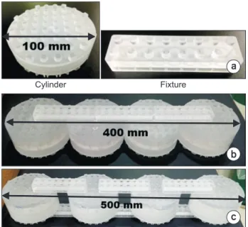
However, the MRAC map is affected by the magnetic field inhomogeneity of MRIs. This study aims to evaluate the characteristics of MRAC maps of physical phantoms on PET/MRI images. Phantom measurements were performed using the Siemens Biograph mMR. The modular type physical phantoms that provide assembly versatility for phantom construction were scanned in a four- channel Body Matrix coil. The MRAC map was generated using the two-point Dixon-based segmentation method for whole-body imaging. The modular phantoms were scanned in compact and non-compact assembly configurations. In addition, the phantoms were scanned repeatedly to generate MRAC maps. The acquired MRAC maps show differently assigned values for void areas.
An incorrect assignment of a void area was shown on a locally compact space between phantoms.
The assigned MRAC values were distorted using a wide field-of-view (FOV). The MRAC values also differed after repeated scans. However, the erroneous MRAC values appeared outside of phantom, except for a large FOV. The MRAC map of the phantom was affected by phantom configuration and the number of scans. A quantitative study using a phantom in a PET/MRI system should be performed after evaluation of the MRAC map characteristics.
Keywords: Phantom study, Artifact, PET/MRI, Attenuation correction map
Copyright © 2020 Korean Society of Medical Physics
CC