Volume 2013, Article ID 313510,12pages http://dx.doi.org/10.1155/2013/313510
Research Article
Attenuation of 𝛽-Amyloid-Induced Oxidative Cell Death by Sulforaphane via Activation of NF-E2-Related Factor 2
Chan Lee,
1Gyu Hwan Park,
2Seong-Ryong Lee,
1and Jung-Hee Jang
11
Department of Pharmacology, School of Medicine, Keimyung University, Daegu 704-701, Republic of Korea
2
Research Institute of Pharmaceutical Sciences, College of Pharmacy, Kyungpook National University, Daegu 702-701, Republic of Korea
Correspondence should be addressed to Jung-Hee Jang; [email protected] Received 11 January 2013; Revised 19 May 2013; Accepted 22 May 2013 Academic Editor: Mi-Kyoung Kwak
Copyright © 2013 Chan Lee et al. This is an open access article distributed under the Creative Commons Attribution License, which permits unrestricted use, distribution, and reproduction in any medium, provided the original work is properly cited.
𝛽-amyloid peptide (A𝛽), a major component of senile plaques, plays important roles in neuropathology of Alzheimer’s disease (AD).
An array of in vitro and in vivo data indicates that A𝛽-induced neuronal death is mediated by oxidative stress. In this study, we aimed to investigate effects of sulforaphane (SUL), an isothiocyanate in cruciferous vegetables, on A𝛽-induced oxidative cell death in SH- SY5Y cells. Cells treated with A𝛽
25–35exhibited decreased cell viability and underwent apoptosis as determined by MTT assay and TUNEL, respectively. A𝛽
25–35-induced cytotoxicity and apoptotic characteristics such as activation of c-JNK, dissipation of mitochondrial membrane potential, altered expression of Bcl-2 family proteins, and DNA fragmentation were effectively attenuated by SUL pretreatment. The antiapoptotic activity of SUL seemed to be mediated by inhibition of intracellular accumulation of reactive oxygen species and oxidative damages. SUL exerted antioxidant potential by upregulating expression of antioxidant enzymes including 𝛾-glutamylcysteine ligase, NAD(P)H:quinone oxidoreductase-1, and heme oxygenase-1 via activation of NF-E2-related factor 2(Nrf2). The protective effect of SUL against A𝛽
25–35-induced apoptotic cell death was abolished by siRNA of Nrf2. Taken together, the results suggest that pharmacologic activation of Nrf2 signaling pathway by SUL might be a practical prevention and/or protective treatment for the management of AD.
1. Introduction
Alzheimer’s disease (AD) is one of the most common forms of senile dementia, characterized by progressive loss of memory and decline of cognitive functions due to neuronal death in the brain. There are two classical pathological hallmarks of AD [1]. One is extraneuronal accumulation of amyloid plaques composed of 𝛽-amyloid peptide (A𝛽), which is produced by proteolytic cleavage from amyloid precursor protein (APP) with sequential actions of 𝛽- and 𝛾-secretases. The other is intraneuronal deposits of neurofibrillatory tangles (NFT) consisting of hyperphos- phorylated tau protein generated by actions of upstream kinases such as glycogen synthase kinase-3𝛽 (GSK-3𝛽) and cyclin-dependent kinase 5 (CDK5). Therefore, treatments for AD have been developed based on these two molec- ular approaches [2]. A𝛽-based therapies include utiliz- ing 𝛽- or 𝛾-secretases inhibitors, A𝛽 aggregation blockers,
and A𝛽 catabolism inducers. Tau-based therapies make an advantage of upstream kinase inhibitors, microtubule stabi- lizers, and tau catabolism inducers.
However, pathogenesis of AD appears to be multifac- torial events, whereby genetic as well as environmental factors, oxidative stress, depletion of endogenous antioxi- dants, altered ion levels, inflammation, disruption in neuro- transmission, synaptic dysfunction, and neuronal cell death operate sequentially or in parallel [3]. Among them we have focused on A𝛽-induced oxidative damages and neuronal cell death as one of the major causes of AD pathology.
Oxidative stress has been proposed to be an important factor
in the development and progression of AD and contributes
to A𝛽 aggregation and NTF formation as well [4]. Reactive
oxygen species (ROS) can be derived from diverse cellular
sources, among which are enzymatic reactions, mitochon-
drial deterioration, and imbalance in redox transition metal
ions. The excessive production and accumulation of ROS by
A𝛽 can cause functional and structural changes in critical macromolecules leading to lipid peroxidation, protein oxida- tion, and DNA cleavage and altered signal transduction [5].
The levels of molecular markers for lipid peroxidation (HNE, isoprostanes, etc.) and oxidation of proteins (carbonyls) and DNAs (8-OHdG) are reported to be elevated in the brains or cerebrospinal fluid of patients with AD [6].
Given the involvement of A𝛽-induced oxidative stress in the etiology and pathology of AD, one of the promising approaches to preventive interventions for AD includes antioxidant therapy by inhibiting the detrimental effects of excess ROS through induction of endogenous antioxi- dant enzymes. Particularly, many studies highlighted natural phytochemicals derived from medicinal herbs and foods as potential candidates which can protect neurons against various toxic compounds and exert beneficial effects on neuronal cells [7, 8]. Sulforaphane (4-methylsulfinylbutyl isothiocyanate, SUL) is a naturally occurring isothiocyanate present in cruciferous vegetables, such as broccoli, cabbage, and cauliflower and has been shown to exhibit anticar- cinogenic, anti-inflammatory, antioxidant, chemopreventive, and cytoprotective properties [9, 10]. Recently, it has been reported that SUL can penetrate blood brain barrier and exert neuroprotective effects in diverse in vitro cell culture and in vivo animal models of neurological disorders [11, 12]. SUL has been reported to attenuate microglia-induced inflammation in hippocampus of LPS-treated mice and BV-2 microglia cells [13]. In addition, SUL protected against oxidative stress induced by hypoxia-ischemic injury [14], oxygen and glucose deprivation [15], 6-hydroxydopamine (6-OHDA) [16], super- oxide [17], hydrogen peroxide (H
2O
2), and glutamate [18].
However, there has been no direct evidence demonstrating that the protective effect of SUL against A𝛽-induced oxidative damage and cell death as yet.
Therefore, in this study we examined whether SUL can suppress A𝛽
25–35-induced oxidative damage and cell death in human neuroblastoma SH-SY5Y cells via augmentation of antioxidant defense capacity by activation of NF-E2-related factor 2 (Nrf2) and the subsequent expression of antioxidant and phase II detoxification enzymes which play key roles in inhibiting ROS production and oxidative damages.
2. Materials and Methods
2.1. Chemicals and Reagents. A𝛽
25–35and SUL were pur- chased from American Peptide (Sunnyvale, CA, USA) and LKT Laboratories, Inc. (St. Paul, MN, USA), respec- tively. Dulbecco’s modified Eagle’s medium (DMEM), fetal bovine serum (FBS), and penicillin-streptomycin antibi- otic were supplied from Gibco BRL (Grand Island, NY, USA). Tetramethylrhodamine ethyl ester (TMRE) and 2
7
- dichlorofluorescein diacetate (DCF-DA) dyes were the prod- ucts of Invitrogen Co. (Carlsbad, CA, USA). Anti-phos- pho-JNK (p-JNK), anti-JNK, anti-Bcl-2, anti-Bax, anti- 𝛾-glutamylcysteine ligase (GCL), anti-NAD(P)H:quinone oxidoreductase-1 (NQO-1), and anti-Nrf2 antibodies were obtained from Santa Cruz Biotechnology, Inc (Santa Cruz, CA, USA). Anti-heme oxygenase-1 (HO-1) antibody was
provided by Stressgen (Ann Arbor, MI, USA). Anti-4- hydroxynonenal (4-HNE) and anti-phospho-Nrf2 antibod- ies were supplied from Abcam (Cambridge, MA, USA) and Epitomics, Inc. (Burlingame, CA, USA), respectively.
MTT [3-(4,5-dimethylthiazol-2-yl)-2,5 diphenyltetrazolium bromide], anti-actin antibody, and other chemical reagents were purchased from Sigma-Aldrich (St. Louis, MO, USA).
2.2. SH-SY5Y Cell Culture. SH-SY5Y cells were maintained in DMEM media containing 10% FBS, penicillin (10000 U), and streptomycin (100 𝜇g/mL) in a 5% CO
2incubator at 37
∘C under a humidified atmosphere. The media were changed every other day. Cells were seeded at an appropriate density according to the each experimental scale.
2.3. Cell Viability Assay (MTT Dye Reduction Assay). Cyto- toxicity was determined by the conventional MTT dye reduc- tion assay. Cells were seeded in 48-well plate at a density of 5 × 10
4cells/well and incubated with A𝛽
25–35(15 𝜇M) for 24 h with or without 30 min pretreatment of SUL (1, 2, and 5 𝜇M) or N-acetylcysteine (NAC, 0.5 and 1 mM). After treatment, MTT solution (5 mg/mL) was added and further incubated for 2 h at 37
∘C. The formazan crystals formed in viable cells were extracted with 200 𝜇L of dimethylsulfoxide (DMSO) and the absorbance was measured in a microplate reader at 570 nm (Emax, Molecular Device, CA, USA). Relative cytotoxicity was calculated as percentage of viable cells with respect to the optical density (OD) value of the living cells in the control as 100%.
2.4. Measurement of DNA Fragmentation (TUNEL). For detection of DNA fragmentation, terminal deoxynucleotidyl transferase-mediated dUTP nick end labeling (TUNEL) (Roche diagnostics GmbH, Mannheim, Germany) was per- formed in SH-SY5Y cells (8 × 10
4cells/500 𝜇L in 4-well chamber slide) exposed to 15 𝜇M A𝛽
25–35for 24 h in the presence or absence of SUL or NAC pretreatment. The slide was rinsed with phosphate-buffered saline (PBS) three times and fixed in 10% neutral buffered formalin solution for 30 min at room temperature (RT). After incubation with 0.3% H
2O
2in methanol for 30 min at RT to inactivate endogenous peroxidase, the slide was further reacted with a permeabilizing solution (0.1% sodium citrate and 0.1%
Triton X-100) for 2 min at 4
∘C. The cells were treated with TUNEL reaction mixture for 1 h at 37
∘C and then labeled with antidigoxigenin peroxidase for additional 30 min at 37
∘C.
After rinsing with PBS three times, color development was performed with 3,3
-diaminobenzidine (Vector Laboratories, CA, USA). The stained images were examined under a light microscope (Leica Microsystems, Wetzlar, Germany).
2.5. Western Bolt Analysis. Cells extracts were prepared by
washing cells with PBS and centrifugation at 7,000 g for
5 min. The collected cells were lysed with RIPA buffer
(Pierce Biotechnology, Inc., Rockford, IL, USA) and protease
inhibitor cocktail tablet (Roche Diagnostics) on ice for
30 min. Protein concentration was quantified by BCA Protein
Assay (Pierce Biotechnology). Protein samples were boiled in
SDS sample buffer and separated on SDS-PAGE using 10%–
12% acrylamide gels. Subsequently, protein samples were transferred onto polyvinylidene fluoride (PVDF, Roche Diag- nostics) membranes by transblot electrophoretic transfer for 3 h at a constant current of 300 mA. The nonspecific binding of antibodies was blocked using 5% (w/v) nonfat milk in PBS containing 0.1% Tween-20 (PBST) for 1 h at RT. After blocking, the membranes were probed with the primary antibodies overnight at 4
∘C. The membranes were washed in PBST three times for 10 min each. The corresponding secondary antibodies were diluted in PBST and reacted with the membranes for 1 h at RT. Finally, immunoreac- tive bands were visualized by chemiluminescence method (Pierce Biotechnology). The images and relative density of immunoreactive bands were analyzed by using ImageQuant LAS 4000 Multi-Gauge software (Fujifilm, Tokyo, Japan).
2.6. Measurement of Mitochondrial Membrane Potential (MMP). For detection of mitochondrial transmembrane potential in SH-SY5Y cells, TMRE probe was utilized. The cells were seeded at a density of 8 × 10
4cells/500 𝜇L in 4-well chamber slide and treated with 15 𝜇M A𝛽
25–35for 24 h in the absence or presence of SUL or NAC. After treatment, cells were washed with PBS and further incubated with TMRE solution (50 𝜇M in PBS) for 15 min at 37
∘C. The fluorescence images were recorded and quantified by using a fluorescence microscope (Leica Microsystems) with excitation at 540 nm and emission at 590 nm.
2.7. Measurement of Intracellular ROS Accumulation. To monitor the intracellular accumulation of ROS, the fluo- rescent probe DCF-DA was used. After treatment of SH- SY5Y cells (8 × 10
4cells/500 𝜇L in 4-well chamber slide) with A𝛽
25–35(15 𝜇M) in the presence or absence of SUL for 6 h, cells were incubated with DCF-DA solution (50 𝜇M in PBS) at 37
∘C for 15 min. The fluorescence signals inside cells were excited at 488 nm and emission was monitored at 535 nm. The images were recorded with a fluorescence microscope (Leica Microsystems).
2.8. Protein Oxidation. The levels of protein carbonyls were determined by using OxyBlot Protein Oxidation Detection Kit (Millipore, MA, USA) according to the manufacturer’s instruction. Briefly, the protein samples (15 𝜇g) were dena- tured by SDS (6% final concentration) and then derivatized to 2,4-dinitrophenylhydrazone (DNP-hydrazone) by incuba- tion with 2,4-dinitrophenylhydrazine (DNPH) for 15 min at RT. After adding neutralization solution, the samples were electrophoresed on a 10% SDS-PAGE gel and transferred to PVDF membrane. The membrane was incubated with blocking buffer for 1 h to reduce nonspecific binding and then reacted with anti-DNP primary antibody for 1 h at RT. After two times washing with PBST, the membrane was further incubated with HRP-conjugated secondary anti-rabbit anti- body for 1 h at RT. The carbonylation bands were detected by using chemiluminescence method (Pierce Biotechnology).
2.9. Nuclear Protein Extraction. Nuclear protein extracts were prepared by using the Nuclear Extraction Kit (Chemi- con, Inc., MA, USA). After treatment, SH-SY5Y cells were washed with ice cold PBS and harvested by centrifugation.
The harvested cells were resuspended in ice-cold cytoplasmic lysis buffer, incubated on ice for 15 min, and centrifuged at 8,000 g for 20 min at 4
∘C. The pellet was resuspended in ice- cold nuclear extraction buffer, incubated on ice for 60 min using shaker, and centrifuged at 16,000 g for 7 min at 4
∘C.
The supernatant containing nuclear proteins were stored at
−80
∘C for western blot analysis and electrophoretic mobility shift assay (EMSA). Protein concentrations were determined by Bradford assay (BIO-RAD, CA, USA).
2.10. Electrophoretic Mobility Shift Assay (EMSA). The DNA binding activity of Nrf2 to antioxidant response element (ARE) was assessed by LightShift Chemiluminescent EMSA kit according to the procedure provided from Pierce Biotech- nology. The isolated nuclear protein samples were combined with binding mixture (1 𝜇g poly (dI⋅dC), 50% glycerol, 1%
NP-40, 1 M KCl, 100 mM MgCl
2, and 200 mM EDTA (Pierce Biotechnology)) and incubated on ice for 20 min. Subse- quently, biotin-labeled oligonucleotide specific to Nrf2 (5
- TGGGGAACCTGTGCTGAGTCACTGGAG-3
, Panomics, CA, USA) was added to the reaction mixture and additionally incubated for 10 min at RT. The DNA-protein complexes were separated on the 6% nondenaturing polyacrylamide gel at 80 V for 1 h and then transferred to nylon membrane (Pall Co., MI, USA) at 380 mA for 45 min. The membrane was subjected to immediate cross-linking by transilluminator at 312 nm for 10 min. After blocking the membrane with blocking buffer for 15 min at RT, the membrane was incubated with stabilized streptavidin-HRP for 15 min at RT. After three times washing with wash buffer, the DNA-protein complex bands were detected by chemiluminescence method (Pierce Biotechnology).
2.11. Synthetic Small Interfering RNA (siRNA) Transfection.
For the knockdown experiments of Nrf2, SH-SY5Y cells were transiently transfected with siRNA of Nrf2 (Nrf2-siRNA) using DOTAP transfection reagent (Roche Diagnostics GmbH) in accordance with the manufacturer’s protocol. The sequences of the sense and antisense strands of the human Nrf2-siRNA were as follows: 5
-AAG AGU AUG AGC UGG AAA AAC TT-3
(sense) and 5
-GUU UUU CCA GCU CAU ACU CUU TT
-3
(antisense) which were selected by siRNA Target Finder software provided by Invitrogen. After transfection of SH-SY5Y cells with Nrf2- siRNA, cells were further exposed to A𝛽
25–35(15 𝜇M) for 24 h in the presence or absence of SUL (5 𝜇M) pretreatment and then cell viability and molecular markers for apoptotic cell death were examined.
2.12. Statistical Analysis. SPSS software 13.0 (SPSS, Inc,
Chicago, IL, USA) was used for the statistical analysis. All
data represent at least three independent experiments and
are expressed as mean ± SD. Statistical comparisons between
groups were made by one-way analysis of variance (ANOVA)
Cell viability (% control)
0 20 40 60 80 100
##
−
−
−
−− −+ + + +
1 2 5 1 2 5
∗∗ ∗∗
∗∗
A𝛽25–35
SUL (𝜇M)
(a)
(A)
(C) (D)
(B)
Apoptotic cells (%)
0 10 20 30 40 50
SUL
##
−− −+ + + A𝛽25–35
∗∗
(b)
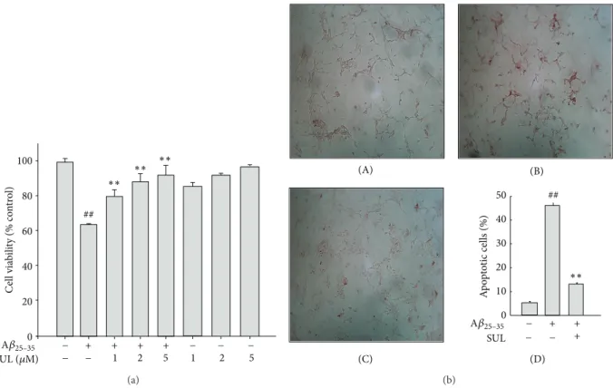
Figure 1: Protective effect of sulforaphane (SUL) on A𝛽
25–35-induced cytotoxicity and apoptotic cell death. SH-SY5Y cells were incubated with 15 𝜇M A𝛽
25–35with or without SUL (1 𝜇M, 2 𝜇M, and 5 𝜇M) for 24 h. (a) Viable cells were determined by MTT dye reduction assay. Cell viability is expressed as the percentage of control. Data are represented as mean ± S.D. (𝑛 = 3).
##𝑃 < 0.01, control versus A𝛽
25–35and
∗∗𝑃 <
0.01, A𝛽
25–35versus A𝛽
25–35+ SUL. (b) DNA fragmentation was measured by TUNEL. (A) Vehicle-treated control; (B) A𝛽
25–35alone (15 𝜇M);
(C) A𝛽
25–35(15 𝜇M) + SUL (5 𝜇M); (D) quantification of apoptotic cell death (%).
followed by Tukey’s test as post hoc analysis to determine indi- vidual group differences. Statistical significance was accepted at value of 𝑃 less than 0.05.
3. Results
3.1. Protective Effect of SUL against A𝛽
25–35-Induced Cyto- toxicity and Apoptotic Cell Death. We have investigated the effect of SUL on A𝛽
25–35-induced cytotoxicity and apop- totic cell death in SH-SY5Y cells by MTT dye reduction assay and TUNEL staining, respectively. Cells were incu- bated with various concentrations of SUL (1 𝜇M, 2 𝜇M, and 5 𝜇M) for 30 min followed by 15 𝜇M A𝛽
25–35treatment for additional 24 h. Pretreatment of SUL protected against A𝛽
25–35-induced cytotoxicity in a concentration-dependent manner (Figure 1(a)). SUL-treated cells exhibited signifi- cantly higher cell viability than A𝛽
25–35-treated group did.
In addition, A𝛽
25–35-induced apoptotic cell death was effec- tively suppressed by the pretreatment with SUL as assessed by TUNEL, which detects DNA fragmentation in situ, a typical marker for apoptosis (Figure 1(b)). SUL significantly reduced the number of TUNEL-positive cells caused by A𝛽
25–35treatment.
We also confirmed the protective effect of SUL against A𝛽
25–35-induced apoptotic cell death by examining pro- or antiapoptotic signals, such as activation of JNK, expression
of Bcl-2 family proteins, and dissipation of mitochondrial membrane potential (MMP). A𝛽
25–35-induced apoptosis of SH-SY5Y cells was accompanied by activation of JNK via phosphorylation (Figure 2(a)) and a decreased Bcl-2 as well as an increased Bax protein levels (Figure 2(b)). How- ever, pretreatment of SUL dramatically reduced A𝛽
25–35- elevated phosphorylation of JNK and expression of pro- apoptotic protein Bax. Moreover, anti-apoptotic protein Bcl- 2 levels were effectively upregulated by SUL pretreatment.
A𝛽
25–35treatment also led to disruption of MMP as assessed by using TMRE cationic probe, which was shown as low fluo- rescence intensity compared with control group (Figure 2(c)).
However, SUL pretreatment effectively restored A𝛽
25–35- decreased TMRE fluorescence intensity up to control levels representing recovery from the dissipation of MMP.
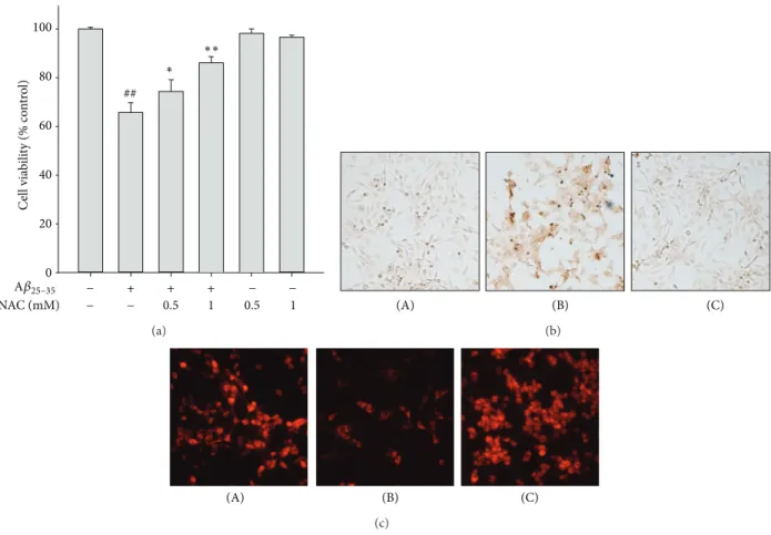
3.2. Inhibitory Effect of SUL on A𝛽
25–35-Induced ROS Produc-
tion and Subsequent Oxidative Damages. It has been reported
that A𝛽
25–35-induced cytotoxicity and apoptotic cell death are
mediated by oxidative stress. In another experiment, A𝛽
25–35-
induced cytotoxicity (Figure 3(a)) and apoptotic cell death
such as DNA fragmentation (Figure 3(b)) and impairment of
MMP (Figure 3(c)) were effectively suppressed by pretreat-
ment with NAC (0.5 mM and 1 mM), a glutathione (GSH)
precursor with strong antioxidant activity. Based on the
involvement of oxidative stress in A𝛽
25–35-induced apoptosis
p-JNK
JNK
0 0.4 0.8 1.2 1.6 2
p-JNK/JNK levels (fold induction)
− + + +
− −
A𝛽25–35
SUL (𝜇M) 2 5
− + + +
− − 2 5
A𝛽25–35
SUL (𝜇M) (a)
Bcl-2
Actin Bax
0 2 4 6 8 10
Bax/Bcl-2 levels (fold induction)
− + + +
− − 2 5
− + + +
− − 2 5
A𝛽25–35
SUL (𝜇M) A𝛽25–35
SUL (𝜇M)
(b)
(B)
(A) (C)
(c)
Figure 2: Protective effect of SUL on A𝛽
25–35-induced pro-apoptotic signals. SH-SY5Y cells were exposed to 15 𝜇M of A𝛽
25–35in the presence or absence of SUL (2 𝜇M and 5 𝜇M) for 24 h. Activation of JNK (a) and expression of Bcl-2 family proteins (b) were assessed by western blot analysis using anti-phospho-JNK, anti-JNK, anti-Bcl-2, anti-Bax, and anti-actin antibodies. Actin levels were monitored to verify equal amount of protein loading. Relative expression levels of p-JNK/JNK and Bax/Bcl-2 were quantified from three independent experiments and are represented on the right panels. (c) Mitochondria membrane potential was measured by immunofluorescence staining using TMRE probe.
The representative images of TMRE fluorescence were shown. (A) Vehicle-treated control; (B) A𝛽
25–35alone (15 𝜇M); (C) A𝛽
25–35(15 𝜇M) + SUL (5 𝜇M).
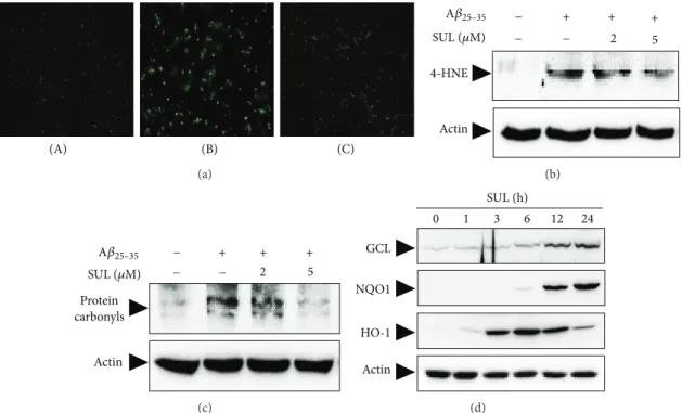
in SH-SY5Y cells, in the next experiment we have examined the effect of SUL on A𝛽
25–35-induced ROS formation. Cells were pretreated with SUL for 30 min before incubation with A𝛽
25–35(15 𝜇M) for additional 6 h. A𝛽
25–35treatment led to intracellular accumulation of ROS, which was attenuated by SUL pretreatment (Figure 4(a)) as assessed by relative fluorescence intensity of DCF-DA dye. The results indicated that SUL could inhibit A𝛽
25–35-induced ROS production in SH-SY5Y cells.
It is well known that ROS can cause oxidative stress to critical cellular macromolecules such as DNA, protein, and lipids. In the present study, treatment of A𝛽
25–35(15 𝜇M) caused oxidative damages to lipids (Figure 4(b)) and proteins in SH-SY5Y cells (Figure 4(c)), which were measured by formation of 4-HNE and protein carbonyls, respectively.
4-HNE and protein carbonyls are indicators of oxidative stress and key markers for oxidation of lipid and protein.
A𝛽
25–35-induced lipid peroxidation (Figure 4(b)) and protein
NAC (mM) 0.5 1 0.5 1
Cell viability (% control)
0 20 40 60 80 100
##
−
−
− + + +
− −
∗∗
∗
A𝛽25–35
(a)
(B)
(A) (C)
(b)
(B)
(A) (C)
(c)
Figure 3: Protective effect of NAC on A𝛽
25–35-induced apoptotic cell death. SH-SY5Y cells were treated with A𝛽
25–35(15 𝜇M) with or without NAC (0.5 mM and 1 mM) for 24 h. (a) Cell viability was measured and calculated by MTT dye reduction assay. Data are shown as mean ± S.D.
(𝑛 = 3).
##𝑃 < 0.01, control versus A𝛽
25–35and
∗𝑃 < 0.05 or
∗∗𝑃 < 0.01, A𝛽
25–35versus A𝛽
25–35+ NAC. (b) Apoptotic cell death was examined by TUNEL staining. (A) Vehicle-treated control; (B) A𝛽
25–35alone (15 𝜇M); (C) A𝛽
25–35(15 𝜇M) + NAC (1 mM). (c) MMP was monitored by relative TMRE fluorescence intensity. (A) Vehicle-treated control; (B) A𝛽
25–35alone (15 𝜇M); (C) A𝛽
25–35(15 𝜇M) + NAC (1 mM).
oxidation (Figure 4(c)) were substantially reduced by pre- treatment of these cells with SUL.
3.3. Augmentation of Cellular Antioxidant Defense Capacity by SUL via Activation of Nrf2. To investigate molecular mech- anisms of neuroprotection exerted by SUL against A𝛽
25–35- induced oxidative cell death, we have assessed expression levels of cellular antioxidant enzymes such as GCS, NQO- 1, and HO-1. SH-SY5Y cells were treated with 5 𝜇M SUL for the indicated time periods, and protein levels of GCS, NQO- 1, and HO-1 were determined by western blot analysis using specific antibodies. As shown in Figure 4(d), the expression of GCS and NQO-1 was increased by SUL treatment in a time- dependent manner which peaked at 24 h. In addition, HO- 1 protein levels increased from 3 h after SUL treatment and were maintained up to 12 h (Figure 4(d)). All these results indicated that SUL could induce the expression of antioxidant enzymes to protect cells from oxidative damages caused by A𝛽
25–35in SH-SY5Y cells.
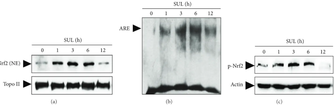
To elucidate upstream regulator for the SUL-induced up- regulation of the antioxidant enzymes, we have focused on the activation of redox-sensitive transcription factor Nrf2.
When SH-SY5Y cells were treated with 5 𝜇M SUL for the indi- cated times, nuclear translocation (Figure 5(a)), ARE-DNA binding (Figure 5(b)), and phosphorylation (Figure 5(c)) of Nrf2 were assessed by western blot analysis and EMSA.
Treatment of SH-SY5Y cells with SUL increased nuclear levels of Nrf2 (Figure 5(a)) and Nrf2 binding to ARE pro- moter sequence (Figure 5(b)) with similar kinetic patterns.
Moreover, SUL treatment increased phosphorylation of Nrf2 at Ser-40 residue as well (Figure 5(c)), which is known to facilitate the dissociation of Nrf2 from Keap1 rendering its translocation to nucleus.
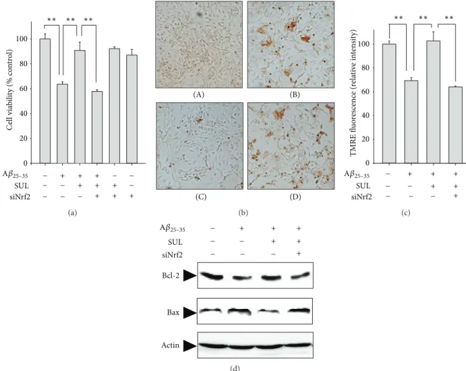
To further verify the direct role of Nrf2 in mediating the cytoprotective effect of SUL against A𝛽
25–35-induced oxida- tive cell death, we have downregulated the Nrf2 expression by transient transfection of SH-SY5Y cells with Nrf2-siRNA. The cellular protection of SUL on A𝛽
25–35-induced cytotoxicity (Figure 6(a)) and DNA fragmentation (Figure 6(b)) were abolished by knockdown of Nrf2 gene with Nrf2-siRNA.
Moreover, the protective effect of SUL on A𝛽
25–35-mediated
proapoptotic signals such as decreased MMP (Figure 6(c))
and increased Bax/Bcl-2 ratio (Figure 6(d)) and subsequent
oxidative damages to lipids determined by 4-HNE formation
(data not shown) were substantially abrogated by Nrf-siRNA
(B)
(A) (C)
(a)
Actin 4-HNE
−
− −
+ + +
A
2 5
𝛽25–35
SUL (𝜇M)
(b)
Protein carbonyls
Actin
−
− −
+ + +
A
2 5
𝛽25–35
SUL (𝜇M)
(c)
Actin GCL
SUL (h)
NQO1
HO-1
0 1 3 6 12 24
(d)
Figure 4: Inhibitory effect of SUL on the A𝛽
25–35-induced intracellular accumulation of ROS and oxidative stress. (a) SH-SY5Y cells were treated with 15 𝜇M A𝛽
25–35for 6 h with or without SUL pretreatment for 30 min. Intracellular ROS levels were monitored by using DCF-DA fluorescence dye. (A) Vehicle-treated control; (B) A𝛽
25–35alone (15 𝜇M); (C) A𝛽
25–35(15 𝜇M) + SUL (5 𝜇M). ((b)-(c)) SH-SY5Y cells were exposed to 15 𝜇M A𝛽
25–35in the presence or absence of SUL (2 𝜇M and 5 𝜇M) for 24 h. Molecular markers for oxidative damages such as lipid peroxidation (b) and protein oxidation (c) were determined by western blot analysis and protein carbonyl assay as described in Section 2. (d) The protein expression of antioxidant enzymes for example GCS, NQO-1, and HO-1 was evaluated by western blotting using their specific antibodies. Actin levels were assessed to confirm the equal amount of protein loaded.
transfection. These results suggest a critical role of Nrf2 in SUL-mediated protection against A𝛽
25–35-induced apoptotic cell death.
4. Discussion
In this study, we have examined the protective effect and molecular mechanism of SUL against A𝛽-induced oxidative and apoptotic cell death. The results from the MTT assay and apoptotic analysis (TUNEL) provided a direct evidence demonstrating that SUL could protect SH-SY5Y cells from A𝛽
25–35-induced toxicity through increasing cell viability as well as inhibiting the apoptotic cell death. We also have assessed the effect of SUL on the A𝛽
25–35-induced pro- apoptotic signals such as activation of JNK and increased ratio of Bax to Bcl-2. Pretreatment of SUL elevated the anti- apoptotic Bcl-2 protein levels, decreased the pro-apoptotic Bax protein expression, and attenuated JNK activation via inhibition of its phosphorylation.
It has been reported that A𝛽
25–35-induced cytotoxicity was mediated by oxidative stress. The excessive produc- tion of ROS by A𝛽
25–35and exhaustion of the endogenous antioxidant defense system including GSH, catalase, super- oxide dismutase, and glutathione metabolizing enzymes can cause oxidative damages to critical cellular macromolecules,
mitochondrial dysfunction, and altered cellular signal trans- duction cascades. In the present study, A𝛽
25–35treatment led to intracellular accumulation of ROS in SH-SY5Y cells, which was effectively inhibited by pretreatment with SUL.
Moreover, SUL could alleviate A𝛽
25–35-induced oxidative damages including formation of 4-HNE and protein car- bonyls through decreasing ROS production. Dissipation of MMP reflects the opening of the mitochondrial permeability transition pore due to the ROS release from mitochondria [19]. In this study, during the apoptotic cell death induced by A𝛽
25–35, MMP generated by the gradient of ion concen- trations between two sides of the mitochondrial membrane was decreased, whereas SUL pretreatment restored the dissi- pation of MMP. In accordance with our finding, it has been reported that SUL increases the resistance of liver mitochon- dria to redox-regulated permeability transition pore opening and elevates expression of antioxidant proteins involved in mitochondrial defense against oxidative stress [20].
As the accumulation of ROS can trigger imbalance of redox state, neuronal cells have a set of antioxidant defense enzymes that maintain homeostasis between them.
Therefore, one way to render neuronal cells more resistant
to A𝛽-induced oxidative cell death is to potentiate the
endogenous antioxidant defense system, for instance, to up-
regulate an array of antioxidant enzymes. In the present
Nrf2 (NE)
SUL (h)
Topo II
0 1 3 6 12
(a)
ARE
SUL (h)
0 1 3 6 12
(b)
p-Nrf2
Actin
SUL (h)
0 1 3 6 12
(c)
Figure 5: SUL-induced activation of Nrf2 in SH-SY5Y cells. Cells were incubated with SUL (5 𝜇M) for indicated times and nuclear as well as total protein sample extracts were prepared. (a) Nuclear translocation of Nrf2 was monitored by western blotting of nuclear extracts by probing with anti-Nrf2 specific antibody. (b) Nrf2-ARE binding activity was measured by EMSA according to the manufacturer’s instruction using biotin-labeled oligonucleotide specific for Nrf2. (c) Phosphorylation of Nrf2 was assessed by western blot analysis with anti-phospho- Nrf2 (Ser40) antibody. The levels of Topo II (a) and actin (c) were examined to ensure equal amount of nuclear and total protein as loading controls, respectively.
study, treatment of SUL elevated the protein expression of antioxidant enzymes such as GCL, NQO-1, and HO-1. GCL is a rate-limiting enzyme for biosynthesis of GSH which is a representative endogenous antioxidant molecule and plays an important role in cellular defense against oxidative stress [21]. Because homeostasis of GSH and GSH-dependent enzymes are considered to be key determinants of antiox- idant protection, dysregulation of GSH-related antioxidant network might bring about the initiation and progression of neurodegenerative diseases where oxidative stress is one of critical causes [22].
NQO-1 is a cytosolic flavoprotein that catalyzes the two- electron reduction of quinones to the redox-stable hydro- quinones, preventing their redox cycling and eventually gen- erating the ROS [23]. Increasing evidence supports the role of NQO-1 as a safety valve to sequester ROS and prevent severe oxidative damages in various neuronal disorders including AD [23, 24]. HO-1, known as heat shock protein 32, plays a crucial role in endogenous defense against oxidative stimuli- induced brain injuries by decomposing toxic heme into carbon monoxide, iron, and biliverdin [25]. Biliverdin is subsequently converted into bilirubin through the action of biliverdin reductase and these two molecules serve as potent radical scavengers protecting cells from oxidative damages.
The pharmacological up-regulation of HO-1 expression in brain regions showed promising therapeutic effects in the models of neurodegenerative diseases and brain infections [26].
To further elucidate the upstream regulators for the induction of endogenous antioxidant defense enzymes against oxidative stress, we have focused on the Nrf2-ARE signaling pathway. Recently, abundant evidence suggests the protective functions of Nrf2 and Nrf2-regulated gene prod- ucts in diverse neuronal disorders [27, 28]. Considering that Nrf2 mediates general antioxidant responses, Nrf2 could be a potential therapeutic target for neurodegenerative diseases, where cells are suffering from chronic state of oxidative
stress. Under normal quiescent state, Nrf2 is sequestered in the cytoplasm by a cytoskeletal associated specific negative regulator, Kelch-like ECH associating protein 1 (Keap1).
Upon exposure to ROS or xenobiotics, Nrf2 is liberated from Keap1, translocates from the cytosol to the nucleus, heterodimerizes with accessory proteins such as small Maf protein family, and sequentially binds to antioxidant response element (ARE) promoter region. The binding of Nrf2 to ARE induces the production of diverse antioxidant enzyme and phase II detoxifying genes such as GCL, glutathione-S- transferase (GST), UDP-glycosyltransferases (UGTs), HO-1, and NQO1, which protect cells against oxidative stress as well as a wide range of other toxins [27, 28].
In the present study, the cytoprotective effect of SUL against A𝛽
25–35-induced oxidative damage and cell death seemed to be mediated by up-regulation of antioxidant enzymes through Nrf2 activation. SUL has been considered as an indirect antioxidant because of its ability solely to induce many cytoprotective antioxidant enzymes through the Nrf2-ARE pathway [29]. Induction of the Nrf2-ARE pathway by SUL has been reported to prevent cytotoxicity caused by oxygen and glucose deprivation [15, 30], 6-OHDA [16, 31], superoxide [17], H
2O
2and glutamate [18], 5-S- cysteinyl-dopamine [32], or A𝛽
1–42[33] in neuronal cell lines as well as primary cultures. Furthermore, activation of the Nrf2-ARE pathway is able to protect against brain injuries in the animal models of neurodegenerative diseases [34, 35], spinal cord injury [36–38], focal cerebral ischemia [39], hypoxia-ischemic injury [14], traumatic brain injury [40], subarachnoid [41] or intracerebral hemorrhage [42], or epilepsy [43]. According to in vivo studies, strategies to potentiate Nrf2-ARE pathway by SUL were proved to be useful in improving memory impairment and cognitive dysfunction caused by traumatic brain injury [44] or A𝛽 [34].
Conversely, Nrf2 KO mice models of neurological disorders
including Parkinson’s disease [35], spinal cord injury [37],
traumatic brain injury [40], intracerebral hemorrhage [42],
Cell viability (% control)
0 20 40 60 80 100
− − −
−
−
− − −
−+ + +
+ + +
+ + +
SUL siNrf2 A𝛽25–35
∗∗
∗∗
∗∗
(a)
(B) (A)
(C) (D)
(b)
SUL siNrf2 0 20 40 60 80 100
TMRE fluorescence (relative intensity)
−
−− −− −
+ + +
+ +
+
∗∗ ∗∗ ∗∗
A𝛽25–35
(c)
Bcl-2
Actin Bax
−
−
− − −
−
+ + +
+ +
+ SUL
siNrf2 A𝛽25–35
(d)
Figure 6: Effect of Nrf2 gene knock-down on SUL-mediated protection against A𝛽
25–35-induced apoptotic cell death. SH-SY5Y cells were transiently transfected with siRNA of Nrf2 according to the protocol provided by manufacturer and then exposed to A𝛽
25–35(15 𝜇M) in the presence or absence of SUL (5 𝜇M) for 24 h. (a) MTT assay was performed to measure cell viability. Data are represented as mean ± S.D. (𝑛 = 3).
∗∗𝑃 < 0.01, significantly different between groups. (b) TUNEL staining was conducted to verify DNA fragmentation in situ. (A) Vehicle-treated control; (B) A𝛽
25–35alone (15 𝜇M); (C) A𝛽
25–35(15 𝜇M) + SUL (5 𝜇M); (D) A𝛽
25–35(15 𝜇M) + SUL (5 𝜇M) + Nrf2-siRNA. (c) TMRE staining was performed to compare MMP. Data are represented as mean ± S.D. (𝑛 = 3).
∗∗𝑃 < 0.01, significantly different between groups. (d) Protein expression of Bcl-2 and Bax was determined by western blot analysis using specific antibodies. Actin levels were examined to ensure equal amount of protein loading.
and epilepsy [43] exhibited increased susceptibility to neuro- logical oxidative damages but did not maintain any benefits from the protective effects of SUL. In our experiment, the protective effect of SUL against A𝛽
25–35-caused apoptotic cell death was abolished by down-regulation of Nrf2 gene by transient transfection with Nrf2-siRNA.
Although the molecular milieu of SUL-induced Nrf2 activation in SH-SY5Y cells has not been elucidated, two possible mechanisms for the activation of Nrf2-ARE pathway by SUL have been proposed in other types of cells. One is structural change of Keap1 due to the modification of specific cysteine residues by binding of SUL [29, 45]. The other is the phosphorylation of Nrf2 at Ser-40 residue by mitogen-activated protein kinases [46], protein kinase C [46], and phosphatidylinositol 3-kinase/Akt activated by SUL [31, 47, 48]. Nrf2 phosphorylation by aforementioned
kinases triggers the release of Nrf2 from inhibitory Keap1, thereby facilitating the Nrf2 translocation to nucleus. How- ever, phosphorylation of Nrf2 at Tyr-568 residue by GSK- 3𝛽 can promote its nuclear exclusion or proteolysis [46, 49].
Nevertheless, the molecular signaling pathways activating Nrf2 appears to be pleiotropic and dependent on cell types as well as stimuli.
5. Conclusions
In conclusion, a phytochemical SUL attenuates A𝛽
25–35-
induced oxidative stress and pro-apoptotic signals such as
activation of JNK, an increase in pro-apoptotic Bax, and a
decrease in anti-apoptotic Bcl-2, thereby inhibiting apop-
totic neuronal cell death in SH-SY5Y cells. Moreover, SUL
induced the activation of Nrf2-ARE signaling pathway, which
Phosphorylation
Upregulation of antioxidant
enzymes
(e.g., GCL, NQO-1, and HO-1) of Nrf2
SH modification
APP
Activation of Nrf2 SUL
of Keap1 ROS production
Activation of JNK
Increased Bax/Bcl-2 levels
Dissipation of MMP
Apoptosis
Lipid peroxidation
Protein oxidation
Oxidative damages A𝛽
A𝛽 A𝛽
A𝛽 𝛽-/𝛾-secratases H3C–S–CH2–CH2–CH2–CH2–N C
O
S
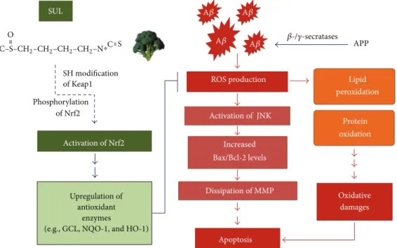
Figure 7: Schematic diagram that describes neuroprotective effects of SUL against A𝛽-induced oxidative cell death in AD. SUL attenuates A𝛽-induced oxidative damages, pro-apoptotic signals, and apoptotic cell death through the activation of Nrf2-ARE signaling pathway, which consequently fortify Nrf2-dependent antioxidant defense capacity.
consequently results in up-regulation of Nrf2-dependent antioxidant capacity, leading to reduction in the A𝛽
25–35- induced oxidative damages (Figure 7). Taken together, the results in the present study suggest pharmacologic activation of the Nrf2 signaling pathway by SUL might be a practical pre- ventative and therapeutic strategy for AD patients. However, further studies are required to obtain more insights into the molecular mechanisms of SUL-induced Nrf2 activation and clinical application of SUL.
Conflict of Interests
The authors declare that they have no conflict of interests.
Authors’ Contribution
Chan Lee and Gyu Hwan Park contributed equally to this work.
Acknowledgments
This work was supported by the National Research Foun- dation of Korea (NRF) grant funded by the Ministry of Education, Science and Technology, Korea government (nos.
2009-0067980 and 2010-0004959).
References
[1] D. M. Barten and C. F. Albright, “Therapeutic strategies for Alzheimer’s disease,” Molecular Neurobiology, vol. 37, no. 2-3, pp. 171–186, 2008.
[2] M. P. Mattson, “Pathways towards and away from Alzheimer’s disease,” Nature, vol. 430, no. 7000, pp. 631–639, 2004.
[3] M. W. Marlatt, P. J. Lucassen, G. Perry, M. A. Smith, and X. Zhu,
“Alzheimer’s disease: cerebrovascular dysfunction, oxidative stress, and advanced clinical therapies,” Journal of Alzheimer’s Disease, vol. 15, no. 2, pp. 199–210, 2008.
[4] R. Sultana, M. Perluigi, and D. A. Butterfield, “Oxidatively modified proteins in Alzheimer’s disease (AD), mild cognitive impairment and animal models of AD: role of Abeta in pathogenesis,” Acta Neuropathologica, vol. 118, no. 1, pp. 131–150, 2009.
[5] D. A. Butterfield, J. Drake, C. Pocernich, and A. Castegna, “Evi- dence of oxidative damage in Alzheimer’s disease brain: central role for amyloid 𝛽-peptide,” Trends in Molecular Medicine, vol.
7, no. 12, pp. 548–554, 2001.
[6] A. Gella and N. Durany, “Oxidative stress in Alzheimer disease,”
Cell Adhesion and Migration, vol. 3, no. 1, pp. 88–93, 2009.
[7] S.-Y. Park, “Potential therapeutic agents against Alzheimer’s disease from natural sources,” Archives of Pharmacal Research, vol. 33, no. 10, pp. 1589–1609, 2010.
[8] P. Williams, A. Sorribas, and M.-J. R. Howes, “Natural products as a source of Alzheimer’s drug leads,” Natural Product Reports, vol. 28, no. 1, pp. 48–77, 2011.
[9] K. L. Cheung and A.-N. Kong, “Molecular targets of dietary phenethyl isothiocyanate and sulforaphane for cancer chemo- prevention,” AAPS Journal, vol. 12, no. 1, pp. 87–97, 2010.
[10] B. M. Kaminski, D. Steinhilber, J. M. Stein, and S. Ulrich, “Phy- tochemicals resveratrol and sulforaphane as potential agents for enhancing the anti-tumor activities of conventional cancer therapies,” Current Pharmaceutical Biotechnology, vol. 13, no. 1, pp. 137–146, 2012.
[11] C. E. Guerrero-Beltr´an, M. Calder´on-Oliver, J. Pedraza-Chaver-
ri, and Y. I. Chirino, “Protective effect of sulforaphane against
oxidative stress: recent advances,” Experimental and Toxicologic Pathology, vol. 64, no. 5, pp. 503–508, 2012.
[12] N. A. Kelsey, H. M. Wilkins, and D. A. Linseman, “Nutraceutical antioxidants as novel neuroprotective agents,” Molecules, vol. 15, no. 11, pp. 7792–7814, 2010.
[13] N. G. Innamorato, A. I. Rojo, ´ A. J. Garc´ıa-Yag¨ue, M. Yamamoto, M. L. De Ceballos, and A. Cuadrado, “The transcription factor nrf2 is a therapeutic target against brain inflammation,” Journal of Immunology, vol. 181, no. 1, pp. 680–689, 2008.
[14] Z. Ping, W. Liu, Z. Kang et al., “Sulforaphane protects brains against hypoxic-ischemic injury through induction of Nrf2- dependent phase 2 enzyme,” Brain Research, vol. 1343, pp. 178–
185, 2010.
[15] C. A. Danilov, K. Chandrasekaran, J. Racz, L. Soane, C. Zielke, and G. Fiskum, “Sulforaphane protects astrocytes against oxida- tive stress and delayed death caused by oxygen and glucose deprivation,” GLIA, vol. 57, no. 6, pp. 645–656, 2009.
[16] A. Siebert, V. Desai, K. Chandrasekaran, G. Fiskum, and M. S. Jafri, “Nrf2 activators provide neuroprotection against 6-hydroxydopamine toxicity in rat organotypic nigrostriatal cocultures,” Journal of Neuroscience Research, vol. 87, no. 7, pp.
1659–1669, 2009.
[17] P. Bergstr¨om, H. C. Andersson, Y. Gao et al., “Repeated transient sulforaphane stimulation in astrocytes leads to prolonged Nrf2- mediated gene expression and protection from superoxide- induced damage,” Neuropharmacology, vol. 60, no. 2-3, pp. 343–
353, 2011.
[18] A. D. Kraft, D. A. Johnson, and J. A. Johnson, “Nuclear factor E2-related factor 2-dependent antioxidant response element activation by tert-butylhydroquinone and sulforaphane occur- ring preferentially in astrocytes conditions neurons against oxidative insult,” Journal of Neuroscience, vol. 24, no. 5, pp. 1101–
1112, 2004.
[19] B. Mignotte and J.-L. Vayssiere, “Mitochondria and apoptosis,”
European Journal of Biochemistry, vol. 252, no. 1, pp. 1–15, 1998.
[20] T. Greco, J. Shafer, and G. Fiskum, “Sulforaphane inhibits mitochondrial permeability transition and oxidative stress,”
Free Radical Biology and Medicine, vol. 51, no. 12, pp. 2164–2171, 2011.
[21] K. Aoyama, M. Watabe, and T. Nakaki, “Regulation of neuronal glutathione synthesis,” Journal of Pharmacological Sciences, vol.
108, no. 3, pp. 227–238, 2008.
[22] W. M. Johnson, A. L. Wilson-Delfosse, and J. J. Mieyal, “Dys- regulation of glutathione homeostasis in neurodegenerative diseases,” Nutrients, vol. 4, no. 10, pp. 1399–1440, 2012.
[23] A. K. Raina, D. J. Templeton, J. C. Deak, G. Perry, and M. A.
Smith, “Quinone reductase (NQO1), a sensitive redox indicator, is increased in Alzheimer’s disease,” Redox Report, vol. 4, no. 1-2, pp. 23–27, 1999.
[24] Y. Wang, K. Santa-Cruz, C. Decarli, and J. A. Johnson,
“NAD(P)H:quinone oxidoreductase activity is increased in hip- pocampal pyramidal neurons of patients with Alzheimer’s disease,” Neurobiology of Aging, vol. 21, no. 4, pp. 525–531, 2000.
[25] A. Jazwa and A. Cuadrado, “Targeting heme oxygenase-1 for neuroprotection and neuroinflammation in neurodegenerative diseases,” Current Drug Targets, vol. 11, no. 12, pp. 1517–1531, 2010.
[26] A. Cuadrado and A. I. Rojo, “Heme oxygenase-1 as a therapeutic target in neurodegenerative diseases and brain infections,”
Current Pharmaceutical Design, vol. 14, no. 5, pp. 429–442, 2008.
[27] G. Joshi and J. A. Johnson, “The Nrf2-ARE pathway: a valuable therapeutic target for the treatment of neurodegenerative dis- eases,” Recent Patents on CNS Drug Discovery, vol. 7, no. 3, pp.
218–229, 2012.
[28] F. L. van Muiswinkel and H. B. Kuiperij, “The Nrf2-ARE signalling pathway: promising drug target to combat oxidative stress in neurodegenerative disorders,” Current Drug Targets, vol. 4, no. 3, pp. 267–281, 2005.
[29] A. T. Dinkova-Kostova and P. Talalay, “Direct and indirect antioxidant properties of inducers of cytoprotective proteins,”
Molecular Nutrition and Food Research, vol. 52, supplement 1, pp. S128–S138, 2008.
[30] L. Soane, W. Li Dai, G. Fiskum, and L. L. Bambrick, “Sul- foraphane protects immature hippocampal neurons against death caused by exposure to hemin or to oxygen and glucose deprivation,” Journal of Neuroscience Research, vol. 88, no. 6, pp.
1355–1363, 2010.
[31] C. Deng, R. Tao, S.-Z. Yu, and H. Jin, “Sulforaphane protects against 6-hydroxydopamine-induced cytotoxicity by increasing expression of heme oxygenase-1 in a PI3K/Akt-dependent manner,” Molecular Medicine Reports, vol. 5, no. 3, pp. 847–851, 2012.
[32] D. Vauzour, M. Buonfiglio, G. Corona et al., “Sulforaphane pro- tects cortical neurons against 5-S-cysteinyl-dopamine-induced toxicity through the activation of ERK1/2, NrF-2 and the upregulation of detoxification enzymes,” Molecular Nutrition and Food Research, vol. 54, no. 4, pp. 532–542, 2010.
[33] H.-M. Park, J.-A. Kim, and M.-K. Kwak, “Protection against amyloid beta cytotoxicity by sulforaphane: role of the protea- some,” Archives of Pharmacal Research, vol. 32, no. 1, pp. 109–115, 2009.
[34] H. V. Kim, H. Y. Kim, H. Y. Ehrlich, S. Y. Choi, D. J. Kim, and Y.
Kim, “Amelioration of Alzheimer’s disease by neuroprotective effect of sulforaphane in animal model,” Amyloid, vol. 20, no. 1, pp. 7–12, 2012.
[35] A. Jazwa, A. I. Rojo, N. G. Innamorato, M. Hesse, J. Fern´andez- Ruiz, and A. Cuadrado, “Pharmacological targeting of the transcription factor NRf2 at the basal ganglia provides disease modifying therapy for experimental parkinsonism,” Antioxi- dants and Redox Signaling, vol. 14, no. 12, pp. 2347–2360, 2011.
[36] A. Jazwa, A. I. Rojo, N. G. Innamorato, M. Hesse, J. Fernandez- Ruiz, and A. Cuadrado, “Pharmacological targeting of the transcription factor Nrf2 at the basal ganglia provides disease modifying therapy for experimental parkinsonism,” Antioxi- dants & Redox Signaling, vol. 14, no. 12, pp. 2347–2360, 2011.
[37] L. Mao, H. Wang, X. Wang, H. Liao, and X. Zhao, “Transcription factor Nrf2 protects the spinal cord from inflammation pro- duced by spinal cord injury,” Journal of Surgical Research, vol.
170, no. 1, pp. e105–e115, 2011.
[38] X. Wang, J. P. d. R. Vaccari, H. Wang et al., “Activation of the nuclear factor E2-related factor 2/antioxidant response element pathway is neuroprotective after spinal cord injury,” Journal of Neurotrauma, vol. 29, no. 5, pp. 936–945, 2012.
[39] J. Zhao, N. Kobori, J. Aronowski, and P. K. Dash, “Sulforaphane reduces infarct volume following focal cerebral ischemia in rodents,” Neuroscience Letters, vol. 393, no. 2-3, pp. 108–112, 2006.
[40] Y. Hong, W. Yan, S. Chen, C. R. Sun, and J. M. Zhang, “The
role of Nrf2 signaling in the regulation of antioxidants and
detoxifying enzymes after traumatic brain injury in rats and
mice,” Acta Pharmacologica Sinica, vol. 31, no. 11, pp. 1421–1430,
2010.
[41] G. Chen, Q. Fang, J. Zhang, D. Zhou, and Z. Wang, “Role of the Nrf2-ARE pathway in early brain injury after experimental subarachnoid hemorrhage,” Journal of Neuroscience Research, vol. 89, no. 4, pp. 515–523, 2011.
[42] X. Zhao, G. Sun, J. Zhang et al., “Transcription factor Nrf2 protects the brain from damage produced by intracerebral hemorrhage,” Stroke, vol. 38, no. 12, pp. 3280–3286, 2007.
[43] A. I. Rojo, P. Rada, J. Egea, A. O. Rosa, M. G. L´opez, and A.
Cuadrado, “Functional interference between glycogen synthase kinase-3 beta and the transcription factor Nrf2 in protection against kainate-induced hippocampal celldeath,” Molecular and Cellular Neuroscience, vol. 39, no. 1, pp. 125–132, 2008.
[44] P. K. Dash, J. Zhao, S. A. Orsi, M. Zhang, and A. N. Moore, “Sul- foraphane improves cognitive function administered following traumatic brain injury,” Neuroscience Letters, vol. 460, no. 2, pp.
103–107, 2009.
[45] C. Hu, A. L. Eggler, A. D. Mesecar, and R. B. Van Breemen,
“Modification of Keap1 cysteine residues by sulforaphane,”
Chemical Research in Toxicology, vol. 24, no. 4, pp. 515–521, 2011.
[46] Y.-S. Keum, “Regulation of the Keap1/Nrf2 system by chemo- preventive sulforaphane: implications of posttranslational mod- ifications,” Annals of the New York Academy of Sciences, vol. 1229, no. 1, pp. 184–189, 2011.
[47] E. Leoncini, M. Malaguti, C. Angeloni, E. Motori, D. Fabbri, and S. Hrelia, “Cruciferous vegetable phytochemical sulforaphane affects phase II enzyme expression and activity in rat cardiomy- ocytes through modulation of Akt signaling pathway,” Journal of Food Science, vol. 76, no. 7, pp. H175–H181, 2011.
[48] L. Wang, Y. Chen, P. Sternberg, and J. Cai, “Essential roles of the PI3 kinase/Akt pathway in regulating Nrf2-dependent antioxidant functions in the RPE,” Investigative Ophthalmology and Visual Science, vol. 49, no. 4, pp. 1671–1678, 2008.
[49] M. Salazar, A. I. Rojo, D. Velasco, R. M. De Sagarra, and A.
Cuadrado, “Glycogen synthase kinase-3𝛽 inhibits the xenobi-
otic and antioxidant cell response by direct phosphorylation
and nuclear exclusion of the transcription factor Nrf2,” Journal
of Biological Chemistry, vol. 281, no. 21, pp. 14841–14851, 2006.
Submit your manuscripts at http://www.hindawi.com
Stem Cells International
Hindawi Publishing Corporation
http://www.hindawi.com Volume 2014
Hindawi Publishing Corporation
http://www.hindawi.com Volume 2014
INFLAMMATION
Hindawi Publishing Corporation
http://www.hindawi.com Volume 2014
Behavioural Neurology
Endocrinology
International Journal ofHindawi Publishing Corporation
http://www.hindawi.com Volume 2014
Hindawi Publishing Corporation
http://www.hindawi.com Volume 2014
Disease Markers
Hindawi Publishing Corporation
http://www.hindawi.com Volume 2014
BioMed
Research International
Oncology
Journal ofHindawi Publishing Corporation
http://www.hindawi.com Volume 2014
Hindawi Publishing Corporation
http://www.hindawi.com Volume 2014
Oxidative Medicine and Cellular Longevity
Hindawi Publishing Corporation
http://www.hindawi.com Volume 2014
PPAR Research The Scientific World Journal
Hindawi Publishing Corporation
http://www.hindawi.com Volume 2014
Immunology Research
Hindawi Publishing Corporation
http://www.hindawi.com Volume 2014
Journal of
Obesity
Journal ofHindawi Publishing Corporation
http://www.hindawi.com Volume 2014
Hindawi Publishing Corporation
http://www.hindawi.com Volume 2014
Computational and Mathematical Methods in Medicine
Ophthalmology
Journal ofHindawi Publishing Corporation
http://www.hindawi.com Volume 2014
Diabetes Research
Journal ofHindawi Publishing Corporation
http://www.hindawi.com Volume 2014
Hindawi Publishing Corporation
http://www.hindawi.com Volume 2014
Research and Treatment
AIDS
Hindawi Publishing Corporation
http://www.hindawi.com Volume 2014
Gastroenterology Research and Practice
Hindawi Publishing Corporation
http://www.hindawi.com Volume 2014
Parkinson’s Disease
Evidence-Based Complementary and Alternative Medicine
Volume 2014 Hindawi Publishing Corporation
http://www.hindawi.com