Korean J Anesthesiol Vol. 49, No. 6, December, 2005
INTRODUCTION
There has been a revival of interest in low-flow anesthesia (LFA) techniques over the past decade or so, primarily driven by the introduction of newer anesthetic agents with low solubility and relatively higher costs. The advantages of LFA include a reduction in the consumption of anesthetic gases, reduced operating room and environmental pollution, and the conservation of heat and humidity within the respiratory tract of patients.
1-3)Historically, most anesthesiologists have been trained to use high-flow an- esthesia (HFA), and the anesthetic equipment and available anesthetic agents have not been suitable for LFA. HFA techniques, however, have the potential for inducing hypoxia and/or hyper- capnia, administration of an over- or underdosage of anesthetic gases, and the accumulation of potentially toxic degradation
products.
4-7)Laparoscopic cholecystectomy (LC) with intra-abdominal CO
2insufflation has a special anesthetic consideration in that it may induce hypercapnia, acidemia, and depressed hemodynamics.
8)A number of studies have been published evaluating the use of LFA in a wide variety of surgical procedures, including gastrointestinal, gynechological and general surgery.
9-11)Despite many advantages of the LFA as mentioned above, few studies have actually been done using LFA with sevoflurane in LC. One likely explanation is the potential for hypercapnia with LFA resulting from rebreathing exhaled CO
2coupled with an increase in arterial CO
2from the intraperitoneal CO
2insufflation.
The purpose of this study was to investigate whether the LFA is safe and compatible with the LC involving abdominal CO
2insufflation.
MATERIALS AND METHODS
After obtaining Institutional Review Board approval and written informed consent from each patient, 80 patients, ASA I or II status, who were to undergo elective LC under general anesthesia were
Low-flow Sevoflurane Anesthesia in Laparoscopic Cholecystectomy
Department of Anesthesiology and Pain Medicine, School of Medicine, Keimyung University, Daegu, Korea
Young Ho Jang, M.D., and Sue Rung Oh, M.D.
Background: Anesthetists participating in laparoscopic cholecystectomy (LC) with CO2 pneumoperitoneum has been cautious about adapting low-flow anesthesia (LFA). We investigated the efficacy of LFA compared to high-flow anesthesia (HFA) in LC.
Methods: Eighty patients undergoing LC were randomly assigned to one of the two groups (n = 40 each). In LFA, 1 L/min (50% O2 and N2O) of the total fresh gas flow (FGF) was used, whereas 4 L/min of the total FGF was used for HFA. Inspiratory and expiratory concentrations of O2, N2O, CO2, and sevoflurane were serially measured. Subjects were monitored for heart rate, blood pressure, and any procedural complications.
Results: None of the patients experienced any episodes of hypoxia, hypercapnia, and arrhythmia in both groups. The maximal end-tidal CO2 was 40.9 ± 3.9 mmHg in LFA and 38.2 ± 3.6 mmHg in HFA, respectively. The minimal O2 saturation was 98.3 ± 0.6% in LFA and 98.8 ± 0.7% in HFA, respectively. The inspiratory CO2 concentrations in both groups were all less than 1 mmHg throughout the anesthesia.
Conclusions: In conclusion, LFA with sevoflurane using FGF of 1 L/min with setting of 50% O2 and N2O for LC could be performed safely without the risk of complications like hypercapnia, hypoxia, or arrhythmia compared to HFA. (Korean J Anesthesiol 2005; 49: S 1∼5)
ꠏꠏꠏꠏꠏꠏꠏꠏꠏꠏꠏꠏꠏꠏꠏꠏꠏꠏꠏꠏꠏꠏꠏꠏꠏꠏꠏꠏꠏꠏꠏꠏꠏꠏꠏꠏꠏꠏꠏꠏꠏꠏꠏꠏꠏꠏꠏꠏꠏꠏꠏꠏꠏꠏꠏꠏꠏꠏꠏꠏꠏꠏꠏꠏꠏꠏꠏꠏꠏꠏꠏꠏꠏꠏꠏꠏꠏꠏꠏꠏꠏꠏꠏꠏꠏꠏꠏꠏꠏꠏꠏꠏꠏꠏꠏꠏꠏꠏꠏꠏꠏꠏꠏꠏꠏꠏꠏꠏ Key Words: fresh gas flow, laparoscopy, sevoflurane.
Received:June 26, 2005
Corresponding to:Young Ho Jang, Department of Anesthesiology and Pain Medicine, School of Medicine, Keimyung University, 194 Dongsan-dong, Jung-gu, Daegu 700-712, Korea. Tel: 82-53-250-7287, Fax: 82-53-250-7240, E-mail: [email protected]
enrolled. The patients were randomly assigned to one of two groups, LFA or HFA (n = 40 in each group), depending on fresh gas flow (FGF) settings. Patients with cardiopulmonary disease, previous adverse reactions to inhaled agents, intravenous anesthesia, obesity, smoking history, and where the procedure was subsequently changed to open cholecystectomy were excluded from the study.
Age, gender, and weight were recorded for all subjects. There was no statistically difference in patient demographics (including age, gender, weight, and anesthetic duration) between the two groups (Table 1).
All anesthetic procedures were performed by one of the two authors, who was experienced in the application of the LFA. All patients were premedicated with midazolam (7.5 mg orally), the night before the operation, and intramuscular 2-2.5 mg midazolam and 0.2 mg glycopyrrolate, 30 min before entering the operating room (OR).
After arrival in the OR, baseline hemodynamic parameters were recorded after a 5 min stabilization period on the operating table.
Before anesthetic induction, all patients breathed 100% O
2for 3 min, FGF 6 L/min, using a face mask connected to a semiclosed breathing circuit (Datex Ohmeda 7,000 ventilator, Datex-Ohmeda, USA). After preoxygenation, 2 mg/ kg propofol, 0.6 mg/kg rocuronium bromide, and 1-2μg/kg fentanyl were administered intravenously. Tracheal intubation was followed by mechanical ventilation with a tidal volume of 10 ml/kg at 10 breaths/min.
Inspiratory/expiratory ratio was set as 1:2. These respiratory parameters were not modified during the procedures. All procedures were performed using the same circle breathing system and vaporizer (Vapor 2000, Drager, Germany) with sevoflurane under standard operating room conditions. The accuracy of the flowmeters was verified by passing the FGF through a dry gas meter (Parkinson-Cowan, Birmingham, UK).
Lead II of an electrocardiogram on the patient's back and
saturation via pulse oximetry (SpO
2) at patient's finger were con-
tinuously monitored. An automated blood pressure cuff was applied to the right arm. To ensure uniform conditions, same patient monitoring equipment was used in all cases. Electrocar- diography, oscillatory blood pressure, and pulse oximetry was mo- nitored using Agilent M1205A (Agilent technologies Co., Germany). Peak airway pressure was measured using anesthesia machine (Datex Ohmeda 7,000 ventilator, Datex-Ohmeda, USA).
Tidal volume and minute ventilation were measured using 5420 volume monitor (Ohmeda, USA). We also serially measured inspiratory and expiratory fractional concentrations of the O
2, N
2O, CO
2, and sevoflurane using a multi-gas monitor (Capnomac Ultima, Datex-Ohmeda, Finland). The accuracy of multi-gas monitor was tested before the study (accuracy for ± 10%). Fresh soda lime (Sodasorb
Ⓡ, W.R. Grace & Company, USA) was used during each individual anesthesia procedure.
Variables were measured immediately prior to CO
2pneumo- peritoneum and every 5 minutes thereafter until the end of surgery but were presented 10 (T1), 20 (T2), 30 (T3) minutes after CO
2pneumoperitoneum and immediately before emergence from anesthesia (T4). Neuromuscular transmission was monitored by train-of-four stimulation (TOF Watch, Organon Teknika, Nether- lands).
After tracheal intubation, O
2and N
2O flow was 2 L/min and 2 L/min, respectively. These parameters were maintained throughout the duration of anesthesia in HFA group. The initial inspiratory sevoflurane concentration was 2.6 vol% in HFA group.
In LFA group, initial FGF was 4.5 L/min, 1.5 L/min O
2and 3.0 L/min N
2O, and an inspiratory sevoflurane concentration was 2.6 vol%. This initial high flow phase lasted 10 min to allow for initial rapid uptake of N
2O and elimination of nitrogen. Thereafter, FGF was reduced to 1 L/min, 500 ml/ min O
2and 500 ml/min N
2O, and the vaporizer dial of sevoflurane was set at 3.0 vol%. During the CO
2pneumoperitoneum, the intra-abdominal pressure was maintained between 12 and 15 mmHg by CO
2insufflator.
In both groups, sevoflurane concentration was adjusted throug- hout the anesthesia to maintain systemic arterial blood pressure and heart rate (HR) within ± 30% of baseline values. Light anesthesia was defined as tachycardia (HR > + 30% of baseline values or HR > 110 beats/min) or hypertension (mean arterial pressure [MAP] > + 30% of baseline values or MAP > 100 mmHg).
The occurrence of hypoxia and hypercapnia was noted throughout the anesthesia. Hypoxia and hypercapnia were defined as SpO
2less than 90% and end- tidal CO
2(ETCO
2) more than 50 mmHg, respectively.
Whenever the inspiratory concentration of O
2was noted to be
Table 1. Patient Demographicsꠚꠚꠚꠚꠚꠚꠚꠚꠚꠚꠚꠚꠚꠚꠚꠚꠚꠚꠚꠚꠚꠚꠚꠚꠚꠚꠚꠚꠚꠚꠚꠚꠚꠚꠚꠚꠚꠚꠚꠚꠚꠚꠚꠚꠚꠚꠚꠚꠚꠚꠚꠚꠚꠚꠚ
LFA HFA
(n = 40) (n = 40) ꠏꠏꠏꠏꠏꠏꠏꠏꠏꠏꠏꠏꠏꠏꠏꠏꠏꠏꠏꠏꠏꠏꠏꠏꠏꠏꠏꠏꠏꠏꠏꠏꠏꠏꠏꠏꠏꠏꠏꠏꠏꠏꠏꠏꠏꠏꠏꠏꠏꠏꠏꠏꠏꠏꠏ
Age (yr) 50.0 ± 11.5 48.9 ± 12.3
Sex (m/f) 18/22 20/20
Weight (kg) 64.8 ± 9.5 61.9 ± 10.4
Anesthetic duration (min) 67.0 ± 11.3 70.4 ± 12.3 ꠏꠏꠏꠏꠏꠏꠏꠏꠏꠏꠏꠏꠏꠏꠏꠏꠏꠏꠏꠏꠏꠏꠏꠏꠏꠏꠏꠏꠏꠏꠏꠏꠏꠏꠏꠏꠏꠏꠏꠏꠏꠏꠏꠏꠏꠏꠏꠏꠏꠏꠏꠏꠏꠏꠏ Data are mean ± SD except sex (number). LFA: low-flow anes- thesia, HFA: high-flow anesthesia.
low 30%, O
2flow was increased by 10% of the total FGF, while reducing the N
2O flow by the same amount. At the end of surgery, the vaporizer dial of sevoflurane was turned off and the O
2flow was increased to 6 L/min along with discontinuation of N
2O administration. Residual neuromuscular block was antag- onized at the start of skin closure by the administration of pyridostigmine and glycopyrrolate. Patients were extubated upon awakening.
Prior to the study, a power analysis was performed to determine population size based on the 3% incidence of hypercapnia during CO
2pneumoperitoneum as reported by Qureshi EA.
12)We presumed that a ± 10% incidence of hypercapnia in LFA group would be as effective and safe as the HFA group. This required recruitment of 36 patients in each group, as noted above.
Data was presented as mean ± standard deviation (SD). Data analysis was performed using the SPSS statistical Package pro- gram version 11.0 (SPSS Inc., Chicago, USA). Statistical analysis for demographics, maximal ETCO
2, and minimal SpO
2was done by using independent sample t-test. We used GLM (General Linear Model) repeated measures procedures to analyze the changes in hemodynamics and concentrations of O
2, N
2O, CO
2and sevo- flurane between two groups. Statistical significance was set at five percent level.
RESULTS
Maximal ETCO
2was 40.9 ± 3.9 mmHg in LFA group and 38.2
± 3.6 mmHg in HFA group. The minimal SpO
2was 98.3 ± 0.6%
(range 97-100%) in LFA group and 98.8 ± 0.7% (range 98- 100%) in HFA group (P = 0.005). In both groups, the inspiratory O
2concentration never dropped below 30% on the multi-gas monitor. None of the patients experienced episodes of hypoxia, hypercapnia, and arrhythmia requiring treatment in either group.
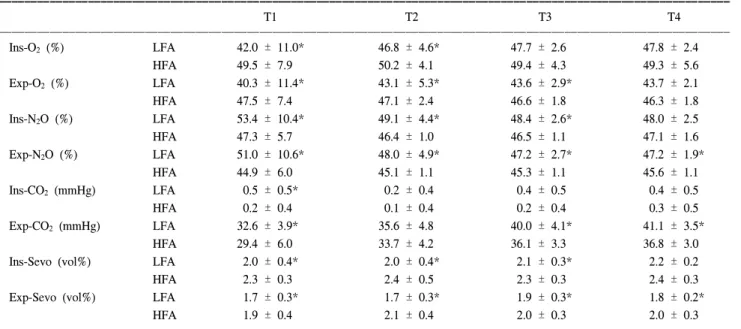
The changes in concentration of gases are presented in Table 2. The inspiratory O
2concentrations were significantly lower in LFA group than in HFA group at T1 and T2 (P = 0.004). There were no statistical differences in inspiratory O
2concentrations at T3 and T4 between two groups. The inspiratory N
2O con- centrations in LFA group were significantly higher in LFA group than in HFA group at T1, T2, and T3 (P = 0.007, 0.003 and 0.001, respectively).
The inspiratory CO
2concentration in LFA group was signifi- cantly higher than in HFA group at T1 (P = 0.032). However, the inspiratory CO
2concentrations in both groups were all less than 1 mmHg throughout the duration of anesthesia. The ETCO
2in LFA group were significantly higher in LFA group than in HFA group at T1, T3, and T4 (P = 0.016, 0.000 and 0.000). The highest
Table 2. Changes in Inspiratory and Expiratory Gases during CO2 Pneumoperitoneum
ꠚꠚꠚꠚꠚꠚꠚꠚꠚꠚꠚꠚꠚꠚꠚꠚꠚꠚꠚꠚꠚꠚꠚꠚꠚꠚꠚꠚꠚꠚꠚꠚꠚꠚꠚꠚꠚꠚꠚꠚꠚꠚꠚꠚꠚꠚꠚꠚꠚꠚꠚꠚꠚꠚꠚꠚꠚꠚꠚꠚꠚꠚꠚꠚꠚꠚꠚꠚꠚꠚꠚꠚꠚꠚꠚꠚꠚꠚꠚꠚꠚꠚꠚꠚꠚꠚꠚꠚꠚꠚꠚꠚꠚꠚꠚꠚꠚꠚꠚꠚꠚꠚꠚꠚꠚꠚꠚꠚꠚꠚꠚꠚꠚꠚꠚ
T1 T2 T3 T4
ꠏꠏꠏꠏꠏꠏꠏꠏꠏꠏꠏꠏꠏꠏꠏꠏꠏꠏꠏꠏꠏꠏꠏꠏꠏꠏꠏꠏꠏꠏꠏꠏꠏꠏꠏꠏꠏꠏꠏꠏꠏꠏꠏꠏꠏꠏꠏꠏꠏꠏꠏꠏꠏꠏꠏꠏꠏꠏꠏꠏꠏꠏꠏꠏꠏꠏꠏꠏꠏꠏꠏꠏꠏꠏꠏꠏꠏꠏꠏꠏꠏꠏꠏꠏꠏꠏꠏꠏꠏꠏꠏꠏꠏꠏꠏꠏꠏꠏꠏꠏꠏꠏꠏꠏꠏꠏꠏꠏꠏꠏꠏꠏꠏꠏꠏ
Ins-O2 (%) LFA 42.0 ± 11.0* 46.8 ± 4.6* 47.7 ± 2.6 47.8 ± 2.4
HFA 49.5 ± 7.9 50.2 ± 4.1 49.4 ± 4.3 49.3 ± 5.6
Exp-O2 (%) LFA 40.3 ± 11.4* 43.1 ± 5.3* 43.6 ± 2.9* 43.7 ± 2.1
HFA 47.5 ± 7.4 47.1 ± 2.4 46.6 ± 1.8 46.3 ± 1.8
Ins-N2O (%) LFA 53.4 ± 10.4* 49.1 ± 4.4* 48.4 ± 2.6* 48.0 ± 2.5
HFA 47.3 ± 5.7 46.4 ± 1.0 46.5 ± 1.1 47.1 ± 1.6
Exp-N2O (%) LFA 51.0 ± 10.6* 48.0 ± 4.9* 47.2 ± 2.7* 47.2 ± 1.9*
HFA 44.9 ± 6.0 45.1 ± 1.1 45.3 ± 1.1 45.6 ± 1.1
Ins-CO2 (mmHg) LFA 0.5 ± 0.5* 0.2 ± 0.4 0.4 ± 0.5 0.4 ± 0.5
HFA 0.2 ± 0.4 0.1 ± 0.4 0.2 ± 0.4 0.3 ± 0.5
Exp-CO2 (mmHg) LFA 32.6 ± 3.9* 35.6 ± 4.8 40.0 ± 4.1* 41.1 ± 3.5*
HFA 29.4 ± 6.0 33.7 ± 4.2 36.1 ± 3.3 36.8 ± 3.0
Ins-Sevo (vol%) LFA 2.0 ± 0.4* 2.0 ± 0.4* 2.1 ± 0.3* 2.2 ± 0.2
HFA 2.3 ± 0.3 2.4 ± 0.5 2.3 ± 0.3 2.4 ± 0.3
Exp-Sevo (vol%) LFA 1.7 ± 0.3* 1.7 ± 0.3* 1.9 ± 0.3* 1.8 ± 0.2*
HFA 1.9 ± 0.4 2.1 ± 0.4 2.0 ± 0.3 2.0 ± 0.3
ꠏꠏꠏꠏꠏꠏꠏꠏꠏꠏꠏꠏꠏꠏꠏꠏꠏꠏꠏꠏꠏꠏꠏꠏꠏꠏꠏꠏꠏꠏꠏꠏꠏꠏꠏꠏꠏꠏꠏꠏꠏꠏꠏꠏꠏꠏꠏꠏꠏꠏꠏꠏꠏꠏꠏꠏꠏꠏꠏꠏꠏꠏꠏꠏꠏꠏꠏꠏꠏꠏꠏꠏꠏꠏꠏꠏꠏꠏꠏꠏꠏꠏꠏꠏꠏꠏꠏꠏꠏꠏꠏꠏꠏꠏꠏꠏꠏꠏꠏꠏꠏꠏꠏꠏꠏꠏꠏꠏꠏꠏꠏꠏꠏꠏꠏ Values are mean ± SD. LFA: low-flow anesthesia, HFA: high-flow anesthesia, Ins: inspiration, Exp: expiration, Sevo: sevoflurane. T1, T2, and T3: 10, 20, and 30 minutes after carbon dioxide pneumoperitoneum, T4: immediately before emergence of anesthesia. *: P < 0.05 compared to HFA.
ETCO
2level was observed at T4 in both groups. The inspiratory and expiratory sevoflurane concentrations were lower in LFA group than in HFA group throughout the duration of anesthesia (P
< 0.05).
DISCUSSION
The LFA was first introduced by Foldes et al.
13)in 1952. Since then, the technique has been variously defined by different authors.
1,14)The purpose of this study was to examine the efficacy of LFA with sevoflurane in LC with CO
2pneumoperitoneum.
Traditionally, anesthetists participating in LC have been quite cautious about adapting LFA due to the potential for inducing hy- poxia, hypercapnia, and accumulation of toxic degradation products such as compound A by sevoflurane due to the exhaled CO
2rebreathing and administration of small amount of oxygen less than 500 ml/min.
4-7)However, it has several benefits, including the reduced consumption of anesthetic gases, reduced environmental pollution, and conservation of heat and humidity within the respiratory tract.
1-3)In the present study, a semiclosed-circuit with a soda lime ab- sorbent was used as the CO
2absorbent. All our patients were operated as the first case of the day. Therefore, the fresh soda lime absorbent was used during each anesthesia. The inspiratory CO
2concentrations in LFA and HFA groups were less than 1 mmHg throughout the anesthesia and there in no case did the ETCO
2increase more than 50 mmHg in LFA group. The duration of anesthesia was short in all our patients (mean 67.0 min in LFA and 70.4 min in HFA). These findings suggest that the clinically significant CO
2rebreathing is not developed in patients undergoing LC with a brief period of anesthesia with intraperitoneal CO
2insufflation if fresh soda lime is used in the circuit.
One another important barrier to LFA acceptance is the possi- bility of hypoxia resulting from inspiration of low O
2concen- trations during rebreathing. This may be one of the major disadvantages associated with the use of LFA. In our present study, the minimal SpO
2in LFA group was 98.3 ± 0.6% and this result was similar to that of HFA group (98.8 ± 0.7%), although it was noted to be statistically significant. No patients experienced hypoxia defined as decrease in SpO
2less than 90% in the LFA group.
Sajedi et al.
15)reported that the LFA could be used with relative safety in anesthetic m anagement of patients during LC. However, they used halothane as an inhalation agent. Sevoflurane has a number of properties that allow for the full realisation of the
benefits of LFA. The lower solubility of sevoflurane compared to halothane results in more rapid alveolar equilibration and tissue elimination, and this property is particularly well suited for use during LFA. However, the potentially toxic degradation products may be accumulated during the LFA with sevoflurane.
16)The flow rate is one of the most important factors in determining the rate at which Compound A is produced. Although we did not directly check the Compound A concentrations in our present study, none of our patients revealed any postoperative renal dysfunction clinically significant or postoperative blood urea nitrogen con- centrations that would suggest potentially toxic Compound A production.
We presumed the hypercapnia as ETCO
2without direct checking the arterial CO
2tension (PaCO
2). The diversity has been shown between PaCO
2and ETCO
2during LC.
17)However, Bhavani- Shankar et al.
18)demonstrated that the difference between PaCO
2and ETCO
2was only 2.6 ± 1.2 mmHg during CO
2pneumo- peritoneum in 8 pregnant women undergoing LC. They concluded that capnography is adequate to measure CO
2status and to guide ventilation during laparoscopic surgery. This was our justification not to directly measure PaCO
2in our patients.
In conclusion, LFA with sevoflurane using FGF of 1 L/min with setting of 50% O
2and N
2O for LC is safe without any significant likelihood of risks such as hypercapnia, hypoxia, and arrhythmia.
REFERENCES
1. Baum JA, Aitkenhead AR: Low-flow anaesthesia. Anaesthesia 1995;
50: S37-44.
2. Imberti R, Preseglio I, Imbriani M, Ghittori S, Cimino F, Mapelli A, et al: Low flow anaesthesia reduces occupational exposure to inhalation anaesthetics: environmental and biological measurements in operating room personnel. Acta Anaesthesiol Scand 1995; 39:
586-91.
3. Kleemann PP: Humidity of anaesthetic gases with respect to low flow anaesthesia. Anaesth Intensive Care 1994; 22: 396-408.
4. Liu J, Laster MJ, Eger EI 2nd, Taheri S: Absorption and degra- dation of sevoflurane and isoflurane in a conventional anesthetic circuit. Anesth Analg 1991; 72: 785-9.
5. Hargasser S, Hipp R, Breinbauer B, Mielke L, Entholzner E, Rust M, et al: A lower solubility recommends the use of desflurane more than isoflurane, halothane, and enflurane under low-flow conditions. J Clin Anesth 1995; 7: 49-53.
6. Aldrete JA: Compound A concentrations during sevoflurane anes- thesia in children depend on fresh gas flow. Anesthesiology 1996;
85: 684.
7. Meakin GH: Low-flow anaesthesia in infants and children. Br J Anaesth 1999; 83: 50-7.
8. Holzman M, Sharp K, Richards W: Hypercarbia during carbon di- oxide gas insufflation for therapeutic laparoscopy: a note of cau- tion. Surg Laparosc Endosc 1992; 2: 11-4.
9. Bito H, Ikeda K: Closed-circuit anesthesia with sevoflurane in hu- mans. Effects on renal and hepatic function and concentrations of breakdown products with soda lime in the circuit. Anesthesiology 1994; 80: 71-6.
10. Bito H, Ikeda K: Degradation products of sevoflurane during low- flow anaesthesia. Br J Anaesth 1995; 74: 56-9.
11. Lu CC, Ho ST, Wang JJ, Wong CS, Tsai CS, Chang SY, et al:
Minimal low-flow isoflurane-based anesthesia benefits patients undergoing coronary revascularization via preventing hyperglyce- mia and maintaining metabolic homeostasis. Acta Anaesthesiol Sin 2003; 41: 165-72.
12. Qureshi FA: Anesthesia related complications of laparoscopic cho- lecystectomy. J Coll Physicians Surg Pak 2003; 13: 369-71.
13. Foldes FF, Ceravolo AJ, Carpenter SL: The administration of nit- rous oxide-oxygen anesthesia in closed systems. Ann Surg 1952;
136: 978-81.
14. Gregorini P: Effect of low fresh gas flow rates on inspired gas composition in a circle absorber system. J Clin Anesth 1992; 4:
439-43.
15. Sajedi P, Naghibi K, Soltani H, Amoshahi A: A randomized, pro- spective comparison of end-tidal CO2 pressure during laparoscopic cholecystectomy in low and high flow anesthetic system. Acta Anaesthesiol Sin 2003; 41: 3-5.
16. Morio M, Fujii K, Satoh N, Imai M, Kawakami U, Mizuno T, et al: Reaction of sevoflurane and its degradation products with soda lime. Toxicity of the byproducts. Anesthesiology 1992; 77:
1155-64.
17. Bures E, Fusciardi J, Lanquetot H, Dhoste K, Richer JP, Lacoste L, et al: Ventilatory effects of laparoscopic cholecystectomy. Acta Anaesthetiol Scand 1996; 40: 566-73.
18. Bhavani-Shankar K, Steinbrook RA, Brooks DC, Datta S: Arterial to end-tidal carbon dioxide pressure difference during laparoscopic surgery in pregnancy. Anesthesiology 2000; 93: 370-3.