외사시 수술 후 정위를 보인 환자에서의 감각상태
: 외사시 수술 후 정위를 보인 환자의 감각상태를 확인하고 개선된 감각상태에 영향을 미치는 술 전 요인들을 조 사하였다.
: 외사시 진단 하에 수술받고 6개월 이상 경과 관찰 후 시행한 교대 프리즘가림 검사상 정위 혹은 4 프리 즘이내의 외편위를 보인 45명의 환자를 전향적으로 조사하였다. 수술 전, 후에 시행한 감각기능 검사는 워쓰 4점 검 사, 티트무스 검사(원), 바골리니 렌즈검사였고, 예후와 관련하여 조사한 인자는 수술 시 연령, 약시 유무, 사시각의 크 기, 감각기능 검사 결과, 굴절부등, 술 전 융합상태 등이었다.
: 술 후 감각기능 검사에서 정상을 보인 경우는 워쓰 4점 검사가 75.6%(34/45), 티트무스 검사가 77.8%(35/45), 바골리니 렌즈검사가 73.3%(33/45)였고 술 후 2가지 이상의 감각기능 검사에서 호전된 경우가 38명(84.4%)이었다.
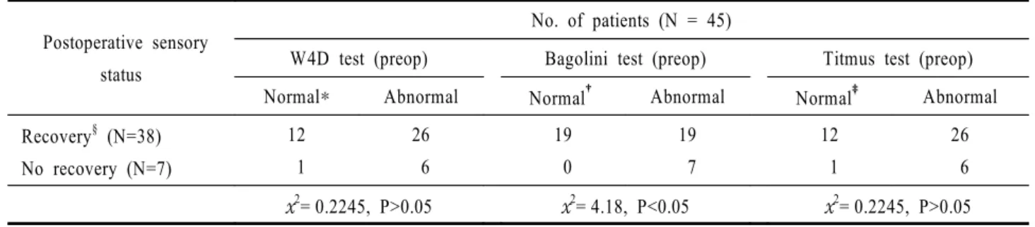
술 전에 비정상을 보인 군에서 술 후 정상을 보인 경우는 워쓰 4점 검사에서 65.6%(21/32), 바골리니 렌즈검사에서 53.8%(14/26), 티트무스 검사에서 68.8%(22/32)로 나타났다. 술 후 양안시 기능의 회복은 술 전 바골리니 렌즈검사 결과만이 술 후 양안시 기능회복의 예측과 상관관계가 있었으며( 2=4.18, P<0.05), 다른 인자에서는 유의한 차이를 보 이지 않았다(P>0.05).
: 수술 후 정위 및 4프리즘이하의 운동융합능력을 보이는 외편위 환자에서 감각기능 검사는 술 전에 비해 호전된 결과를 보였으며 술 전 바골리니 렌즈검사만이 수술 후의 감각기능회복의 예후를 판단하는데 도움이 되었다.
<한안지 44(1):128-133, 2003>
Table 2. Clinical results of Worth 4 dot test, Bagolini test & Titmus stereoacuity test No. of patients (%)
W4D test Bagolini test Titmus test
Normal Abnormal Normal Abnormal Normal Abnormal
Postoperative status Preoperative status
34 (75.6) 13 (28.9)
11 (24.4) 32 (71.1)
33 (73.3) 19 (42.2)
12 (26.7) 26 (57.8)
35 (77.8) 13 (28.9)
10 (22.2) 32 (71.1) , : defined neither suppression nor ARC at 33 cm
: defined as stereoacuity less than 100 second of arc (circle method) at 40 cm
Figure 1. Number of patients showing normal response at postoperative & preoperative examination.
Table 1. Age and sex distribution
Age No. of patients
Male Female Total
5 10 years Over than 10 years
7 8
17 13
24 21
Total 15 30 45
34
13 33
19 35
13
0 5 10 15 20 25 30 35
No. of pts.
W4D test Bagolini test Titmus test
Postoperative Preoperative
Table 4-1. Evaluation of factors influencing sensory recovery after operation
Postoperative sensory status
No. of patients (N = 45)
W4D test (preop) Bagolini test (preop) Titmus test (preop)
Normal Abnormal Normal Abnormal Normal Abnormal
Recovery§ (N=38) No recovery (N=7)
12 1
26 6
19 0
19 7
12 1
26 6
2= 0.2245, P>0.05 2= 4.18, P<0.05 2= 0.2245, P>0.05 , : defined neither suppression nor ARC at 33 cm
: defined as stereoacuity less than 100 second of arc (circle method) at 40 cm
§ : defined as normal response in more than 2 tests after operation
Table 3. Relationships between preoperative sensory status & postoperative sensory status
Preoperative examination (N = 45)
W4D test Bagolini test Titmus test
Normal (N=13)
Abnormal (N=32)
Normal (N=19)
Abnormal (N=26)
Normal (N=13)
Abnormal (N=32) Postoperative
examination
Normal Abnormal
13 0
21 11
19 0
14 12
13 0
22 10 38.2% (13/34)§
65.6% (21/32)
57.6% (19/33)§ 53.8% (14/26)
37.1% (13/35)§ 68.8% (22/32) , : defined neither suppression nor ARC at 33 cm
: defined as stereoacuity less than 100 second of arc (circle method) at 40 cm
§: No. of pts showing preop normal response / No. of pts showing postop normal response : No. of pts showing postop normal response / No. of pts showing preop abnormal response
Table 4-3. Evaluation of factors influencing sensory recovery after operation
Postoperative sensory status
No. of patients (N = 45) Amount of exodeviation
20-29 30-39 >40
Recovery (N=38) No recovery (N=7)
11 1
17 1
10 5
Total 12 18 15
P>0.05
: defined as normal response in more than 2 tests after operation Table 4-2. Evaluation of factors influencing sensory recovery after operation
Postoperative sensory status
No. of patients (N = 45)
Age Amblyopia Anisometropia
5-10 yrs >10 yrs Yes no yes no
Recovery (N=38) No recovery (N=7)
21 3
17 4
5 1
33 6
4 2
34 5
2= 0.037, P>0.05 2= 1.306, P>0.05 2= 0.786, P>0.05 : defined as normal response in more than 2 tests after operation
1) von Noorden GK. Binocular Vision and ocular motility, 5th ed. St. Louis: The C.V. Mosby Company, 1996;349.
2) . , . , 1999; 199.
3) von Noorden GK. Binocular Vision and ocular motility, 5th ed.
St. Louis: The C.V. Mosby Company, 1996; chap.2, chap. 13.
4) Rosenbaum AL, Stathacopoulos RA. Subjective and objec- tive criteria for recommending surgery in intermittent exotropia. Am Orthopt J 1992;42:41-51.
5) von Noorden GK. Binocular Vision and ocular motility, 5th ed. St. Louis: The C.V. Mosby Company, 1996;351-2.
6) Abroms AD, Monhey BG, Rush DP, Parks MM. Timely surgery in intermittent and constant exotropia for superior sensory outcome. Am J Ophthalmol 2000;131:111-6.
7) . , . , 1999; 283.
8) , . .
1999;40:538-4.
9) , , , .
. 1999;40:3174-9.
10) Stathacopoulos RA, Rosenbaum AL, Zanoni D. Distance stereoacuity. Assessing control in intermittent exotropia.
Ophthalmology 1993;100:495-500.
11) Yildrim C, Multu FM, Chen Y, Altinsoy HI. Assessment of central and peripheral fusion and near and distance stereoacuity in intermittent exotropic patients before and after strabismic surgery. Am J Ophthalmol 1999;128:220-30
12) , , .
. 2000;41:758-63.
13) Walsh LA, Laroche GR, Tremblay F. The use of binocular visual acuity in the assessment of intermittent exotropia.
JAAPOS 2000;4:154-7.
Sensory Status in Patients Showing Orthophoria After Strabismus Surgery in Exotropes
Kyeong Wook Lee, M.D.1, Se Youp Lee, M.D.2, Young Chun Lee, M.D.1
Department of Ophthalmology, College of Medicine, The Catholic University of Korea, Seoul1 Department of Ophthalmology, College of Medicine, Keimyung University2
Purpose : The purpose of this study is to evaluate sensory function and stereoacuity in patients showing orthophoria after surgery in exotropes, and to elucidate factors influencing postoperative improved sensory status Methods : We prospectively studied 45 patients showing orthophoria or exophoria within 4 prism diopters after strabismic surgery in exotropes. Three tests (Worth-4-dot test, Bagolini striated lens test and Titmus test) were performed at pre and postoperative period. We studied the effect of different variables (age at surgery, visual acuity, angle of deviation, Bagolini striated lens test, Titmus test, anisometropia, and fusional status) with suspected clinical influence on the sensory and motor outcomes.
Results : In Worth-4-dot test, 34 patients (75.6%) showed improvement after surgery. Thirty-three patients (73.3%) and 35 patients (77.8%) showed improvement respectively in the Bagolini striated lens test and Titmus test. Thirty-eight patients showed improvement in 2 or more tests after surgery and 7 patients showed no change.
Of patients showing preoperative abnormal response in each test, 21 patients (65.6%) in Worth-4-dot test, 14 patients (53.8%) in Bagolini striated lens test, and 22 patients (68.8%) in Titmus test showed normal response at postoperative 6 months. Of 3 tests and other factors, Bagolini striated lens test was the only factor predicting postoperative sensory recovery ( 2=4.18, P<0.05).
Conclusions : In patients showing orthophoria or exophoria within 4 prism diopters after strabismic surgery in exotropes, Bagolini striated lens test was the only meaningful factor predicting postoperative sensory recovery J Korean Ophthalmol Soc 44(1):128-133, 2003
Key Words : orthophoria, exotropes, Worth-4-dot test, Titmus test, Bagolini striated lens test