약학회 지 제 4 4 권 제 2 호 149-154(2000^
Yakhak Hoeji Vol. 44, No. 2
Flavin mononucleotide (1,4-butanediamine) Pt(II) Complex 오 Cisplatin 의 세포주기에 대한 유세포 분석 및 ICR 계 생쥐에서의
신장득성에 대한 생화학적 분석
권 영 이 * • 황 규 자 • 김 안 근 • 김 국 환 * • 김 원 규 ** • 안 동 춘 * * * 숙명 여 자대 학교 약학대 학, *동덕 여 자대 학교 약학대 학,
**한양대학교 의과대학, ***강원대학교 동물자원과학대학
(Received November 25, 1999)Flow cytometry of cell-cycle on Flavin mononucleotide (1,4-butanediamine) Pt(II) Complex and Cisplatin and Their Biochemical Analysis of Nephrotoxicity in ICR Mice
Y. E. Kwon*, K. J. Whang, A. K. Kim, K. H. Kim*, W K. K im ** and D. C. A hn***
College of Pharmacy, Sookmyung Women's University, Seoul 140-742, Korea
^College of Pharmacy, Dongduk Women's University, Seoul 136-714, Korea
**School of Medicine, Hanyang University, Seoul 133-791, Korea
***Department of Veterynary Medicine, Kangwon National University, Chunchon, Kangwon, 200-701, Korea
Abstract — — Flavin mononucleotide (1,4-butanediamine) Pt(H) complex (7FMN) was synthesized and screened anticancer activity \ J. Pharm. Soc. Korea 43(6), 762-770 (1999)]. 7FMN have good water solubility and moderate anticancer activity. In this paper, ceil-cycle specificity and nephrotoxicity were studied. Interaction of DNA with cisplatin and synthesized 7FMN was analyzed by flow cytometry, and showed G2 arrest in L1210 cell line. It means that cell-cycle on L1210 was inhibit in S phase by cisplatin and 7FMN. In order to biochemically analyze nephrotoxicity of cisplatin and 7FMN, after injecting each agent intraperitoneally, blood was exsanguinated after
6hours, 1 day, 3 days and 7 days, respectively. Then, serum was separated from the blood. The serum level of BUN, creatinine and uric acid in cisplatin and 7FMN administrated mice (25—35 g, ICR strain, a dose each
8, 12 and 16 times of the IC
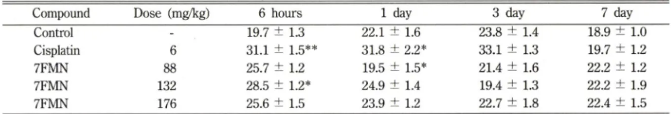
50value, cisplatin; 7 times) were determined by autochemistry analyzer In cisplatin- administered mice group, BUN level was elevated than normal control group at 3rd day and repaired at 7th day.
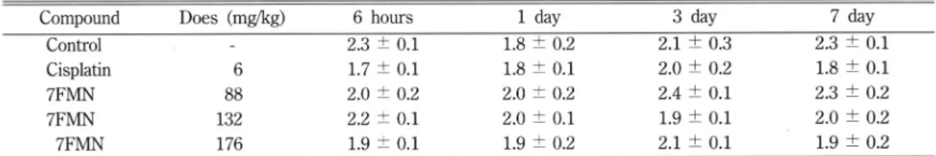
In 7FMN administrated group was not elevated. Creatinine and uric acid level were no difference with the nor
mal control group. Therefore synthesized 7FMN is less toxic than cisplatin in nephrotoxicity.
Keywords □ Flavin mononucleotide (1,4-butanediamine) Pt(II) complex, anticancer activity, flow cytom
etry; nephrotoxicity.
세포주기에 대한 이해가 점차로 발전함에 따라 항암 지만 각각의 단계에서 소비되는 시간은 다밍한데 일단 제와 세포주기와의 관계룰 새로이 조명해 보고 세포주 세포분열이 시작되면 새로이 시작되거나 멈춤이 없이 기 조절인자에 대한 연구가 최근 매우 활발히 진행되
고 있 다 . 모 든 세포는 기본적인 세포주기률 갖고 있
후본 논문에 관한 문의는 이 저■ 자식
1게로 (전화) 02-760-1838 (팩스) 02-762-5178
계속 자라고 계속 분열된다 . 암세포는 여러가지 면에
서 정상세포와 다른데 그 중에서 가장 근본적인 처의
는 암세포의 세포분열이 조절되지 않는다는데 있다 . 항
암제에는 세포주기중 특정 phase 에 있는 세포에만 작
용하는 cell cycle(phase)-specific drug(CCPS )이 있
150 권영이 • 황규자 • 김인근 • 김국환 • 김원규 • 안동춘
NHa ^ Cl /
관\
N H 3 C I
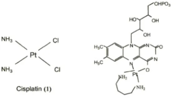
C isplatin (1)
F M N ( 1 ,4 - b u t a n e d ia m in e ) P t ( ! l) c o m p le x (2 )
Fig. 1 - Structures of Cisplatin (1) and 7FMN (2).
위하여는 정상 세포에 대한 득성이 적어야 한다.
Cisplatin은 이미 잘 알려진 바와 같이 다른 항암제로 치료가 불7}능한 고형암이나 전럽선암등에도 치료효과 롤 나타낸 우수한 함우>제 이 나 심 각 한 득성, 특히 신장득성이 가장 큰 문제가 되고었어 투여시 많은 제한을 받고있는 실정이다. 따라서 저자등이 신장득성 이 적을 것으로 에상되는 분자를 설계하여 합성한 7FMN에 대한 신장득성을 혈청생화학적인 방법으로 시스폴라틴과 비교, 연구하였다.
고, 세포주기에 관계없이 작용하는 cell cycle (phase)- nonspecific drug (CCPNS)이 있다. 모든 진핵세포들은 각각의 세포주기(cell cycle)가 다른 비복제 상태 (nonreplicating state)인 G0/G1 phase와 DNA합성기인 S phase, 그리고 분열기인 G2/M phase로 구성되어 었으며 각 단계마다 일정한 세포내 DNA content를 함유하고 있다. 따라서 암세포에 함"y-제룰 f 여한후 세 포주기의 번화가 어떻게 일어나는 지를 밝혀내고 이 정 보를 토대로 하여, 새로운 세포주기 조절인자률 발견하 여 여기에 적용할 수 있다면 이률 이용한 암의 정복이 가능할 것으로 보인다. 현재 임상쉬1서 우수한 항암제로 사용되고 있는 cisplatin (1) (Fig. 1)의 경우 암세포마다 다른 경향을 나타내고 있다고 보고된 논문이 었고,^
G2 arrest를 나타낸다고 주장하는 논문도 있다.패> 함 제가 세포주기의 어느 단계의 DNA와 결합하여 세포증 식의 억제를 나타내는지에 대한 기전을 밝히는 것은 매 우 중요한 의머틀 가지는데 그것은 세포사멸(apoptosis) 과 직접적인 연관을 가지기 때문미다.®> 이에 저자등어 합성한 leaving ligand로 flavin mononucleotide(FMN) 를, carrier ligand로 1,4-bu-tanediamine을 도입하여 합성한 백금착체인 FMN(1,4-butanediamine) Pt(II) complex(이하 7FMN) (2) (Fig. 1)의 경우 cisplatin보 다 항암활성은 떨어지지만 수용성이 매우 좋고, 현재 임상에서 우수한 항암제로 쓰이고 있는 carboplatin의 ICjo는 일반적으로 시스폴라틴의 10배정도이나 7FMN 의 ICjo는 시스폴라틴의 약 6배로 상당히 유의성었는 항암칠성을 보여주었으므로*®^ 인체에 적용시 유리하고 득성이 적을 것으로 추정된다. 따라서 cisplatin과 비교 하여 같은 암세포주에서 세포주기와 어떤 상관관계가 있는지 그 작용기전을 밝히기 위하여 유세포분석법 (flow cytometry)을 이용하여 연구하였다.
또한 합성된 함 •성 화합물이 신약후보물질이 되기
실험방법
Flow cytometry 에 의한 세포주기 특어성 분석 시약, 기구 및 기기 - 사 M t 암세포주는 부유세포로 서 생쥐의 백혈병 세포인 L1210 세포주이며 한국세포 주은행에서 냉동세포를 분양받아 배양하여 시용하였다.
RPMI 1640배지, penidllin-streptomydne 1000 unit/
m/, fetal bovin serum(FBS), phosphate buffer saline (PBS)는 Gibco에서, cisplatin, RNase와 propium iodide는 Sigma에서 구입하였다. CO2 가스는 동아가스 사에서 구입하식 사용하였고, 그외에 사용된 시약은 세 포배<^0당 또는 특급시약을 시용하였다. 세포수 계수는 hematocytometer를 이용하여 광학현미경 (Olympus CK2)으로 관찰하여 실시하였다. 5 m/ flow cytome- try용 tube는 Falcon #2052률 사용하였고, 세포주기 특이성에 대한 DNA content의 분석은 서울대학교 의 과대 학 임상의학연구소에서 FACSCalibur(Bectone &
Dickinson, USA)를 이용하여 유세포 분석법으로 실시 하였다.
배앙 및 유세포분석 -37°C, 5% CO2 존재하에 L1210 2X10^ 개/m/인 세포주 6 m/롤 T25-flask에 취하고 cisplatin과 합성된 백금착체인 7FMN에 대한 세포주기 특이성을 분석하기 위하쉬 각각 세포의 증식 이 약 40%와 약 80% 억제되는 농도로 처러하였다.
(Table I) 대조군 세포에는 배지만 6 ml 가하여 주었
다. 48시간 배양후 1200 rpm, 10분 원심분리하여 배
지를 제거하고 PBS용액 3 m/률 가하여 세포룰 세척
하였다. 5 ml flow cytometry 용 tube에 세포률 1
X 10® 개 취§1여 PBS로 2회 세척하였다. 세포를 고정
하기 위하쉬 70% 에틴을 1 m/률 서서히 가하고 4°C
에서 24시간 방치하였다. 원심분리하여 에탄을을 제거
하고 냉각시킨 PBS-2 mM EDTA용액으로 세포률 2
7FMN 파 cisplatin 의 유세포 ^ 및 신장득성 151
Table I - Serum levels of BUN in mice after a single i.p. administration of cisplatin and 7FMN
Compound Dose (mg/kg) 6 hours 1 day 3 day 7 day
Control
-19.7 ± 1.3 22.1 ± 1.6 23.8 ± 1.4 18.9 ± 1.0
Cisplatin 6 31.1 ± 1.5** 31.8 ± 2.2* 33.1 ± 1.3 19.7 ± 1.2
7FMN 88 25.7 ± 1.2 19.5 ± 1.5* 21.4 ± 1.6 22.2 ± 1.2
7FMN 132 28.5 ± 1.2* 24.9 ± 1.4 19.4 ± 1.3 22.2 ± 1.9
7FMN 176 25.6 ± 1.5 23.9 ± 1.2 22.7 ± 1.8 22.4 ± 1.5
Control group were administered 6.6 mi/kg of PBS by i.p., Each value represents the means ± S.E. of 5-6 IC R mice male Each unit is mg/dL, Significantly different from control: *P<0.05, **P<0.001
회 세척후 PBS-2 mM EDTA용액 800 |j/률 가하였 다. RNased mg/m/) 용액을 100 tj/가하고 상온에서 30분 방치후 형광물질을 DNA에 도입하기 위하식 PI (400 ^1£/111/)용액 100 |i/가하여 유세포분석 (flow cytometry)를 실시하였다.
신장득성에 대한 생화학적 분석
실험동물-실험동물은 체중 약 25 g내외의 수컷 ICR계 생쥐룰 대한실험동물센터에서 구입하여 사용하 였다. 실험 기간중 동물은 한잉=대학교 의과대학 해부 학교실내 청정동물실에서 사육하였으며 사료(삼양사) 와 물은 무제한 공급하였다. 실험동물은 대조군과 cisplatin 투여군, 7FMN 투여 3군으로 분류하고, 각군 에 대하여 20- 22마러씩 배정하였으며, 이것을 다시 6 시간, 24시간, 3일, 7일 처리군으로 나누어 한 군에 각각 5~6마리씩 약물처러하였다.
실험동몰의 처치 - 체중 1 kg당 투여한 약물용량은 cisplatin 6 mgCICjo 7), 7FMN 88 mg(IC5o 8), 132 mgdCjoX 12), 176 mgdCjoX 16)으로 하였다. 각 각 해당량만큼 취하식 7FMN은 PBS를 가하여 6.6 ml로, cisplatin은 DMSO 200 |a/에 녹인후 PBS를 가하여 6.6 m/로 하였다. DMSO의 함량은 마우스 1 마리당(30 g) 적용될수 있는 20 \il 보다 훨씬 적은 양으로*®> 실험에 꺼치는 영향이 없도록 하였다. PBS 에 녹인 약물들을 0.2 |im 주사기용 필터로 여과하식 멸균하식 복강내 주사하고 주사일을 0일로하여 6^1간, 1일, 3일 및 7일 경과후 5~6마리썩 심장채혈 후 경 추탈구법으로 희생시켰다.
BUN, creatinine, uric acid의 측정 一Cisplatin과 7FMN이 투여된 생쥐듬로부터 얻은 혈액을 4,500 rpm, 15분 원심분리하여 혈청만 취하여 BUN, creatinine, uric add를 죽정시까지 -2(fC 이하에서 보관하였다. BUN, creatinine, uric acid는 생화학 자
동분석기 (Olympus reply, Japan)로 즉정하였으며 각 동물의 평균치와 표준편차로 나타내었다.
실 험 결 과 및 고 찰
Flow cytometry에 의한 세포주기 특이성 분석 - L1210 세포주에 cisplatin파 합성된 7FMN을 세포의 증식이 약 40%억제되는 농도인 IC40는 cisplatin이 1.5 |iM/m/, 7FMN은 10 이며, 80%억제되는 농도인 ICgo는 cisplatin 5 \iMJml, 7FMN이 30 jiM/
m/이다. 이들 농도로 각각 처리하여 배양한 후 이들 각 세포주기 단계에 있는 세포들을 측정하기 위하쉬 DNA 형광명색 시약인 propidium iodide(PI)로 염색하 였다. PI는 세포내 DNA content에 따라 결합하므로 유세포분석기 (flow cytometry)로 즉정하여 발산되는 형광강도에 따라 DNA 영역을 구분하였다. 약물처러하 지 않은 L1210 세포주 대조군의 DNA content는 Diploid률 100%로 보았을 때 G0/G1 phase의 세포가 33.56%, G2/M phase의 세포가 15.26%, S phase의 세포가 51.18%였다(Fig. 2). Cisplatin (1)을 처리한 세포주에서 세포증식이 40%정도 억제될 때부터 G2
D iplo id 100%
G O /G l 33.56%
G 2/M 15.26%
S 51.18%
0 200 400 600 800 1000
FL2-A 800
C ontrol C ells
Fig. 2 - DNA content of cellcyde analysis of L1210
control cells.
152 권영이 ■ 황규자 • 김인근 • 김국환 • 김원규 • 안동춘
g
S
FL2-A FL2-A
C is p la tin - T r e a te d C e lls
slunoo
wl mu o o